. 2014 Nov 14;289(46):31905-31913.
doi: 10.1074/jbc.M114.599662. Epub 2014 Sep 17.

Crystal structure of a lipoxygenase in complex with substrate: the arachidonic acid-binding site of 8R-lipoxygenase

Affiliations

Crystal structure of a lipoxygenase in complex with substrate: the arachidonic acid-binding site of 8R-lipoxygenase

David B Neau et al. J Biol Chem. .

Abstract

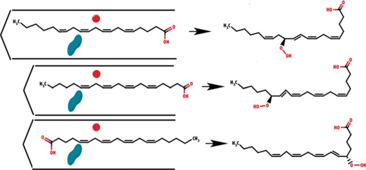
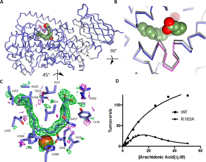
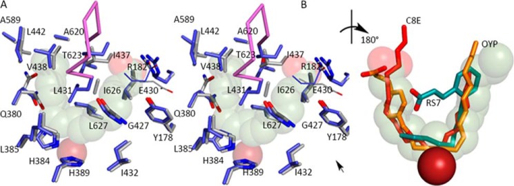
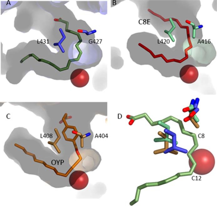
Lipoxygenases (LOX) play critical roles in mammalian biology in the generation of potent lipid mediators of the inflammatory response; consequently, they are targets for the development of isoform-specific inhibitors. The regio- and stereo-specificity of the oxygenation of polyunsaturated fatty acids by the enzymes is understood in terms of the chemistry, but structural observation of the enzyme-substrate interactions is lacking. Although several LOX crystal structures are available, heretofore the rapid oxygenation of bound substrate has precluded capture of the enzyme-substrate complex, leaving a gap between chemical and structural insights. In this report, we describe the 2.0 Å resolution structure of 8R-LOX in complex with arachidonic acid obtained under anaerobic conditions. Subtle rearrangements, primarily in the side chains of three amino acids, allow binding of arachidonic acid in a catalytically competent conformation. Accompanying experimental work supports a model in which both substrate tethering and cavity depth contribute to positioning the appropriate carbon at the catalytic machinery.

Keywords: Arachidonic Acid (AA) (ARA); Eicosanoid Biosynthesis; Lipid Signaling; Lipoxygenase Pathway; Protein Structure; X-ray Crystallography.


Figures

FIGURE 1.
FIGURE 1.
Schematic of the current model for product specificity in lipoxygenases. Cavity depth and substrate orientation combine to confer regio- and stereo- specificity. The substrate (AA shown here) must align between the catalytic iron (red sphere) and O2 channel (blue peanut). Differences in cavity depth and head-to-tail orientation determine the hydroperoxyeicosatetraenoic acid generated.
FIGURE 2.
FIGURE 2.
8R-LOX with AA. A, ribbon drawings of 8R-LOX with (blue) and without (gray) AA in the active site. Fe2+ is shown as a red sphere. The helical insertion described in all LOX structures is shown in violet. B, detail of the outward shift of the arched helix observed in the presence of AA. C, an omit map contoured +2/−2σ (green/magenta) reveals clear electron density for AA. D, the steady state rate dependence on substrate concentration of 8R-LOX and the mutant R182A. 8R-LOX data were fit to the Michaelis-Menten equation, whereas the R182A data were fit to Equation 1. Equation 1 describes cooperative substrate inhibition for the fit, n = 2.4 ± 0.3.
FIGURE 3.
FIGURE 3.
The AA-binding site. A, the substrate adopts a horseshoe shape in the U-shaped channel (stereo). The side chains of highly conserved amino acids line the base of the active site, along with Gly-427. Glu-430, part of an inter-helical charge cluster that includes the substrate carboxylate, is shown in line rendering. The Fe2+ (transparent dark red sphere) is positioned behind the substrate. B, detail of the superposition of inhibitors (rotated ∼180° with respect to A) observed in 15-LOX-2 (red) 12-LOX (gold, C; red, O) and 15-LOX-1 (teal, C; red, O). The 15-LOX-2 and 12-LOX inhibitors conform to the AA placement, whereas the 15-LOX-1 inhibitor overlaps partially. The Fe2+, solid dark red sphere, is in front of the substrate.
FIGURE 4.
FIGURE 4.
The antarafacial relationship between Fe2+ and a putative O2 access channel. A, in 8R-LOX the direction of the channel is set by Gly-427 and the orientation of the shielding amino acid Leu-431. B and C, similar channels in 15-LOX-2 and 12-LOX. Note the presence of an Ala pushes the tubular opening deeper into the body of the enzyme. D, detail of the relationship between the Gly/Ala switch and the Leu-431 shielding residues. Note the absence of the side chain in the 8R-enzyme (blue) and positioning of Leu-431 leave C8 (C-2) of the AA unprotected. Blue, 8R-LOX; green, 15-LOX-2; beige, 12-LOX. In contrast, in the 15-LOX-2 and 12-LOX enzymes, the same carbon would be shielded by the Ala and the invariant Leu no longer shields C12 (C+2) at the opposite end of the pentadiene.

References

    1. Yamamoto S. (1992) Mammalian lipoxygenases: molecular structures and functions. Biochim. Biophys. Acta 1128, 117–131 - PubMed
    1. Brash A. R. (1999) Lipoxygenases: occurrence, functions, catalysis, and acquisition of substrate. J. Biol. Chem. 274, 23679–23682 - PubMed
    1. Ivanov I., Heydeck D., Hofheinz K., Roffeis J., O'Donnell V. B., Kuhn H., Walther M. (2010) Molecular enzymology of lipoxygenases. Arch. Biochem. Biophys. 503, 161–174 - PubMed
    1. Schneider C., Pratt D. A., Porter N. A., Brash A. R. (2007) Control of oxygenation in lipoxygenase and cyclooxygenase catalysis. Chem. Biol. 14, 473–488 - PMC - PubMed
    1. Andreou A., Feussner I. (2009) Lipoxygenases: structure and reaction mechanism. Phytochemistry 70, 1504–1510 - PubMed

Publication types

Associated data

LinkOut - more resources