. 2014 Oct 31;289(44):30511-30524.
doi: 10.1074/jbc.M114.605527. Epub 2014 Sep 17.

Translational roles of elongation factor 2 protein lysine methylation

Affiliations

Translational roles of elongation factor 2 protein lysine methylation

Maria C Dzialo et al. J Biol Chem. .

Abstract

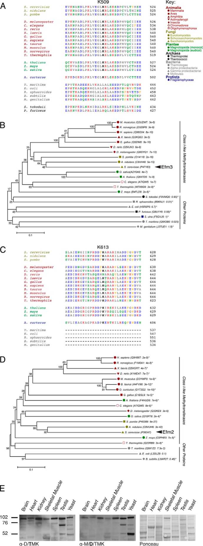
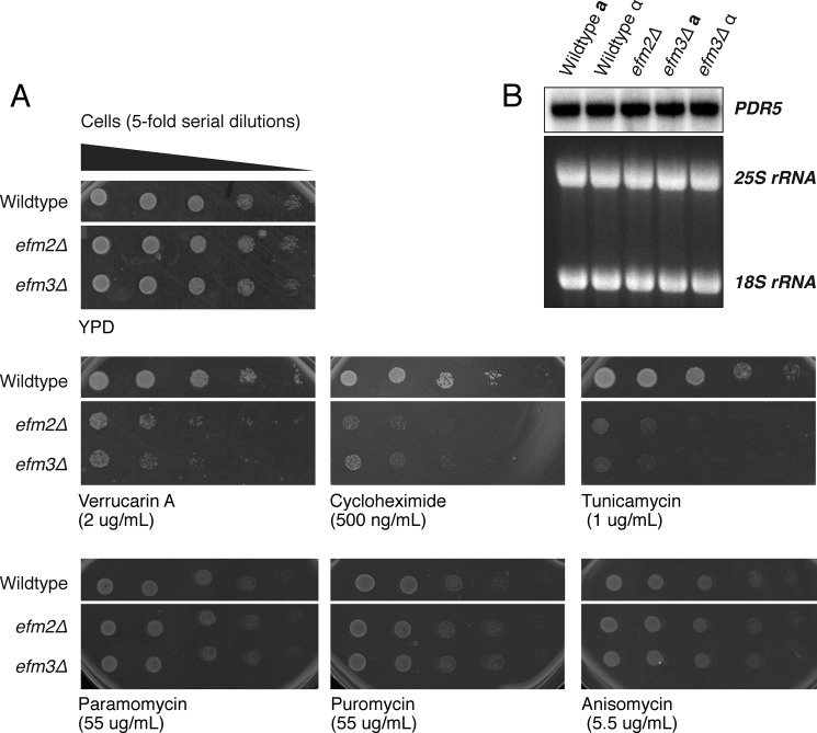
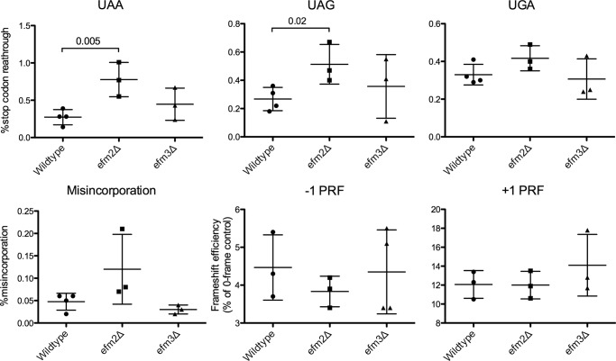
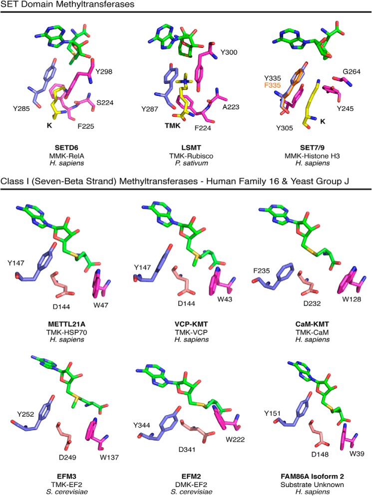
Methylation of various components of the translational machinery has been shown to globally affect protein synthesis. Little is currently known about the role of lysine methylation on elongation factors. Here we show that in Saccharomyces cerevisiae, the product of the EFM3/YJR129C gene is responsible for the trimethylation of lysine 509 on elongation factor 2. Deletion of EFM3 or of the previously described EFM2 increases sensitivity to antibiotics that target translation and decreases translational fidelity. Furthermore, the amino acid sequences of Efm3 and Efm2, as well as their respective methylation sites on EF2, are conserved in other eukaryotes. These results suggest the importance of lysine methylation modification of EF2 in fine tuning the translational apparatus.

Keywords: Lysine Methylation; Protein Methylation; Protein Synthesis; Ribosome Function; S-Adenosylmethionine (SAM); Translation Elongation Factor.


Figures

FIGURE 1.
FIGURE 1.
Deletion of EFM3/YJR129C results in loss of trimethylated lysines in 100-kDa polypeptides in S. cerevisiae. Immunoblots were performed on whole cell lysates as described under “Experimental Procedures” with various antibodies directed against methylated lysine residues. The positions of molecular weight markers are shown on the left. A, polypeptides from lysates of strains with deletions of candidate methyltransferase genes were probed with anti-di-/trimethyllysine (Upstate Biotechnology, 07-756). Arrows indicate the distinct signals at 100 and 50 kDa, correlating to EF2/EF3 and EF1A, respectively. B, polypeptides from known and putative EFM knock-out strains were immunoblotted with multiple methyllysine antibodies. The same membrane was probed, stripped, and reprobed with anti-di-/trimethyllysine (left; Upstate Biotechnology, 07-756), anti-dimethyllysine (middle left; Abcam, Ab7315), or anti-trimethyllysine (middle right; Immunechem, ICP0602). The Ponceau-stained membrane is shown on the right as a protein loading control. The type of methyllysine recognized by each antibody is indicated below each blot with the boldface and underlined letter representing the strongest specificity. C, comparison of wild type and EFM knock-out strains, including efm3Δ, in both mating type backgrounds. All samples were run on the same gel; spaces indicate where non-relevant lanes were removed.
FIGURE 2.
FIGURE 2.
Deletion of EFM2 results in loss of dimethylated lysine, and deletion of EFM3 results in loss of trimethylated lysine in 100-kDa polypeptides. Lysates from in vivo radiolabeled cells were fractionated by SDS-PAGE, and the 100 kDa gel region was excised. Gel slices were acid-hydrolyzed as described under “Experimental Procedures.” The resulting hydrolysates were loaded onto a high resolution cation exchange column with standards of methylated lysine derivatives. A, using a pH 3.8 elution buffer, radiolabeled methylated lysine derivatives were separated. The position of the standards, detected by ninhydrin reactivity, is shown by the dashed line. Due to a tritium isotope effect, the radiolabeled derivatives elute slightly before the non-labeled standards (58). Each trace is representative of three independent experiments. B, separation of hydrolysates from radiolabeled wild type and efm3Δ cells in both mating type backgrounds. A pH 4.5 elution buffer was used. The ninhydrin profiles are shown for methylated standards run in a separate experiment due to interference from the large amounts of ammonium ion present in the gel hydrolysates. Each trace is representative of two independent experiments. C, the amount of di- and trimethyllysine radioactivity as a percentage of the total radioactivity in the hydrolysate is shown, with error bars reflecting the S.D. p values from Student's t test are shown when less than 0.05.
FIGURE 3.
FIGURE 3.
Deletion of EFM3 results in loss of trimethyllysine 509 on elongation factor 2. The 100-kDa protein bands from wild type and knock-out lysates were excised, in-gel trypsin-digested, and analyzed by LC-MS/MS. A Mascot search identified EF2 as the top hit (score = 13,763.99) for the 100 kDa band. Known trimethylation at Lys-509 (TMK509) (A) and dimethylation at Lys-613 (DMK613) (B) were identified in EF2 peptides LVEGLKR and DDFKAR, respectively, in wild type lysate. Dimethylation at Lys-509 (DMK509) and monomethylation at Lys-613 (MMK613) were also observed in EF2 from wild type lysate. C, the effect of efm2Δ and efm3Δ on the methylation of Lys-509 and Lys-613 was examined by comparison of EF2 from wild-type lysate with that from knock-out lysates. PRM-MS with extracted precursor → product ion transitions (within 10 ppm) was used to identify methylated peptides in each sample condition (WT, efm2Δ, and efm3Δ).
FIGURE 4.
FIGURE 4.
Conservation of EF2 methylation sites and methyltransferases among six kingdoms of life. The region corresponding to the Lys-509 trimethylation site (A) and the Lys-613 dimethylation site (C) in S. cerevisiae is shown for representative organisms. The methylated lysine residue is shown in boldface type. Aliphatic residues are shown in gray, acidic residues in blue, basic residues in red, polar residues in green, and aromatic residues in orange. In C, Archaea were removed from alignment due to significant sequence differences. B and D, phylogenetic trees depicting evolutionary conservation of Efm3 (B) and Efm2 (D). The UniProt ID of the top ranking alignment for each organism is indicated, along with their respective E values. The percentage of replicate trees in which the associated taxa clustered together in the bootstrap test (500 replicates) is shown beside the branches. The evolutionary distances are in units of the number of amino acid differences per site. The tree is drawn to scale, with branch lengths in the same units as those of the evolutionary distances used to infer the phylogenetic tree. All proteins were mutual best hits except for those indicated with an asterisk. In B, Amphidinium carterae was removed because no significant homolog to Efm3 was found. Synedra ulna was used as a representative Protista but had no homolog for EF2. In D, Protista, Archaea, Rhodobacter sphaeroides, and Mycoplasma genitalium were completely removed due to no significant homology to Efm2. E, polypeptides from cytosolic extracts from various mouse tissues were probed by immunoblotting with anti-di-/trimethyllysine antibodies (left; Upstate Biotechnology, 07-756). The membrane was then stripped and reprobed with anti-dimethyllysine (middle; Abcam, ab7315). The membrane was Ponceau-stained to ensure equal loading (right).
FIGURE 5.
FIGURE 5.
efm2Δ and efm3Δ cells have altered sensitivity to translational inhibitors. A, wild type, efm2Δ and efm3Δ cells were cultured to an A600 of 0.5 and 5-fold serial diluted in water. 3 μl from each dilution was spotted onto a YPD agar plate with or without inhibitors at the indicated concentrations. Images shown are representative of at least three individual replicates. Cells shown are on the same antibiotic plate with other tested strains; only strains relevant to this study are displayed. YPD without inhibitor ensures equal cell loading between strains. The number of days each plate was incubated is indicated below each panel. B, a Northern blot against the multidrug transporter Pdr5 transcript was performed as described (8). The 25 and 18 S ribosomal RNAs are shown as loading controls.
FIGURE 6.
FIGURE 6.
efm2Δ but not efm3Δ cells have increased stop codon read-through. Dual luciferase assays were utilized to measure the percentage of stop codon read-through, amino acid misincorporation, and frameshifting. Titles of each panel indicate the stop codon analyzed, the misincorporation of lysine, or the direction of programmed ribosomal frameshift (PRF). Values for three or four replicates with S.D. are shown as error bars. p values are displayed where differences were less than 0.05.
FIGURE 7.
FIGURE 7.
Similarity in the modeled catalytic centers of Efm2 and Efm3 with SET domain lysine methyltransferases and human Family 16 Class I methyltransferases. Top, the SET domain enzymes, SETD6 (PDB entry 3QXY), LSMT (PDB entry 2H2J), and SET7/9 (PDB entries 3M53 (wild type) and 4J71 (Y335F)) have a catalytically required tyrosine residue (blue) that is positioned between the methyl group and adenine ring of AdoMet (green). Alignment of SET7/9 Y335F showed that the phenylalanine (orange) is positioned almost identically to the wild type tyrosine. Residues implicated in substrate binding are shown in pink (41, 47). The substrate-binding tyrosine (pink) and a nearby side chain carbonyl help balance the full negative charge of the substrate lysine (yellow). The aromatic plane of the tyrosine residue is ∼4.4, 4.2, and 3.5 Å away (in SETD6, LSMT, and SET7/9, respectively) from the substrate lysine ϵ-nitrogen atom, suggesting a cation-π interaction (49). Bottom panels, the catalytic sites on crystal structures of the Class I human METTL21A (PDB entry 4LEC), VCP-KMT (PDB entry 4LG1), and CaM-KMT (PDB entry 4PWY) methyltransferases as well as those of the Phyre2 models of S. cerevisiae Efm2 and Efm3 and human FAM86A are displayed in a similar orientation as the three SET domain enzymes shown above. The Class I enzymes have a tyrosine or phenylalanine residue (blue) oriented similarly as the catalytic tyrosine in the SET enzymes. The aspartate residue (light pink), previously shown to be catalytically required in VCP-KMT (38), could serve a similar purpose as the tyrosine-carbonyl pairings in the SET domain enzymes in stabilizing the substrate lysine. A conserved tryptophan residue (pink) in the Class I enzymes is well suited to form cation-π interactions in a manner similar to the phenylalanine residue in the SET domain methyltransferases.

Similar articles

Cited by

References

    1. Graille M., Figaro S., Kervestin S., Buckingham R. H., Liger D., Heurgué-Hamard V. (2012) Methylation of class I translation termination factors: structural and functional aspects. Biochimie 94, 1533–1543 - PubMed
    1. Jackman J. E., Alfonzo J. D. (2013) Transfer RNA modifications: nature's combinatorial chemistry playground. Wiley Interdiscip. Rev. RNA 4, 35–48 - PMC - PubMed
    1. Liu J., Jia G. (2014) Methylation modifications in eukaryotic messenger RNA. J. Genet. Genomics 41, 21–33 - PubMed
    1. Motorin Y., Helm M. (2011) RNA nucleotide methylation. Wiley Interdiscip. Rev. RNA 2, 611–631 - PubMed
    1. Polevoda B., Sherman F. (2007) Methylation of proteins involved in translation. Mol. Microbiol. 65, 590–606 - PubMed

Publication types

MeSH terms

Substances