. 2014 Nov 15;25(23):3779-97.
doi: 10.1091/mbc.E13-10-0604. Epub 2014 Sep 17.

TBC1D9B functions as a GTPase-activating protein for Rab11a in polarized MDCK cells

Affiliations

TBC1D9B functions as a GTPase-activating protein for Rab11a in polarized MDCK cells

Luciana I Gallo et al. Mol Biol Cell. .

Abstract

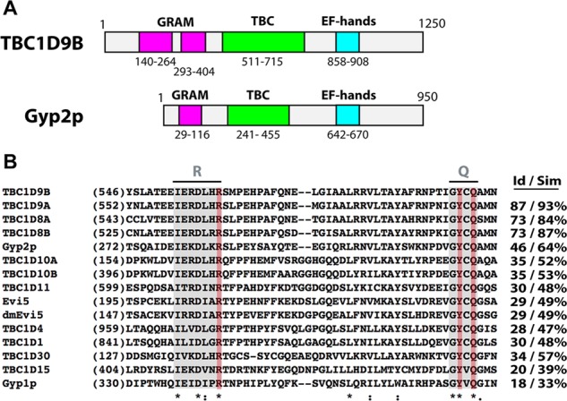
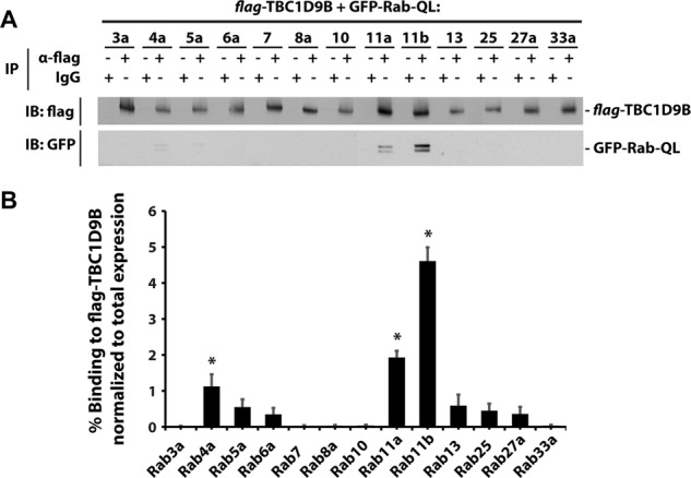
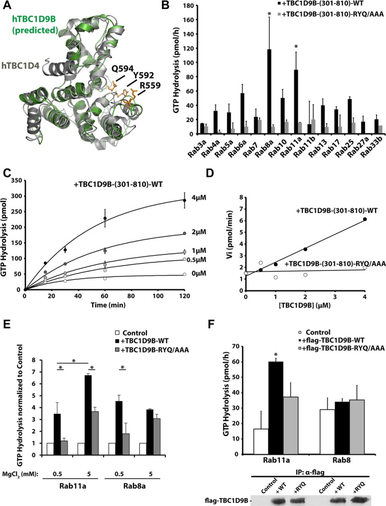
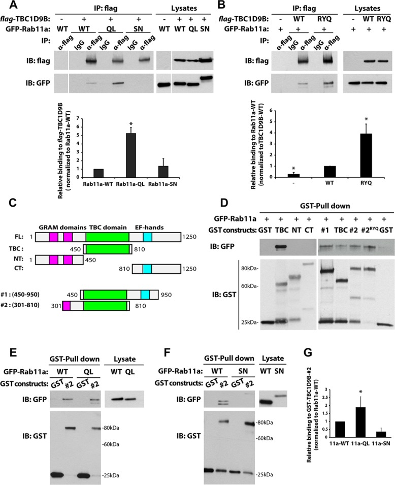
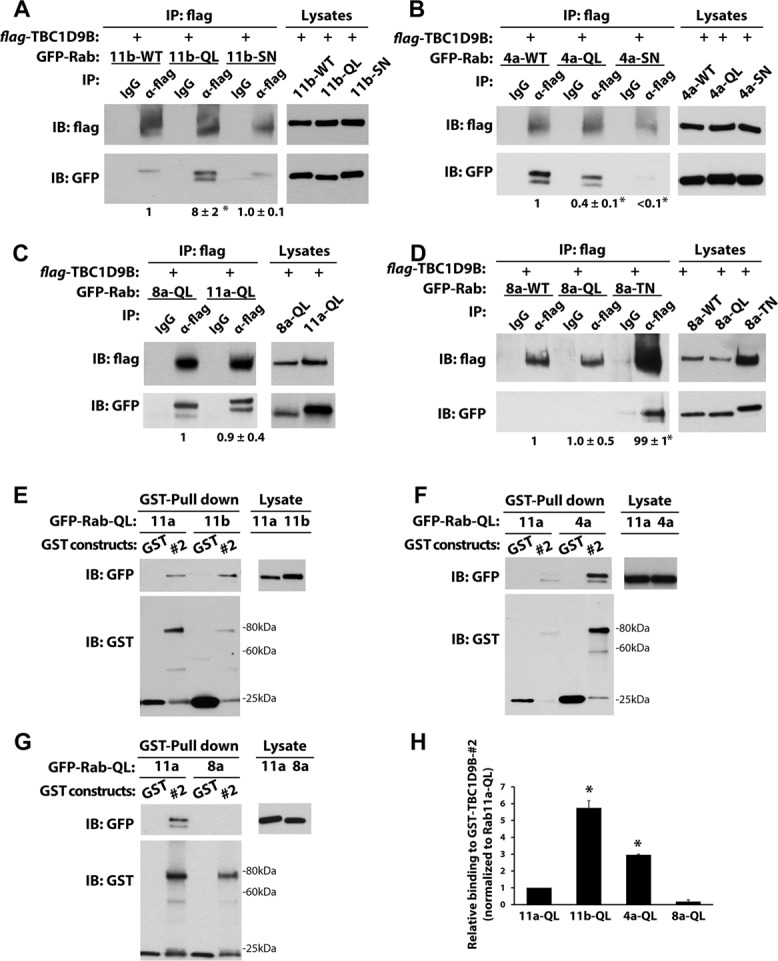
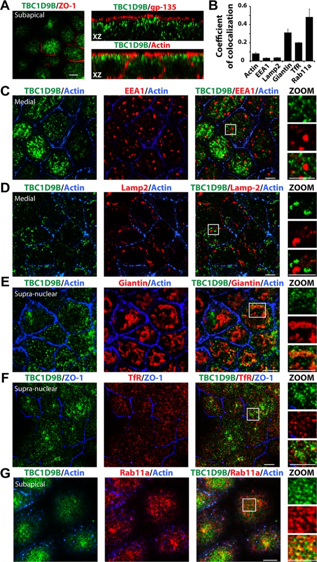
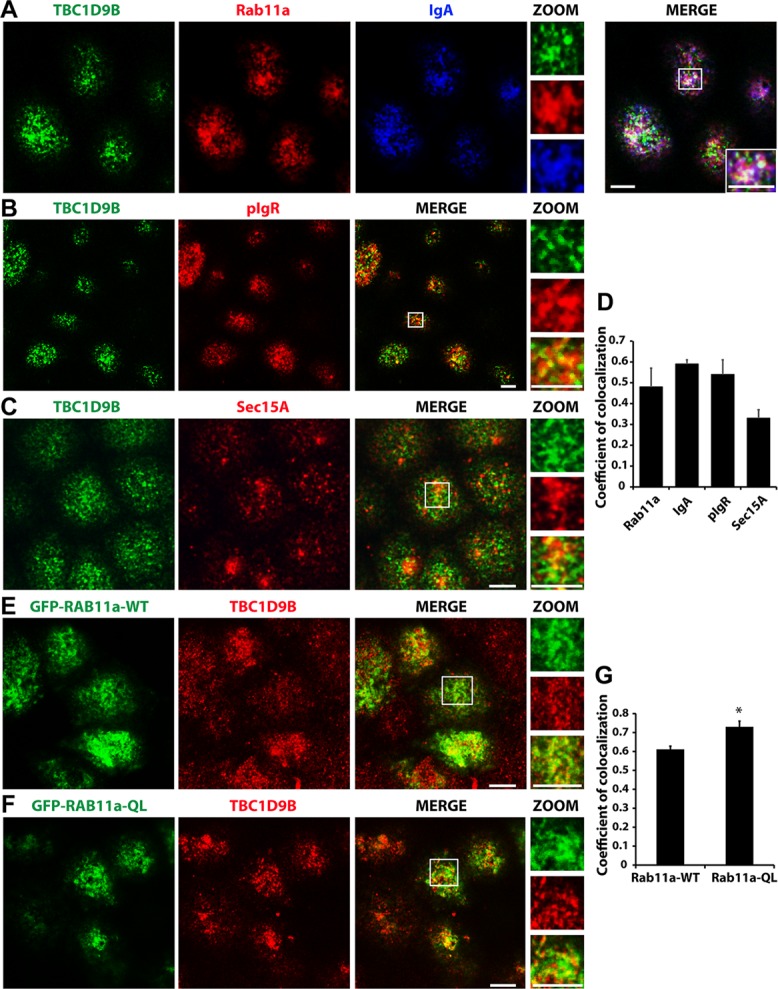
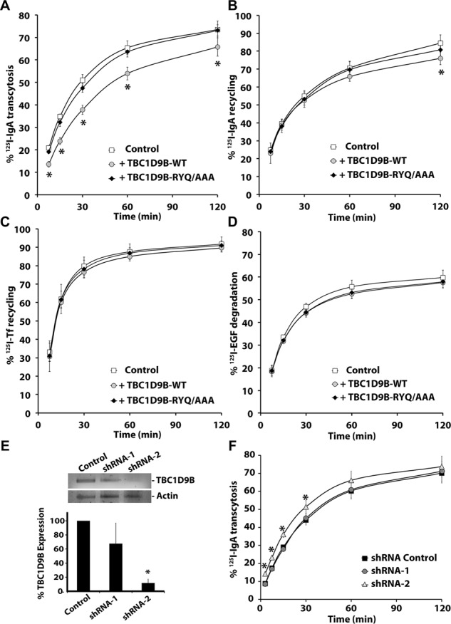
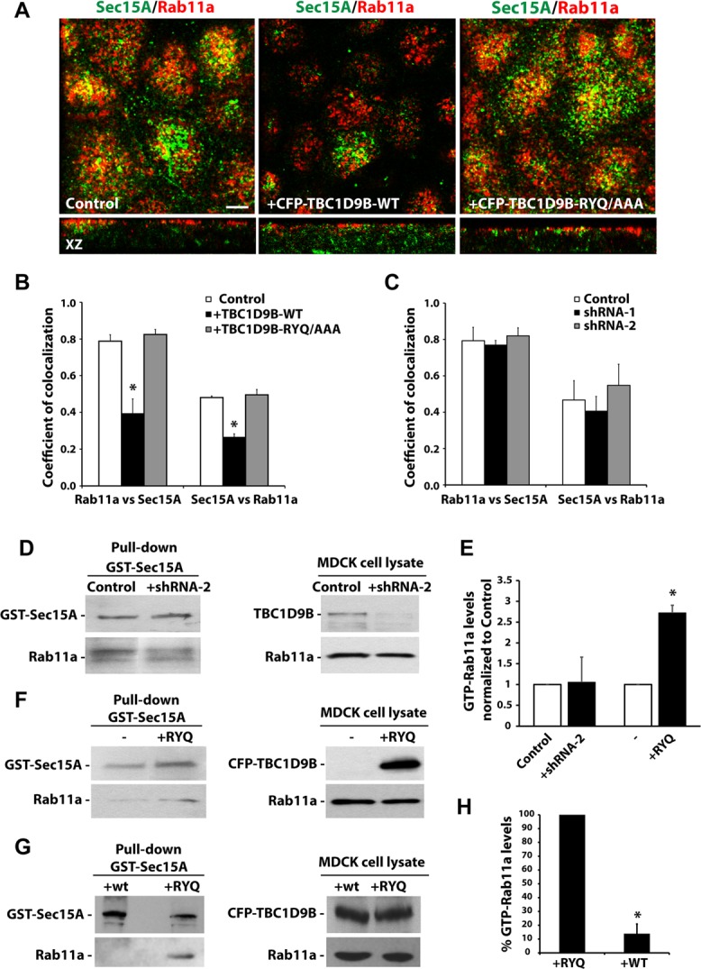
Rab11a is a key modulator of vesicular trafficking processes, but there is limited information about the guanine nucleotide-exchange factors and GTPase-activating proteins (GAPs) that regulate its GTP-GDP cycle. We observed that in the presence of Mg(2+) (2.5 mM), TBC1D9B interacted via its Tre2-Bub2-Cdc16 (TBC) domain with Rab11a, Rab11b, and Rab4a in a nucleotide-dependent manner. However, only Rab11a was a substrate for TBC1D9B-stimulated GTP hydrolysis. At limiting Mg(2+) concentrations (<0.5 mM), Rab8a was an additional substrate for this GAP. In polarized Madin-Darby canine kidney cells, endogenous TBC1D9B colocalized with Rab11a-positive recycling endosomes but less so with EEA1-positive early endosomes, transferrin-positive recycling endosomes, or late endosomes. Overexpression of TBC1D9B, but not an inactive mutant, decreased the rate of basolateral-to-apical IgA transcytosis--a Rab11a-dependent pathway--and shRNA-mediated depletion of TBC1D9B increased the rate of this process. In contrast, TBC1D9B had no effect on two Rab11a-independent pathways--basolateral recycling of the transferrin receptor or degradation of the epidermal growth factor receptor. Finally, expression of TBC1D9B decreased the amount of active Rab11a in the cell and concomitantly disrupted the interaction between Rab11a and its effector, Sec15A. We conclude that TBC1D9B is a Rab11a GAP that regulates basolateral-to-apical transcytosis in polarized MDCK cells.


Figures

FIGURE 1:
FIGURE 1:
Domain structure of TBC1D9B and sequence comparisons of its TBC domain with other TBC domain–containing proteins. (A) Schematic of human TBC1D9B and Saccharomyces cerevisiae Gyp2p domain structure. Predicted GRAM domains are colored pink, TBC domains are colored green, and EF hands are colored light blue. (B) Alignment of the indicated region of the TBC domains using MAFFT software (Larkin et al., 2007; Katoh and Standley, 2013). Sequences including the arginine (R) and glutamine (Q) fingers are shaded gray; key amino acids in the R and Q fingers are shaded red. Identical residues are indicated with an asterisk, conserved amino acid substitutions by a colon, and semiconserved substitutions by a period. The percentage of identity (Id) or similarity (Sim) between the TBC domain of TBC1D9B and those of the indicated proteins is given in the right-hand columns.
FIGURE 2:
FIGURE 2:
TBC1D9B interacts with Rab GTPases. (A) Flag-tagged TBC1D9B, along with the indicated GFP-Rabs containing an activating Q-to-L mutation, were coexpressed in HEK cells. Flag-TBC1D9B was recovered using an anti-flag tag antibody and coimmunopreciptated GFP-tagged Rab-QL detected using an anti-GFP antibody. IgG was used as a nonspecific control. (B) Quantification of the percentage GFP-Rab-QL coimmunoprecipitated with TBC1D9B normalized to the total amount of GFP-Rab-QL in the lysate. Data were obtained from three independent experiments, and the mean ± SEM is shown. Values significantly different from the group means, assessed by ANOVA, are indicated (*p < 0.05).
FIGURE 3:
FIGURE 3:
TBC1D9B in vitro GAP activity. (A) TBC1D4 (PDB: 3QYB) was used as a template (gray) to model the 3D structure of the TBC1D9B TBC domain (green). (B) GTP hydrolysis by the indicated wild-type, GST-tagged Rab loaded with [γ-32P]GTP and incubated with 2 μM of either wild-type or mutant TBC1D9B-(301-810) for 60 min at 30°C. Data are corrected for reactions lacking the TBC1D9B fragment. Those values significantly different from the group means, assessed by ANOVA, are indicated (*p < 0.05). (C) Kinetics of Rab11a GTP hydrolysis loaded with [γ-32P]GTP and incubated with 0, 0.5, 1, 2, or 4 μM recombinant TBC1D9B-(301-810). (D) Initial rates of Rab11a GTP hydrolysis plotted against the concentration of wild-type or mutant TBC1D9B-(301-810). (E) In vitro GAP assays performed in the presence of Rab11a or Rab8a. In these reactions, Mg2+ mixed at a 1:1 M ratio with GTP, was added at a final concentration of 0.5 mM. Alternatively, the reaction was supplemented with 5 mM MgCl2. Reactions were incubated for 30 min at 30°C. Data were normalized to control reactions in which no TBC1D9B-(301-810) was added. Values for TBC1D9B-(301-810) that were significantly different (p < 0.05) from matched incubations performed in the presence of TBC1D9B-RYQ/AAA (301-810) or between the indicated reactions are identified with an asterisk. (F) Top, full-length flag-TBC1D9B-WT or flag-TBC1D9B-RYQ/AAA was immunoprecipitated from HeLa cell lysates and a GAP assay performed using Rab11a or Rab8a loaded with [γ-32P]GTP and incubated for 60 min at 30°C. Lysates from nontransfected cells were used as controls. Bottom, Western blot of a 5-μl aliquot of the immunoprecipitates used in the in vitro assay was detected using an anti-flag antibody. (B–F) Data were obtained from three or more independent experiments performed in duplicate, and the mean ± SEM is shown. In E and F, values significantly different between TBC1D9B-WT and TBC1D9B-RYQ/AAA are indicated with an asterisk (p < 0.05).
FIGURE 4:
FIGURE 4:
Rab11a binds to the TBC domain of TBC1D9B. (A) Left, coimmunoprecipitation of flag-tagged TBC1D9B with GFP-Rab11a-wild-type (WT), GTP-locked GFP-Rab11aQ70L (QL), or GDP-locked GFP-Rab11aS25N (SN) coexpressed in HEK cells. Anti-flag antibody was used to recover flag-TBC1D9B, and coimmunopreciptated GFP-Rab11a was detected using an anti-GFP antibody. Controls included use of IgG instead of the flag-tag antibody and expression of GFP-Rab11a alone (first lane). Right, 2% of each lysate was resolved by SDS–PAGE and the indicated proteins detected by Western blot. Bottom, quantification of coimmunoprecipitations. Values were normalized to the total expression of each protein first and then to the values obtained in experiments using the Rab11a-WT lysate. (B) HEK cells were cotransfected with GFP-Rab11a and either flag-TBC1D9B-wild-type (WT) or the flag-TBC1D9B-RYQ/AAA mutant (RYQ). Flag-tagged TBC1D9B was recovered by immunoprecipitation, and the amount of GFP-Rab11a was quantified. Control reactions were performed for cells that only expressed GFP-Rab11a, or in some cases IgG was substituted for the anti-flag antibody. Right, 2% of each lysate was resolved by SDS–PAGE and the indicated proteins detected by Western blot. Bottom, amount of coimmunoprecipitated GFP-Rab11a normalized to the amount recovered from the TBC1D9B-WT lysate. (C) Fragments of TBC1D9B used in pull-down studies. (D) Top, GST-TBC1D9B fragments, labeled according to C, were used to affinity capture GFP-Rab11a in HEK cell lysates. Here #2RYQ denotes the use of fragment #2 with the RYQ/AAA mutations. GST alone was used as a control. Bottom, GST constructs were resolved by SDS–PAGE and proteins blotted with anti-GST antibody. (E, F) Top left, GST-TBC1D9B-#2 fragment was used to affinity capture GFP-Rab11aQ70L (QL) or GFP-Rab11aS25N (SN) from HEK cell lysates. Right, 2% of each lysate was resolved by SDS–PAGE and GFP-Rab11a detected by Western blot using anti-GFP antibody. Bottom left, GST constructs were resolved by SDS–PAGE and proteins detected using an anti-GST antibody. (G) Quantification of data from E and F. Values were normalized to total expression of the protein of interest and then to the values obtained for the GFP-Rab11a-WT pull down. For A, B, and G, data are from at least three independent experiments, and the mean ± SEM is shown. Values significantly different from the group means, as assessed by ANOVA, are indicated (*p < 0.05).
FIGURE 5:
FIGURE 5:
TBC1D9B interacts with Rab4a and Rab11b. (A) Left, coimmunoprecipitation of flag-tagged TBC1D9B with GFP-Rab11b-WT (11b-WT), GFP-Rab11b-Q70L (11b-QL), or GDP-locked GFP-Rab11bS25N (11b-SN) coexpressed in HEK cells. Anti-flag antibody was used to immunoprecipitate flag-TBC1D9B, and anti-GFP antibody was used to detect the coimmunoprecipitated GFP-tagged Rab protein. IgG was used as a nonspecific control. Right, 2% of each lysate was resolved by SDS–PAGE and overexpressed proteins detected by Western blot. Values were normalized to the total expression of each protein and then normalized to the amount of Rab11b-WT lysate. (B) Same as in A, but HEK cells coexpressed flag-TBC1D9B and GFP-Rab4a-WT (4a-WT), GFP-Rab4aQ72L (4a-QL), or GFP-Rab4aS27N (4a-SN). (C) Same as A, but the cells expressed Rab8aQ67L (8a-QL) or Rab11aQ70L (11a-QL), and after lysis, the incubations were performed in the absence of MgCl2. (D) Same as in A, but HEK cells coexpressed flag-TBC1D9B with GFP-Rab8a-WT (8a-WT), GFP-Rab8a-Q67L (8a-QL), or GFP-Rab8a-T22N (8a-TN). (E–G) Top left, GST alone or GST-TBC1D9B-(301-810) (#2 fragment) was used to affinity capture GFP-Rab11a-QL and GFP-Rab11b-QL (E), GFP-Rab4a-QL (F). or GFP-Rab8a-QL (G). Right, 2% of each lysate was resolved by SDS–PAGE and GFP-Rab-QL detected by Western blot using anti-GFP antibody. Bottom left, GST constructs were resolved by SDS–PAGE and proteins detected on Western blots using an anti-GST antibody. (H) Quantification of data from E–G. Values are normalized to the total expression of the protein first and then to the values obtained for the GFP-Rab11a-WT pull down. Data were obtained from at least three independent experiments, and the mean ± SEM is shown. Values significantly different from the group means, as assessed by ANOVA, are indicated (*p < 0.05).
FIGURE 6:
FIGURE 6:
Subcellular localization of TBC1D9B in MDCK cells. Filter-grown MDCK cells were immunolabeled to show the distribution of the following endogenous proteins. (A) TBC1D9B and the tight junction–associated protein ZO-1 (xy-section, left), the apical membrane protein gp-135 (xz-section, top right), or actin (xz-section, bottom right). (B) Coefficient of colocalizations for endogenous TBC1D9B and the following markers: (C) TBC1D9B, actin, and EEA1 (an early endosome marker); (D) TBC1D9B, actin, and Lamp2 (a late endosome/lysosome marker); (E) TBC1D9B, actin, and giantin (a Golgi marker); (F) TBC1D9B, ZO-1, and TfR (a marker of the common recycling endosome and basolateral early endosomes); and (G) TBC1D9B, actin, and Rab11a (which is associated with the apical recycling endosomes in these cells). In A and C–G, single optical sections from the region just under the apical membrane (subapical), just above the nucleus (supranuclear), or at the level of the nucleus along the lateral membranes (medial) are shown. Boxed regions are magnified in the images below the word ZOOM. Scale bar, 5 μm. In B, data are mean ± SEM from three experiments (n > 100 cells).
FIGURE 7:
FIGURE 7:
Localization of TBC1D9B at sites of active Rab11a function. (A) Filter-grown MDCK cells were pulse labeled with 200 μg/ml IgA at the basolateral surface for 20 min at 18°C and chased for 20 min at 37°C in the presence of apical Cy5-labeled anti-IgA antibodies. The cells were fixed and then immunolabeled to detect endogenous TBC1D9B (green), endogenous Rab11a (red), and transcytosed IgA (blue). In the merged panel, the boxed region is magnified in the bottom right inset. Magnified views of the individual channels are found in the insets under ZOOM. (B, C) MDCK cells were stained for endogenous TBC1D9B and either the (B) pIgR or (C) endogenous Sec15A. (D) Coefficient of colocalization for endogenous TBC1D9B and the indicated proteins. (E, F) MDCK cells were transfected with cDNA encoding GFP-Rab11a-WT (E) or GFP-Rab11a-QL (F) and after 72 h fixed and then stained for endogenous TBC1D9B. (G) Coefficient of colocalization for TBC1D9B and the indicated Rab11a construct. In (B, C, E, and F), the boxed region is magnified in the panels under the label ZOOM. Scale bar, 5 μm. In D and G, data were obtained from at least three independent experiments, and the means ± SEM are shown (n ≥ 150 cells). In G, a Student's t test was used to show that difference between these two sample groups was significantly different (*p < 0.05).
FIGURE 8:
FIGURE 8:
TBC1D9B regulates basolateral-to-apical transcytosis of IgA. The fraction of basolaterally internalized [125I]IgA that was transcytosed (A), the fraction of apically internalized [125I]IgA that was recycled (B), the fraction of basolaterally internalized [125I]Tf that was recycled (C), or the fraction of basolaterally internalized [125I]EGF that was degraded (D) was evaluated in filter-grown MDCK cells expressing CFP-TBC1D9B-WT or CFP-TBC1D9B-RYQ/AAA. (E) Top, RT-PCR analysis of MDCK cells expressing scrambled shRNA or specific shRNAs (shRNA1/2) that targeted canine TBC1D9B expression. Bottom, data are quantified. (F) Basolateral-to-apical transcytosis of [125I]IgA was measured in MDCK cells expressing either scrambled shRNA (shRNA Control) or shRNA-1/shRNA-2. In A–F, data were obtained from three independent experiments performed in triplicate, and the means ± SD are shown. Values different from the control, assessed by ANOVA, are indicated (*p < 0.05).
FIGURE 9:
FIGURE 9:
TBC1D9B has GAP activity in cellula. (A) Localization of endogenous Rab11a and endogenous Sec15A in cells transduced with adenovirus encoding CFP-TBC1D9B-WT or the inactive mutant (-RYQ/AAA). Top, from the subapical region of the cell; bottom, xz-sections. (B, C) Coefficient of colocalization for the fraction of Rab11a that colocalizes with Sec15A (or vice versa) in MDCK cells transduced with adenovirus encoding CFP-TBC1D9B-WT or -RYQ/AAA (B) or in cells expressing shRNA-1 or shRNA-2 against canine TBC1D9B (C). In B, GFP expression was used as control, and in C, scrambled shRNA was used as control. (D) GST-Sec15A–mediated pull down of activated Rab11a in MDCK cells expressing scrambled shRNA (Control) or shRNA-2 specific for canine TBC1D9B. Left, immunoprecipitate from the pull down was analyzed by Western blot, using anti-GST and anti-Rab11a antibodies. Right, 2% of each lysate was analyzed by Western blotting, using anti-TBC1D9B and anti-Rab11a antibodies. (E) Data from D and F are quantified and normalized to control incubations. (F) GST-Sec15A pull down of activated Rab11a in MDCK cells expressing CFP-TBC1D9B-RYQ/AAA (+RYQ) compared with control cells expressing endogenous TBC1D9B (–). Left, the precipitate from the pull down was analyzed by Western blot, using anti-GST and anti-Rab11a antibodies. Right, 2% of each lysate was analyzed by Western blot, using anti-GFP and anti-Rab11a antibodies. (G) GST-Sec15A pull down of activated Rab11a in MDCK cells expressing CFP-TBC1D9B-WT (+WT) or -RYQ/AAA (+RYQ). CFP-TBC1D9B-WT and -RYQ/AAA were detected using anti-GFP antibody, endogenous Rab11a was detected using anti-Rab11a antibody, and GST-Sec15A was detected using GST antibody. (H) Data from G are quantified. In B, C, E, and H, data were obtained from at least three independent experiments, and mean ± SEM is shown. Values different from the control, assessed by ANOVA, are indicated (*p < 0.05).

Similar articles

Cited by

References

    1. Adari H, Lowy DR, Willumsen BM, Der CJ, McCormick F. Guanosine triphosphatase activating protein (GAP) interacts with the p21 ras effector binding domain. Science. 1988;240:518–521. - PubMed
    1. Albert S, Will E, Gallwitz D. Identification of the catalytic domains and their functionally critical arginine residues of two yeast GTPase-activating proteins specific for Ypt/Rab transport GTPases. EMBO J. 1999;18:5216–5225. - PMC - PubMed
    1. Apodaca G, Katz LA, Mostov KE. Receptor-mediated transcytosis of IgA in MDCK cells is via apical recycling endosomes. J Cell Biol. 1994;125:67–86. - PMC - PubMed
    1. Azouz NP, Matsui T, Fukuda M, Sagi-Eisenberg R. Decoding the regulation of mast cell exocytosis by networks of Rab GTPases. J Immunol. 2012;189:2169–2180. - PubMed
    1. Bacallao R, Stelzer EH. Preservation of biological specimens for observation in a confocal fluorescence microscope and operational principles of confocal fluorescence microscopy. Methods Cell Biol. 1989;31:437–452. - PubMed

Publication types