. 2014 Nov 15;25(23):3798-812.
doi: 10.1091/mbc.E14-03-0818. Epub 2014 Sep 17.

A1 adenosine receptor-stimulated exocytosis in bladder umbrella cells requires phosphorylation of ADAM17 Ser-811 and EGF receptor transactivation

Affiliations

A1 adenosine receptor-stimulated exocytosis in bladder umbrella cells requires phosphorylation of ADAM17 Ser-811 and EGF receptor transactivation

H Sandeep Prakasam et al. Mol Biol Cell. .

Abstract

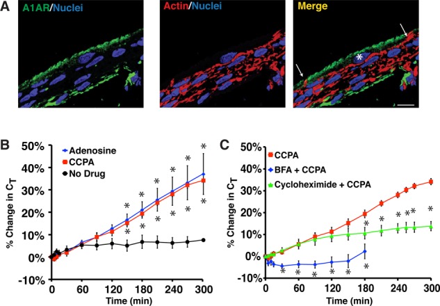
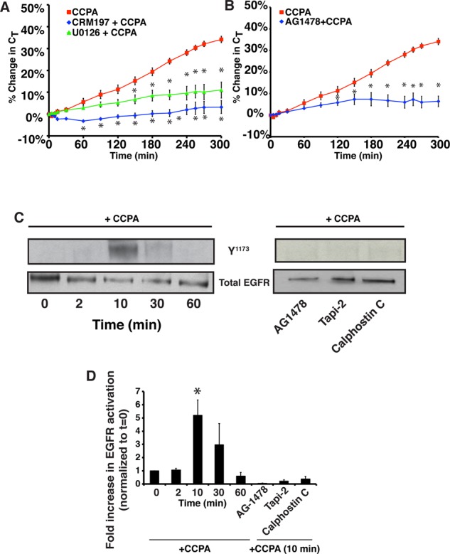
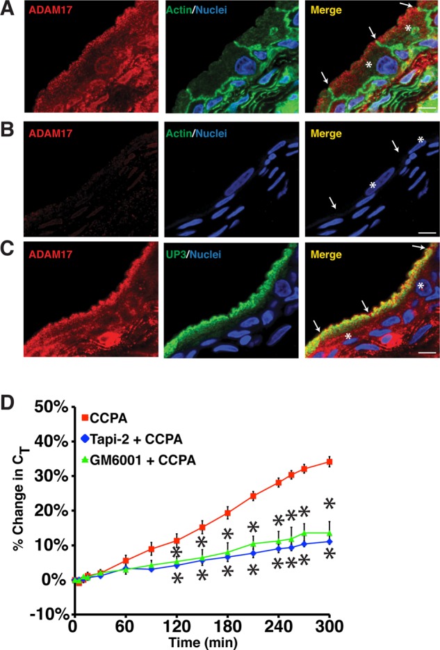
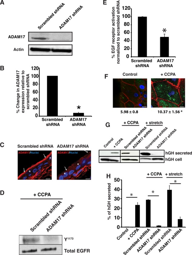
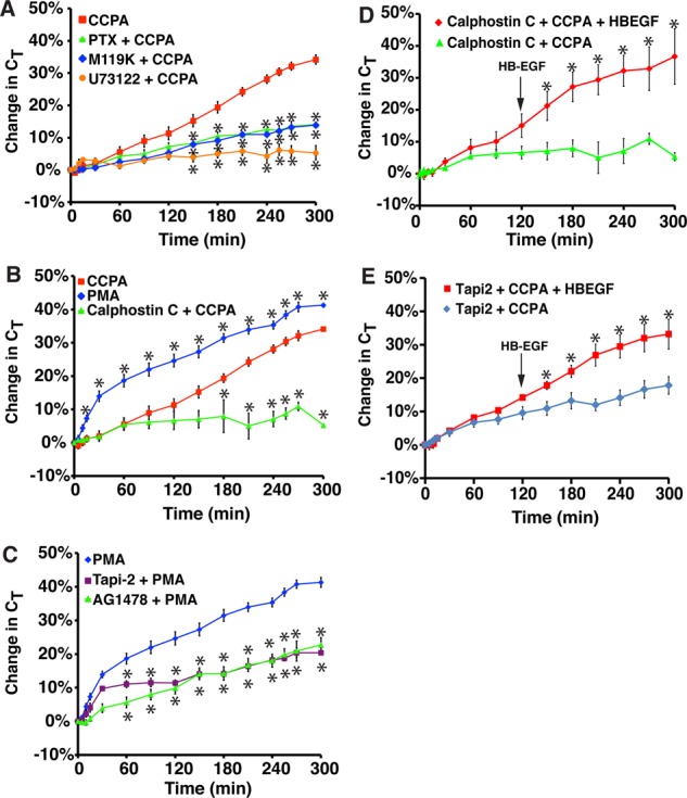
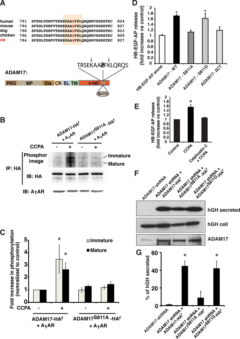
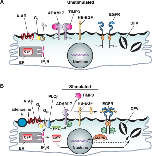
Despite the importance of ADAM17-dependent cleavage in normal biology and disease, the physiological cues that trigger its activity, the effector pathways that promote its function, and the mechanisms that control its activity, particularly the role of phosphorylation, remain unresolved. Using native bladder epithelium, in some cases transduced with adenoviruses encoding small interfering RNA, we observe that stimulation of apically localized A1 adenosine receptors (A1ARs) triggers a Gi-Gβγ-phospholipase C-protein kinase C (PKC) cascade that promotes ADAM17-dependent HB-EGF cleavage, EGFR transactivation, and apical exocytosis. We further show that the cytoplasmic tail of rat ADAM17 contains a conserved serine residue at position 811, which resides in a canonical PKC phosphorylation site, and is phosphorylated in response to A1AR activation. Preventing this phosphorylation event by expression of a nonphosphorylatable ADAM17(S811A) mutant or expression of a tail-minus construct inhibits A1AR-stimulated, ADAM17-dependent HB-EGF cleavage. Furthermore, expression of ADAM17(S811A) in bladder tissues impairs A1AR-induced apical exocytosis. We conclude that adenosine-stimulated exocytosis requires PKC- and ADAM17-dependent EGFR transactivation and that the function of ADAM17 in this pathway depends on the phosphorylation state of Ser-811 in its cytoplasmic domain.


Figures

FIGURE 1:
FIGURE 1:
A1AR-stimulated exocytosis is dependent on protein synthesis and secretion. (A) Cryosection of rat bladder epithelium labeled with an A1AR-specific antibody (green), rhodamine–phalloidin to label the actin cytoskeleton (red), and TOPRO-3 (blue) to label the nucleus. Right, merge. An umbrella cell is marked with an asterisk and the white arrows mark the apicolateral borders of the cell. Scale bar, 12 μm. (B) Rabbit uroepithelium was mounted in Ussing chambers and after equilibration left untreated (no drug), or 1 μM adenosine or 500 nM CCPA was added to the mucosal hemichamber. Percentage changes in CT were monitored. (C) Rabbit tissue was left untreated, pretreated with 5 μg/ml brefeldin A (BFA) for 30 min, or pretreated with 100 ng/ml cyclohexamide for 60 min. CCPA (500 nM) was then added to the mucosal hemichamber and CT recorded. Data for CCPA alone are reproduced from B. (B, C) Mean changes in CT ± SEM (n ≥ 4). Significant differences (p < 0.05), relative to no drug in A or CCPA in B, are indicated with an asterisk.
FIGURE 2:
FIGURE 2:
The A1AR transactivates the EGF receptor. (A) The mucosal surface of rabbit uroepithelium was pretreated with 25 ng/ml CRM197 for 25 min or with 10 μM U0126 for 60 min. CCPA (500 nM) was then added to the mucosal hemichamber, and CT was recorded. (B) Rabbit uroepithelium was pretreated with 1 μM AG1478 for 30 min, and then CCPA (500 nM) was added to the mucosal hemichamber and CT was recorded. (A, B) CCPA control data are reproduced from Figure 1B. Mean changes in CT ± SEM (n ≥ 3). Statistically significant differences (p < 0.05), relative to CCPA treatment alone, are marked with an asterisk. (C, D) Rabbit uroepithelium was either left untreated (left) or treated with AG1478 (1 μM) for 25 min, Tapi-2 (15 μM) for 90 min, or calphostin C (500 nM) for 60 min (right). CCPA (500 nM) was then added to the mucosal hemichamber. Left, cells were lysed at the indicated time points. Right, cells were lysed at the 10-min time point. Equal amounts of proteins were resolved by SDS–PAGE and immunoblots probed with a rabbit anti–EGFR-phospho-Y1173 antibody or rabbit anti-EGFR antibody. (D) Quantification of Y1173 phosphorylation. Data (mean ± SEM, n ≥ 3) are reported as fold increase above untreated tissue samples at t = 0. Statistically significant values (p < 0.05) above t = 0 are marked by an asterisk.
FIGURE 3:
FIGURE 3:
ADAM17 expression in the uroepithelium. (A–C) Cryosections of rat bladder uroepithelium were reacted with antibodies specific for ADAM17 (A), a mixture of antibody and inhibitory peptide (B), or antibodies specific for ADAM17 and uroplakin 3a (C). After incubation with fluorophore-labeled secondary antibodies, the samples were analyzed using confocal microscopy. Where indicated, actin was labeled with phalloidin and nuclei were labeled with TOPRO-3. The umbrella cells are marked with the asterisk in the merged images and the apicolateral junction is indicated by arrowheads; scale bar, 10 μm. (D) Rabbit uroepithelium was pretreated with Tapi-2 (15 μM) or GM6001 (15 μM) for 90 min, CCPA (500 nM) was added, and CT was recorded. Control CCPA data are reproduced from Figure 1B. Data are mean ±SEM (n ≥ 3), and values significantly different (p < 0.05) from CCPA alone are marked with an asterisk.
FIGURE 4:
FIGURE 4:
A1AR-stimulated exocytosis is dependent on ADAM17. (A) Uroepithelial lysates obtained from rat tissues transduced in situ with scrambled shRNA or ADAM17 shRNA were resolved by SDS–PAGE and Western blots probed with rabbit-anti ADAM17 antibody (top) or mouse anti–β-actin (bottom). (B) Quantification of ADAM17 expression in rat tissue treated with scrambled shRNA or ADAM17 shRNA. Data are mean ± SEM (n ≥ 4). The means of the two treatment groups are significantly different (p < 0.05). (C) Immunolocalization of ADAM17 expression (red) in rat tissue treated with scrambled or ADAM17-specific shRNAs. Nuclei (blue) are labeled with TOPRO-3. Scale bar, 13 μm. The cell junctions are marked with arrows and the umbrella cells with an asterisk. (D) CCPA (500 nM) was added to rat tissue treated with scrambled or ADAM17 shRNA and total EGFR or receptor phosphorylated at Y1173 was detected by Western blot. (E) Quantification of effects of scrambled or ADAM17-specific shRNA treatment on EGFR activation. Data are mean ± SEM (n = 4). The two treatment groups were significantly different (p < 0.05). (F) WGA-FITC (50 μg/ml) was added to the mucosal hemichamber of untreated rat tissue (control) or that treated with 500 nM CCPA. After 120 min, the tissue was incubated at 4°C with N-acetylglucosamine to remove surface lectin, fixed, and then processed for immunofluorescence. In these whole-mounted tissues, internalized WGA-FITC is shown in green, phalloidin-labeled actin in red, and TOPRO-3-labeled nuclei in blue. Bar, 15 μm. Quantification of the fluorescence intensity of WGA-FITC below the apical membrane. Data are mean ± SEM (n ≥ 8). The two treatment groups were significantly different (p < 0.05). (G) Rat tissues were transduced with hGH alone (control) or transduced with hGH and either scrambled or ADAM17 shRNAs. The excised bladder tissue was mounted in an Ussing stretch chamber and left untreated (control), treated with CCPA (500 nM), or stretched by filling the mucosal hemichamber. The mucosal fluid was collected and concentrated, the tissues were lysed, and hGH was detected in the secreted fraction (1/20 of total) or tissue lysates (1/13 of total) using Western blot. (H) Quantification of effects of ADAM17-specific shRNAs on hGH secretion. Data are mean ± SEM (n ≥ 3). Statistically significant effects (p < 0.05) are indicated with an asterisk.
FIGURE 5:
FIGURE 5:
Role for Gi, Gβγ, PLC, and PKC in A1AR-stimulated exocytosis. (A) Rabbit tissue was left untreated or pretreated with 100 ng/ml pertussis toxin (PTX) for 90 min, 10 μM M119K for 60 min, or 10 μM U73122 for 60 min. CCPA (500 nM) was added to the mucosal hemichamber, and CT was recorded. (B) Rabbit tissue was left untreated or pretreated with calphostin C (500 nM) for 90 min. Subsequently, tissue was treated with CCPA (500 nM) or PMA (10 nM). (C) Rabbit tissue was pretreated with 15 μM Tapi-2 for 90 min or 1 μM AG1478 for 30 min and then treated with 10 nM PMA. The data for PMA treatment alone were reproduced from B. (A–C) Data for CCPA treatment alone were reproduced from Figure 1B. (D, E) Rabbit tissue was pretreated with calphostin C (D) or Tapi-2 (E) for 90 min, and then at t = 0, CCPA was added to the mucosal hemichamber. After 120 min, HG-EGF (1 nM) was added to the mucosal hemichamber (indicated with an arrow), and the tissue was incubated for additional 180 min. In D, data for calphostin C + CCPA are reproduced from B. (A–E) Data are presented as mean ± SEM (in A–C, n ≥ 3; in D and E, n ≥ 6). In A–C, values that are significantly different from CCPA alone (p < 0.05) are marked with an asterisk. In D and E, values that are significantly different from calphostin C + CCPA or Tapi-2 + CCPA (p < 0.05) are marked with an asterisk.
FIGURE 6:
FIGURE 6:
Effect of Ser-811 phosphorylation on the function of ADAM17. (A) Top, alignment of the C-termini of ADAM17 from different species. Numbers indicate the amino acids involved. The canonical PKC phosphorylation site is shaded, and the position of the conserved Ser residue (Ser-811 in rat) is indicated in red. Bottom, domain structure of ADAM17. CR, cysteine-rich domain; C-tail, cytoplasmic domain; Dis, disintegrin domain; EL, EGF-like; MP, metalloproteinase domain; Pro, propeptide; TM, transmembrane domain. The consensus PKC phosphorylation motif is shown in an expanded view. Ser-811 is shaded, and the critical, flanking basic residues at the −3 and + 2 positions are marked with arrows. (B) HEK-293FT cells expressing the A1AR in combination with ADAM17-HAr or ADAM17S811A-HAr were labeled with 32P-orthophosphate and then left untreated or treated with CCPA (500 nM). HA-tagged ADAM17 constructs were immunoprecipitated and Western blots probed with an anti–HA-HRP- conjugated secondary antibody to detect total ADAM17 (middle) and subsequently exposed to a PhosphorImager screen to detect 32P-labeled ADAM17 (top). Total A1AR was detected by Western blot (bottom). (C) Data (mean ± SEM; n = 3) were quantified, and values significantly different from ADAM17-HAr + CCPA are indicated with an asterisk. (D) HEK-293FT cells were cotransfected with the A1AR, HB-EGF-AP, and ADAM17-HAr, ADAM17S811A-HAr, or ADAM17S811D-HAr. Fold stimulation of HB-EGF-AP release in CCPA-treated cells vs. control, untreated cells. Data are mean ± SEM, n = 5. Data significantly different from HB-EGF-AP alone are indicated with an asterisk. (E) HEK-293FT cells were cotransfected with the A1AR, HB-EGF-AP, and ADAM17-HAr. Cells were pretreated with calphostin C (500 nM) for 60 min, and the fold stimulation of HB-EGF-AP release in CCPA-treated cells vs. control, untreated cells is reported. Data are mean ± SEM, n = 7. Values significantly different from the control, as assessed by ANOVA, are indicated (*p < 0.05). (F) Rat tissues were transduced in situ with hGH and ADAM17 shRNA alone or in combination with ADAM17-HAr, ADAM17S811A-HAr, or ADAM17S811D-HAr. The excised bladder tissue was mounted in an Ussing stretch chamber, and CCPA (500 nM) was added to the mucosal hemichamber. After 60 min, the mucosal fluid was collected and concentrated, the tissues were lysed, and hGH detected using Western blot. (G) Quantification of hGH secretion. Data are mean ± SEM (n = 5). Values that are statistically different from ADAM17 shRNA alone (p < 0.05) are indicated with an asterisk.
FIGURE 7:
FIGURE 7:
Model for function of ADAM17 Ser-811 phosphorylation in A1AR-stimulated EGFR transactivation and exocytosis. See the text for description.

Similar articles

Cited by

References

    1. Amour A, Slocombe PM, Webster A, Butler M, Knight CG, Smith BJ, Stephens PE, Shelley C, Hutton M, Knauper V, et al. TNF-alpha converting enzyme (TACE) is inhibited by TIMP-3. FEBS Lett. 1998;435:39–44. - PubMed
    1. Apodaca G. The uroepithelium: not just a passive barrier. Traffic. 2004;5:1–12. - PubMed
    1. Arribas J, Esselens C. ADAM17 as a therapeutic target in multiple diseases. Curr Pharm Des. 2009;15:2319–2335. - PubMed
    1. Balestreire EM, Apodaca G. Apical epidermal growth factor receptor signaling: regulation of stretch-dependent exocytosis in bladder umbrella cells. Mol Biol Cell. 2007;18:1312–1323. - PMC - PubMed
    1. Bell HL, Gooz M. ADAM-17 is activated by the mitogenic protein kinase ERK in a model of kidney fibrosis. Am J Med Sci. 2010;339:105–107. - PMC - PubMed

Publication types