. 2014 Sep 17;34(38):12678-89.
doi: 10.1523/JNEUROSCI.1183-14.2014.

Regulation of synaptic extracellular matrix composition is critical for proper synapse morphology

Affiliations

Regulation of synaptic extracellular matrix composition is critical for proper synapse morphology

Peri T Kurshan et al. J Neurosci. .

Abstract

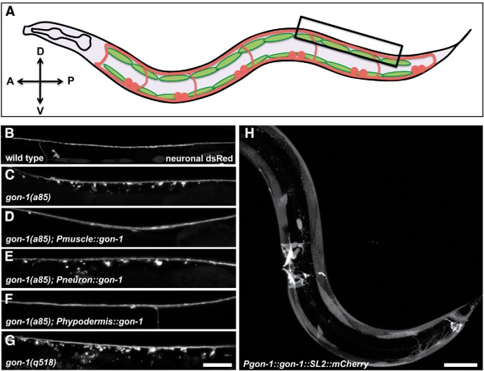
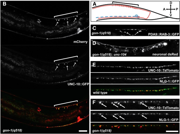
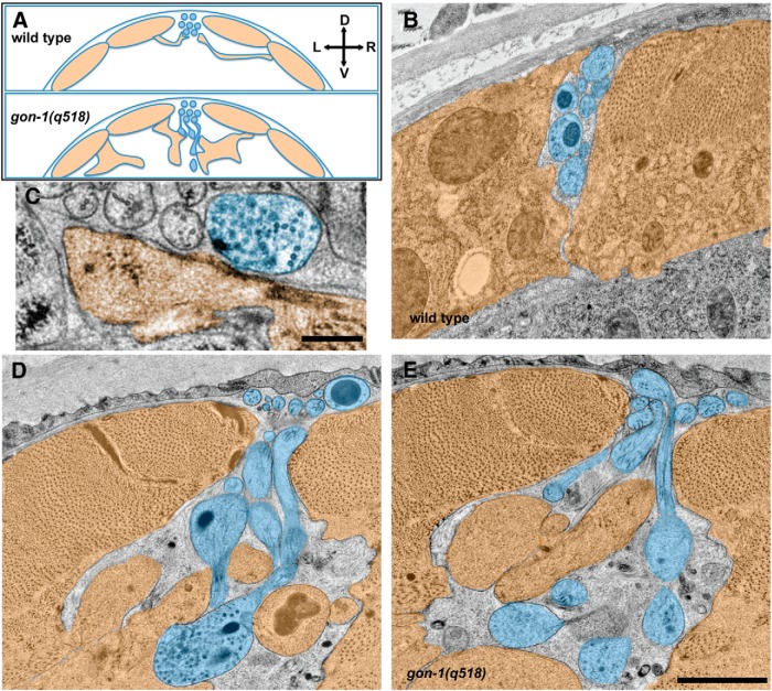
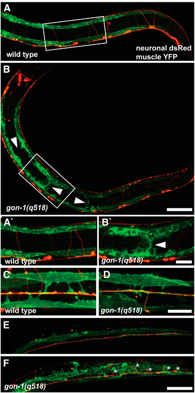
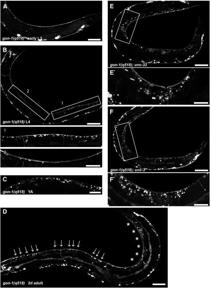
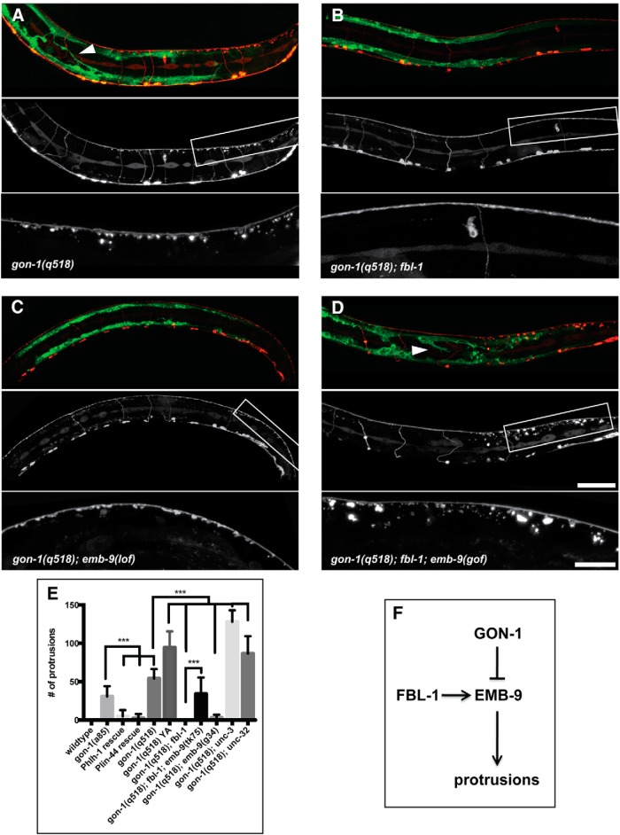
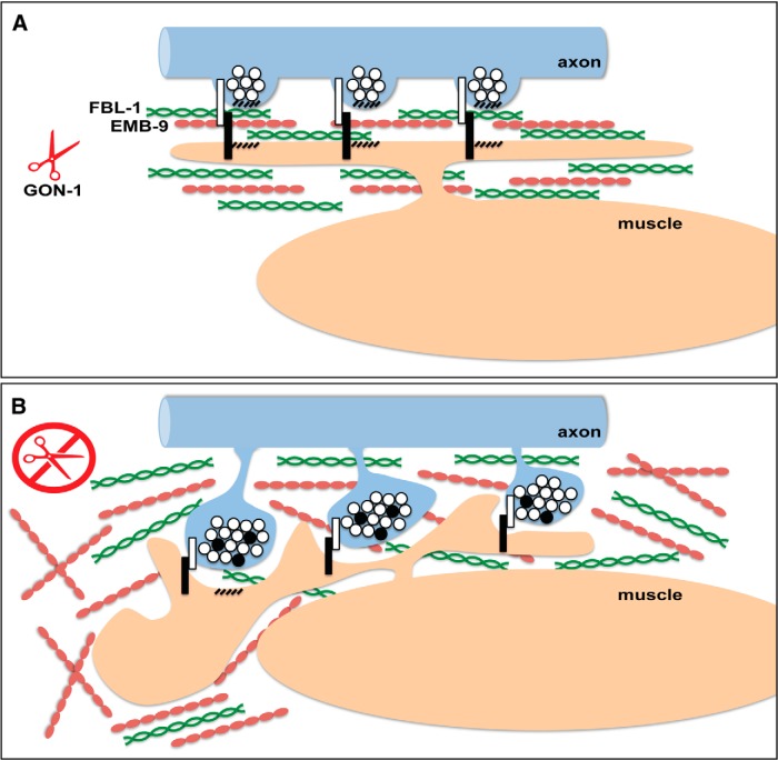
Synapses are surrounded by a layer of extracellular matrix (ECM), which is instrumental for their development and maintenance. ECM composition is dynamically controlled by proteases, but how the precise composition of the ECM affects synaptic morphology is largely unknown. Through an unbiased forward genetic screen, we found that Caenorhabditis elegans gon-1, a conserved extracellular ADAMTS protease, is required for maintaining proper synaptic morphology at the neuromuscular junction. In gon-1 mutants, once synapse formation is complete, motor neuron presynaptic varicosities develop into large bulbous protrusions that contain synaptic vesicles and active zone proteins. A concomitant overgrowth of postsynaptic muscle membrane is found in close apposition to presynaptic axonal protrusions. Mutations in the muscle-specific, actin-severing protein cofilin (unc-60) suppress the axon phenotype, suggesting that muscle outgrowth is necessary for presynaptic protrusions. gon-1 mutants can also be suppressed by loss of the ECM components collagen IV (EMB-9) and fibulin (FBL-1). We propose that GON-1 regulates a developmental switch out of an initial "pro-growth" phase during which muscle arms grow out and form synapses with motor neuron axons. We postulate that this switch involves degradation or reorganization of collagen IV (EMB-9), whereas FBL-1 opposes GON-1 by stabilizing EMB-9. Our results describe a mechanism for regulating synaptic ECM composition and reveal the importance of precise ECM composition for neuronal morphology and synapse integrity.

Keywords: extracellular matrix; metalloproteinase; synapse.


Figures

Figure 1.
Figure 1.
GON-1, normally expressed in muscles, is required non-cell autonomously for proper axon morphology. A, A schematic of the worm from a lateral view. The trIs30 marker, used throughout this study, expresses dsRed in motor neurons (the dorsally projecting DBs and DDs, and the ventrally projecting VDs), and YFP in the lateral muscles. Box indicates region shown in B–G. B, Dorsal cord of a wild-type worm expressing the trIs30 marker. C, gon-1(a85) worms exhibit large protrusions that extend ventrally from the dorsal cord. D, The protrusion phenotype can be rescued by expressing gon-1 in muscle cells using the hlh-1 promoter. E, Protrusions cannot be rescued by expressing gon-1 in neurons using the rab-3 promoter. F, Protrusions can be rescued by expressing gon-1 in tail hypodermal cells using the lin-44 promoter. G, gon-1(q518)-null mutants also exhibit axonal protrusions. Scale bar, 20 μm. H, A recombineered fosmid containing the gon-1 genomic region as well as an SL2 promoter driving mCherry in all cells expressing gon-1 shows that gon-1 is highly expressed in muscles including body wall and vulval and anal depressor muscles. Scale bar, 50 μm. A, anterior; D, dorsal; P, posterior; V, ventral.
Figure 2.
Figure 2.
gon-1 mutant protrusions form specifically at synapses, contain synaptic material, and are apposed to postsynaptic structures. A, Schematic of the DA9 motor neuron (in red). The DA9 cell body resides on the ventral side near the tail, and the DA9 axon projects to the dorsal side and then extends anteriorly, making synapses only in a specific region marked by the bracket. (The DA9 promoter used in B is also active in the adjacent VA12 neuron, shown in blue, which directly sends its axon anteriorly and makes synapses on the ventral side.) B, Pmig-13 driving expression of cytoplasmic mCherry and the synaptic active zone marker unc-10::GFP in gon-1(q518) mutants. Protrusions (arrows) are limited to the synaptic domain of DA9 (bracket) and contain accumulations of UNC-10::GFP. Scale bar, 20 μm. C, Pitr-1 driving expression of the synaptic vesicle marker rab-3::GFP in gon-1(q518) mutants. Protrusions contain accumulations of RAB-3::GFP. D, A loss-of-function mutation in the kinesin unc-104(e1265) does not suppress the protrusion phenotype in gon-1(q518) mutants (trIs30 marker). E, F, Labeling presynaptic DD motor neuron synapses with Punc-47::unc-10::TdTomato and postsynaptic muscles with Punc-129dm::nlg-1::YFP reveals that some postsynaptic material is also mislocalized away from the dorsal cord in gon-1(q518) mutants (F). Scale bar, 20 μm.
Figure 3.
Figure 3.
Ultrastructural analysis confirms that protrusions are filled with vesicles and in close contact with aberrant muscle extensions. A, Schematic showing the relationship between dorsal muscles (orange) and the dorsal nerve cord axons (blue) in cross section. In wild-type worms (top) the dorsal cord sits in between the dorsal muscles, which send thin projections called muscle arms to form synapses along the ventral side of the dorsal cord. This morphology is abnormal in gon-1 mutants (bottom). B, Wild-type dorsal cord and dorsal muscles (muscle arms not shown). C, A normal synapse, containing synaptic vesicles and a presynaptic dense projection, in the gon-1(q518) mutant, formed between an axonal varicosity within the dorsal cord and a muscle arm. Scale bar, 250 nm. D, E, In gon-1(q518) mutants, abnormal vesicle-filled protrusions extend ventrally from the dorsal cord, and are found in close apposition to abnormal muscle overgrowth. Scale bar, 1 μm. D, dorsal; V, ventral; L, left; R, right.
Figure 4.
Figure 4.
gon-1 mutants exhibit gross muscle overgrowth. A, In wild-type worms muscles (green) are restricted to the dorsal and ventral sides and never extend into the middle of the body. B, In contrast, in gon-1(q518) mutants, muscles are disorganized and extend into the body wall cavity (arrowheads). Boxes in A and B indicate areas magnified in A′ and B′. C, Dorsal view of a wild-type worm showing early born muscles arms (green) extending to the dorsal cord (red). D, Dorsal view of gon-1(q518) showing that early born muscle arms still form, although they are often slightly thinner than normal. E, Z-projection of superficial confocal sections from a dorsal view of a gon-1(q518) mutant, showing normal axonal and muscle morphology. F, Deeper confocal sections from the same worm reveal axonal protrusions (in red, asterisks) as well as the surrounding abnormal muscle morphology (in green). Worm shown in E and F has increased muscle GFP expression (wyEx5356 transgene, see Materials and Methods) for better visualization of the posterior muscles. Scale bars: A, B, E, F, 50 μm; A′, B′, C, D, 20 μm.
Figure 5.
Figure 5.
Synaptic protrusions develop after initial synaptogenesis is complete and their location is age and activity dependent. A, An early L3 gon-1(q518) larva exhibiting no synaptic protrusions. B, A mid-L4 gon-1(q518) larva. Synaptic protrusions are largely restricted to the posterior third of the worm (Box 1). Boxes indicate areas of magnification below. C, A young adult gon-1(q518) exhibiting more pronounced synaptic protrusions. D, A 2-d-old adult gon-1(q518) with synaptic protrusions visible along the length of the worm (arrows), although still most pronounced in the most posterior region (asterisks). E, F, Crossing gon-1(q518) into the synaptic activity mutants unc-32(e189) (E) and unc-3(e151) (F) leads to an enhancement of the phenotype such that protrusions are no longer limited to the posterior region. Boxes indicate areas of magnification below. Scale bars: A, 20 μm; B, 50 μm, insets, 20 μm; C, 20 μm; D, 50 μm; E, F, 50 μm, insets, 20 μm.
Figure 6.
Figure 6.
Genetic interactions between gon-1, fbl-1, and emb-9. Loss-of-function mutations in fbl-1 and emb-9 suppress the gon-1 phenotypes, while gain-of-function mutations in emb-9 de-suppresses the fbl-1 suppression. A, gon-1(q518) mutants exhibiting synaptic protrusions and muscle abnormalities (arrowhead). B, Suppression of both synapse and muscle phenotypes in gon-1(q518); fbl-1(q771) double mutants. C, Suppression of both phenotypes in gon-1(q518); emb-9(g34) double mutants transferred from the permissive temperature (16 degrees) after embryonic development and then grown at the restrictive temperature (25 degrees) until the L4 larval stage. These worms exhibit a ruffling of the axons, but this phenotype exists in the emb-9(g34) single mutants, and is therefore independent of the gon-1 mutant protrusion phenotype. D, De-suppression of both phenotypes in gon-1(q518); fbl-1(q771); emb-9(tk75) triple mutants. In A–D, the marker is trIs30: top shows muscles in green and motor neurons in red, the middle motor neurons are shown alone, and the box indicates the area magnified and shown in the bottom. Scale bars: 50 μm, insets, 20 μm. E, Quantification of synaptic protrusion phenotype. Comparisons were made to the appropriate control genetic background. Statistical test: one-way ANOVA and Sidak's multiple-comparisons test. ***p ≤ 0.001. F, Diagram of proposed model in which GON-1 and FBL-1 act in opposition, with GON-1 degrading and FBL-1 stabilizing EMB-9. An overabundance of EMB-9 may lead to synaptic protrusions.
Figure 7.
Figure 7.
Muscle-specific unc-60B/cofilin is required for the gon-1 synaptic phenotype in motor neurons. A, Synaptic protrusions in gon-1(a85). B, Protrusions are suppressed in unc-60B(wy789), an allele isolated from a modifier screen conducted in gon-1(a85). C, Protrusions are suppressed in a canonical allele of unc-60B(e723). D, Muscle-specific expression of unc-60B under the hlh-1 promoter de-suppresses the phenotype in gon-1(a85); unc-60B(wy789) double mutants. Scale bar, 20 μm.
Figure 8.
Figure 8.
A model for GON-1 function in maintaining synapse morphology at the NMJ. A, In wild-type worms, GON-1 may function to degrade collagen IV/EMB-9, which is stabilized by fibulin/FBL-1, or transform it from a pro-growth to a pro-stability conformation. B, In the absence of GON-1, overabundance of pro-growth EMB-9 may lead to an overgrowth of the muscle, including the muscle arms. Since trans-synaptic adhesion molecules reside at synapses (white and black bars), muscle arm overgrowth may pull on presynaptic varicosities causing the presynaptic protrusions exhibited by these mutants. These protrusions are filled with clear-core and dense-core vesicles containing synaptic vesicle and active zone proteins (white and black circles).

Similar articles

Cited by

References

    1. Apte SS. A disintegrin-like and metalloprotease (reprolysin-type) with thrombospondin type 1 motif (ADAMTS) superfamily: functions and mechanisms. J Biol Chem. 2009;284:31493–31497. doi: 10.1074/jbc.R109.052340. - DOI - PMC - PubMed
    1. Bajor M, Kaczmarek L. Proteolytic remodeling of the synaptic cell adhesion molecules (CAMs) by metzincins in synaptic plasticity. Neurochem Res. 2013;38:1113–1121. doi: 10.1007/s11064-012-0919-6. - DOI - PMC - PubMed
    1. Blelloch R, Kimble J. Control of organ shape by a secreted metalloprotease in the nematode Caenorhabditis elegans. Nature. 1999;399:586–590. doi: 10.1038/21196. - DOI - PubMed
    1. Brenner S. The genetics of Caenorhabditis elegans. Genetics. 1974;77:71–94. - PMC - PubMed
    1. Carlson SS, Valdez G, Sanes JR. Presynaptic calcium channels and α3-integrins are complexed with synaptic cleft laminins, cytoskeletal elements and active zone components. J Neurochem. 2010;115:654–666. doi: 10.1111/j.1471-4159.2010.06965.x. - DOI - PMC - PubMed

Publication types

MeSH terms

LinkOut - more resources