. 2014 Sep 17;34(38):12893-903.
doi: 10.1523/JNEUROSCI.1201-14.2014.

Intracellular accumulation of amyloid-β (Aβ) protein plays a major role in Aβ-induced alterations of glutamatergic synaptic transmission and plasticity

Affiliations

Intracellular accumulation of amyloid-β (Aβ) protein plays a major role in Aβ-induced alterations of glutamatergic synaptic transmission and plasticity

Cristian Ripoli et al. J Neurosci. .

Abstract

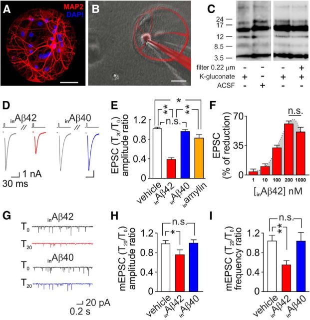
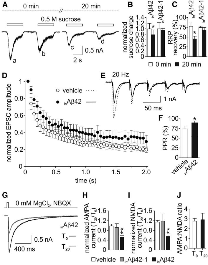
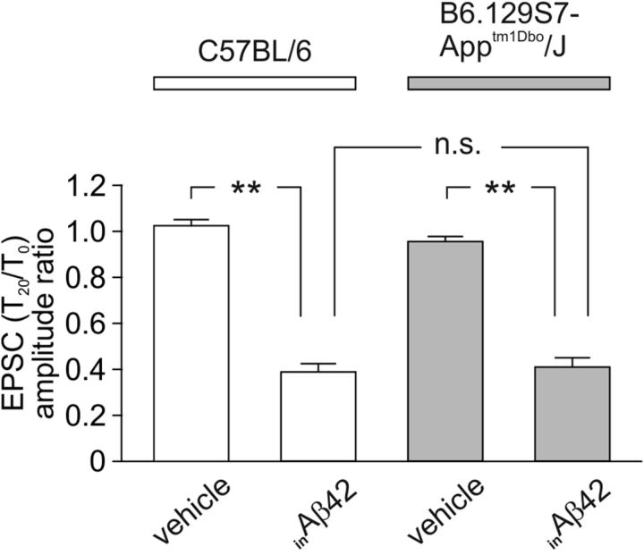
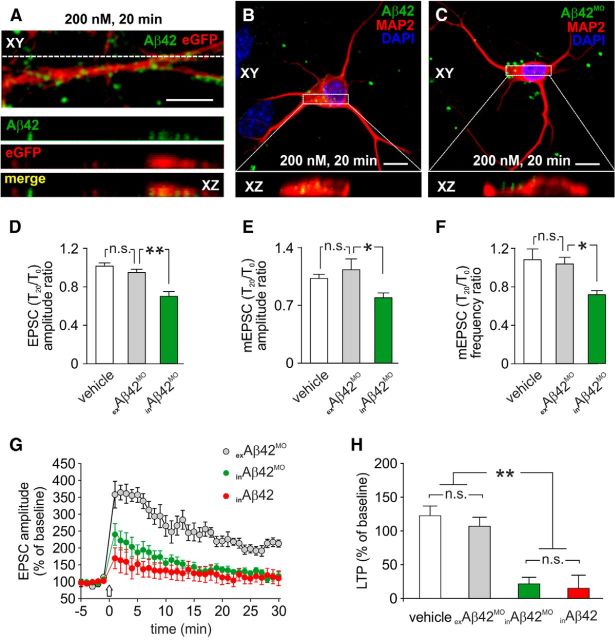
Intracellular accumulation of amyloid-β (Aβ) protein has been proposed as an early event in AD pathogenesis. In patients with mild cognitive impairment, intraneuronal Aβ immunoreactivity was found especially in brain regions critically involved in the cognitive deficits of AD. Although a large body of evidence demonstrates that Aβ42 accumulates intraneuronally ((in)Aβ), the action and the role of Aβ42 buildup on synaptic function have been poorly investigated. Here, we demonstrate that basal synaptic transmission and LTP were markedly depressed following Aβ42 injection into the neuron through the patch pipette. Control experiments performed with the reverse peptide (Aβ42-1) allowed us to exclude that the effects of (in)Aβ depended on changes in oncotic pressure. To further investigate (in)Aβ synaptotoxicity we used an Aβ variant harboring oxidized methionine in position 35 that does not cross the neuronal plasma membrane and is not uploaded from the extracellular space. This Aβ42 variant had no effects on synaptic transmission and plasticity when applied extracellularly, but induced synaptic depression and LTP inhibition after patch-pipette dialysis. Finally, the injection of an antibody raised against human Aβ42 (6E10) in CA1 pyramidal neurons of mouse hippocampal brain slices and autaptic microcultures did not, per se, significantly affect LTP and basal synaptic transmission, but it protected against the toxic effects of extracellular Aβ42. Collectively, these findings suggest that Aβ42-induced impairment of glutamatergic synaptic function depends on its internalization and intracellular accumulation thus paving the way to a systemic proteomic analysis of intracellular targets/partners of Aβ42.

Keywords: 6E10; amyloid-β protein; autaptic hippocampal neurons; intraneuronal accumulation; synaptic transmission; whole-cell LTP.


Figures

Figure 1.
Figure 1.
Accumulation of inAβ plays a major role in Aβ-induced alterations of glutamatergic synaptic function. A, Representative image of a hippocampal autaptic culture. Red staining for MAP2 identifies the single neuron grown onto glial microisland. Blue staining (DAPI) identifies cell nuclei. B, Image depicting intracellular application of 200 nm Aβ42. C, Representative Western blot of Aβ oligomer distribution in ACSF and unfiltered or 0.22 μm filtered K-gluconate solution. None of the above described experimental conditions markedly affected Aβ42 small oligomer distribution. D, Representative traces of EPSC currents at T0 (gray lines) and after 20 min intracellular application of 200 nm Aβ42 (red line) or 200 nm Aβ40 (blue line). Stimulus artifacts for EPSC currents were removed for clarity. E, Bar graphs (mean ± SEM) showing the T20/T0 ratio of EPSC amplitude in autaptic neurons exposed to vehicle (white bar), inAβ42 (red bar), inAβ40 (blue bar), or amylin (orange bar). F, Dose–response relationship of inAβ42 effects on EPSC amplitude. G, Representative traces of mEPSC currents at T0 (gray lines) and T20 with 200 nm inAβ42 (red line) or 200 nm inAβ40 (blue line). Bar graphs (mean ± SEM) showing the T20/T0 ratio of mEPSC amplitude (H) and frequency (I) in autaptic neurons exposed to vehicle (white bars), inAβ42 (red bars), or inAβ40 (blue bars). Scale bars: A, B, 50 μm. *p < 0.05; **p < 0.005; n.s.: p > 0.05.
Figure 2.
Figure 2.
Intracellular application of Aβ42 markedly affects glutamatergic synaptic transmission. A, Representative traces of paired-pulse currents induced by 4 s applications of 0.5 m sucrose (4 s interpulse intervals) at T0 and T20 of inAβ42. Following Aβ42 injection (trace c collected at T20) the sucrose charge was markedly lower than at T0 (trace a). B, Bar graphs (mean ± SEM) showing the normalized sucrose charge at T0 (white bars) and T20 (black bars) in inAβ42- or inAβ42-1-injected neurons. C, Bar graphs (mean ± SEM) showing the RRP recovery rate expressed as the peak amplitude of the second responses normalized to that of the first response at T0 (b/a in A; white bars) and T20 (d/c in A; black bars) in inAβ42- or inAβ42-1-injected neurons. D, EPSC amplitudes normalized to the first response during 2 s trains at 20 Hz. E, Representative traces of the first five responses evoked by trains of stimuli (20 Hz) after 20 min of vehicle or inAβ42 applications. F, Mean PPRs after 20 min of vehicle or inAβ42 application. G, Representative traces of NMDA currents (stimulus artifacts removed for clarity) at T0 (black line) and T20 (gray line) with inAβ42. Bar graphs (mean ± SEM) showing the T20/T0 ratio of AMPA (H) and NMDA (I) currents with vehicle (white bars), inAβ42-1 (gray bars), and inAβ42 (black bars). J, Bar graphs (mean ± SEM) showing the AMPA:NMDA ratio at T0 and T20 with inAβ42. *p < 0.05; **p < 0.01.
Figure 3.
Figure 3.
EPSC amplitude inhibition induced by inAβ42 is independent of its interaction with either APP or APP cleavage products. Bar graphs (mean ± SEM) showing the T20/T0 ratio of EPSC amplitude in autaptic neurons derived from C57BL/6 (white bars) and B6.129S7-Apptm1Dbo/J mice (APP knock-out mice; gray bars) exposed to either vehicle or 200 nm inAβ42. **p < 0.005; n.s.: p > 0.05.
Figure 4.
Figure 4.
Twenty minute injection of Aβ42 does not significantly affect dendritic spine density of CA1 hippocampal neurons. A, B, Representative examples of CA1 neurons filled with biocytin and revealed with avidin conjugated to Alexa Fluor 488. Neuron shown in A was injected with vehicle, whereas neuron shown in B was injected with 200 nm Aβ42 for 20 min. Bottom boxes in A and B show high-magnification images of dendritic segments of cells in A and B, respectively. Scale bar, 3 μm. Alexa Fluor 488 fluorescence intensity (8-bit depth) was represented according to the color scale on the right (bottom = 0, top = 256). C, Bar graphs showing the mean number of dendritic spines per 100 μm. n.s.: p > 0.05.
Figure 5.
Figure 5.
LTP is markedly inhibited by inAβ42. A, Time course of EPSC amplitudes before and after HFS (indicated by arrow) in hippocampal brain slices treated with vehicle (white circles) and inAβ42 (black circles). B, Bar graphs (mean ± SEM) showing the EPSC amplitudes measured during the last 5 min of recording under the conditions described for A and during the last 5 min of inAβ42-1. **p < 0.005; n.s.: p > 0.05.
Figure 6.
Figure 6.
Extracellular Aβ42MO has no effects on synaptic transmission and plasticity but induces synaptic depression and LTP inhibition after patch-pipette dialysis. A, Representative example of a dendrite from eGFP+ hippocampal neuron (red) following 20 min extracellular application of 200 nm IRIS 5-labeled Aβ42 (green). Representative examples of neurons exposed for 20 min to 200 nm IRIS 5-labeled Aβ42 (B) and Aβ42MO (C). Red staining indicates MAP2 immunoreactivity. Bottom boxes in A–C represent X-Z cross sections from the Z-stack acquisitions showing the different neuronal accumulation of Aβ42 analogs after 20 min treatments. Bar graphs comparing the T20/T0 ratio of EPSC amplitude (D), mEPSC amplitude (E), and mEPSC frequency (F) measured in autaptic neurons following application of vehicle (white bars), exAβ42MO (gray bars), and inAβ42MO (green bars). G, Time course of EPSC amplitudes before and after HFS (indicated by arrow) in hippocampal slices treated with exAβ42MO (gray circles), inAβ42MO (green circles), and inAβ42 (red circles). H, Bar graphs (mean ± SEM) showing the EPSC amplitudes measured during the last 5 min of recording under the conditions described for G and during the last 5 min of vehicle. Scale bars: A–C, 10 μm. *p < 0.05; **p < 0.005; n.s.: p > 0.05.
Figure 7.
Figure 7.
Inhibition of hippocampal LTP and basal synaptic transmission induced by exAβ42 likely depends of its ability to be uploaded intraneuronally. A, Time course of EPSC amplitudes before and after HFS (indicated by arrow) in hippocampal slices treated with vehicle (white circles), exAβ42 (gray circles), and exAβ42 + in6E10 (black circles). B, Bar graphs (mean ± SEM) showing the EPSC amplitudes measured during the last 5 min of recording under the conditions described for A and during the last 5 min of in6E10 alone. Bar graphs comparing the T20/T0 ratio of EPSC amplitude (C), mEPSC amplitude (D), and mEPSC frequency (E) measured in autaptic neurons following application of vehicle (white bars), in6E10 (striped bars), exAβ42 + in6E10 (black bars), and exAβ42 (gray bars). *p < 0.05; **p < 0.01; n.s.: p > 0.05.

Similar articles

Cited by

References

    1. Almeida CG, Tampellini D, Takahashi RH, Greengard P, Lin MT, Snyder EM, Gouras GK. Beta-amyloid accumulation in APP mutant neurons reduces PSD-95 and GluR1 in synapses. Neurobiol Dis. 2005;20:187–198. doi: 10.1016/j.nbd.2005.02.008. - DOI - PubMed
    1. Attar A, Ripoli C, Riccardi E, Maiti P, Li Puma DD, Liu T, Hayes J, Jones MR, Lichti-Kaiser K, Yang F, Gale GD, Tseng CH, Tan M, Xie CW, Straudinger JL, Klärner FG, Schrader T, Frautschy SA, Grassi C, Bitan G. Protection of primary neurons and mouse brain from Alzheimer's pathology by molecular tweezers. Brain. 2012;135:3735–3748. doi: 10.1093/brain/aws289. - DOI - PMC - PubMed
    1. Bayer TA, Wirths O. Intracellular accumulation of amyloid-beta-a predictor for synaptic dysfunction and neuron loss in Alzheimer's disease. Front Aging Neurosci. 2010;2:8. doi: 10.3389/fnagi.2010.00008. - DOI - PMC - PubMed
    1. Benilova I, De Strooper B. Neuroscience. Promiscuous Alzheimer's amyloid: yet another partner. Science. 2013;341:1354–1355. doi: 10.1126/science.1244166. - DOI - PubMed
    1. Benilova I, Karran E, De Strooper B. The toxic Aβ oligomer and Alzheimer's disease: an emperor in need of clothes. Nat Neurosci. 2012;15:349–357. doi: 10.1038/nn.3028. - DOI - PubMed

Publication types

MeSH terms

LinkOut - more resources