Review
. 2014 Nov;3(11):1262-8.
doi: 10.5966/sctm.2014-0086. Epub 2014 Sep 17.

Intercellular cooperation and competition in brain cancers: lessons from Drosophila and human studies

Affiliations
Review

Intercellular cooperation and competition in brain cancers: lessons from Drosophila and human studies

Indrayani Waghmare et al. Stem Cells Transl Med. 2014 Nov.

Abstract

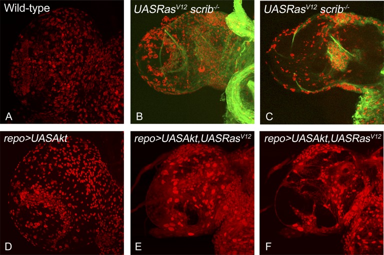
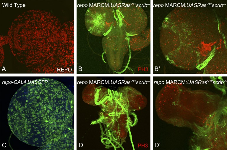
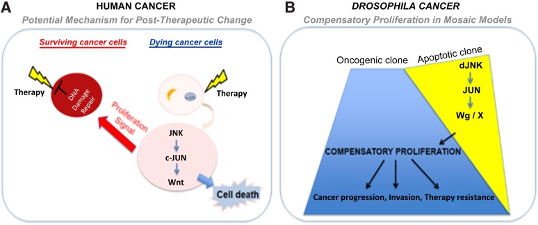
Glioblastoma (GBM) is a primary brain cancer with an extremely poor prognosis. GBM tumors contain heterogeneous cellular components, including a small subpopulation of tumor cells termed glioma stem cells (GSCs). GSCs are characterized as chemotherapy- and radiotherapy-resistant cells with prominent tumorigenic ability. Studies in Drosophila cancer models demonstrated that interclonal cooperation and signaling from apoptotic clones provokes aggressive growth of neighboring tumorigenic clones, via compensatory proliferation or apoptosis induced proliferation. Mechanistically, these aggressive tumors depend on activation of Jun-N-terminal kinase (upstream of c-JUN), and Drosophila Wnt (Wg) in the apoptotic clones. Consistent with these nonmammalian studies, data from several mammalian studies have shown that c-JUN and Wnt are hyperactivated in aggressive tumors (including GBM). However, it remains elusive whether compensatory proliferation is an evolutionarily conserved mechanism in cancers. In the present report, we summarize recent studies in Drosophila models and mammalian models (e.g., xenografts of human cancer cells into small animals) to elucidate the intercellular interactions between the apoptosis-prone cancer cells (e.g., non-GSCs) and the hyperproliferative cancer cells (e.g., GSCs). These evolving investigations will yield insights about molecular signaling interactions in the context of post-therapeutic phenotypic changes in human cancers. Furthermore, these studies are likely to revise our understanding of the genetic changes and post-therapeutic cell-cell interactions, which is a vital area of cancer biology with wide applications to many cancer types in humans.


Figures

Figure 1.
Figure 1.
Drosophila glioma models to study cell-cell signaling interactions. Comparisons of overgrowth and glial cell numbers in the dorsal lobe of Drosophila larval brain from mature third instar larvae are shown for the following genotypes: wild-type (A), w; repo-Gal4[4.3] UAS-mCD8:GFP repo-flp5/yw; +/+; FRT82B Tub-Gal80/ UAS RasV12 FRT82B scrib2 (B, C), UASAkt (D), and yw/UAS Akt; +/UAS RasV12; repo-Gal4 UASGFP/+ (E, F). All samples were stained for antibody against the glial-specific marker Repo (red). The samples in (A, B, D, E) show the surface view, and (C, F) show the medial view through the dorsal lobe of the brain. The glioma in (B, C) were generated using the MARCM approach, resulting in positively marked clones (green fluorescent protein expressing) of glial cells that are mutant for the tumor suppressor gene scribble and simultaneously overexpress oncogenic Ras. The glioma in (E, F) were induced by misexpression of oncogenic Ras and Akt in the glial cells using repo-Gal4. Note that both approaches cause overrepresentation of the glial cells specifically and cause overgrowth in the dorsal brain lobes compared with the normal wild-type controls. All images were scanned at identical magnification. Magnification, ×40. Abbreviation: MARCM, mosaic analysis with a repressible cell marker.
Figure 2.
Figure 2.
The MARCM approach allows tracking of glioma of clonal origin. We established a system to positively mark clones induced specifically in the glial cells in the Drosophila brain using the MARCM approach. This system drives the expression of transgenes (e.g., UAS GFP, UAS RasV12) specifically in the glial cells and also causes recombination specifically in the glial cells because the expression of the flippase enzyme is under the control of the repo promoter (repo-flp). (A): Dorsal lobe of wild-type mature third instar larva stained for antibodies to Repo can be compared with (C): dorsal lobe of repo-Gal4 UAS GFP, which shows the expression of the repo-Gal4 transgene using UAS GFP reporter expression. Images in (A, C) show that the repo-Gal4 driver is capable of driving transgenes in all glial cells (red in [A]) in the brain. (B, D): Examples of positively marked clones are shown. Higher magnification images in (B′) and (D′) show the effects of clones early (day 5) and later (day 7) in development. Magnification, ×40 (A, C, B′, D′), ×20 (B, D). Abbreviation: MARCM, mosaic analysis with a repressible cell marker.
Figure 3.
Figure 3.
Human and Drosophila models of intercellular interactions and their effect on cancer. The cartoon depicts the similarities in cell-cell signaling interactions that can occur in human cancer cells (A) or in Drosophila cancer models (B). (A): The cartoon illustrates how radiation could produce signals that affect the heterogeneous cancer cells such that the cells that die of radiotherapy (radiation sensitive) produce a signal that induces proliferation of the surviving (radiation-resistant) cancer cells. (B): A similar interaction occurs in Drosophila mosaic cancer models, in which dying cells (apoptotic clones induced by the loss of tumor suppressor genes, e.g., scrib−/−) produce signals that can synergize with the neighboring oncogenic clones (induced by activation of oncogene, e.g., RasV12), to promote growth, progression, and therapy resistance of cancer cells. Abbreviations: dJNK, Drosophila Jun-N-terminal kinase; Wg, Drosophila Wnt.

References

    1. Wu M, Pastor-Pareja JC, Xu T. Interaction between Ras(V12) and scribbled clones induces tumour growth and invasion. Nature. 2010;463:545–548. - PMC - PubMed
    1. Rudrapatna VA, Cagan RL, Das TK. Drosophila cancer models. Dev Dyn. 2012;241:107–118. - PMC - PubMed
    1. Ryoo HD, Bergmann A. The role of apoptosis-induced proliferation for regeneration and cancer. Cold Spring Harb Perspect Biol. 2012;4:a008797. - PMC - PubMed
    1. Patel PH, Edgar BA. Tissue design: How Drosophila tumors remodel their neighborhood. Semin Cell Dev Biol. 2014;28:86–95. - PubMed
    1. Igaki T, Pagliarini RA, Xu T. Loss of cell polarity drives tumor growth and invasion through JNK activation in Drosophila. Curr Biol. 2006;16:1139–1146. - PubMed

Publication types

MeSH terms