. 2014 Sep 3:9:77-84.
doi: 10.4137/BMI.S16553. eCollection 2014.

Increased Expression of Phosphorylated FADD in Anaplastic Large Cell and Other T-Cell Lymphomas

Affiliations

Increased Expression of Phosphorylated FADD in Anaplastic Large Cell and Other T-Cell Lymphomas

Suketu Patel et al. Biomark Insights. .

Abstract

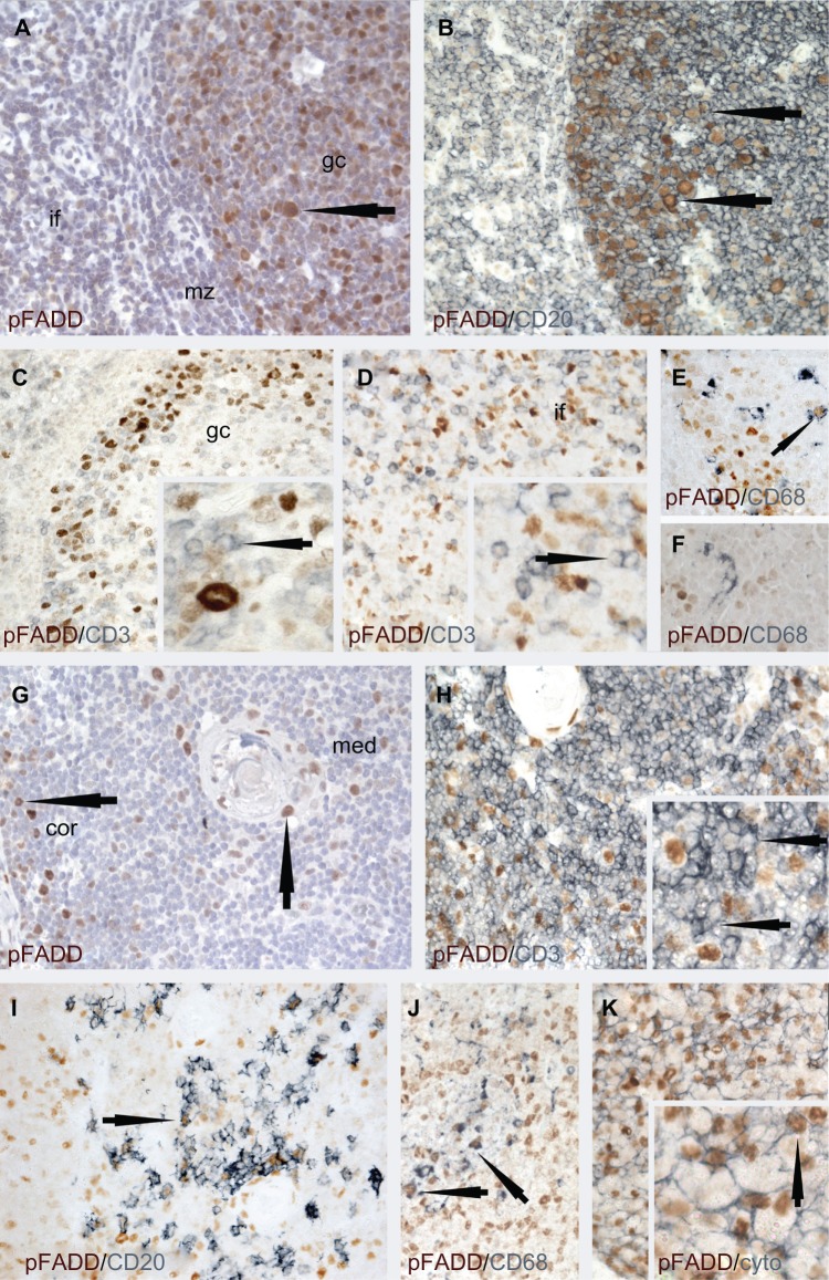
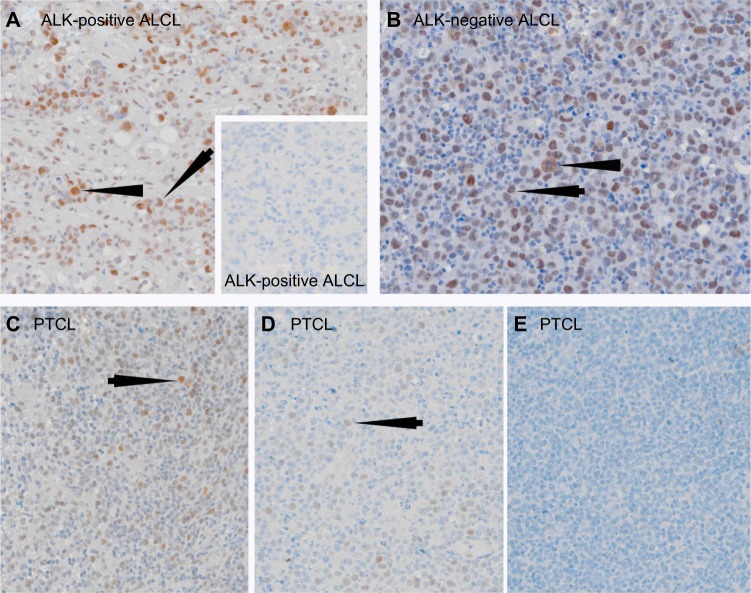
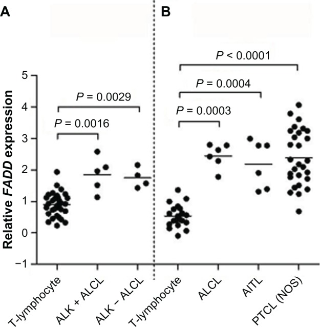
FAS-associated protein with death domain (FADD) is a major adaptor protein involved in extrinsic apoptosis, embryogenesis, and lymphocyte homeostasis. Although abnormalities of the FADD/death receptor apoptotic pathways have been established in tumorigenesis, fewer studies have analyzed the expression and role of phosphorylated FADD (pFADD). Our identification of FADD as a lymphoma-associated autoantigen in T-cell lymphoma patients raises the possibility that pFADD, with its correlation with cell cycle, may possess role(s) in human T-cell lymphoma development. This immunohistochemical study investigated pFADD protein expression in a range of normal tissues and lymphomas, particularly T-cell lymphomas that require improved therapies. Whereas pFADD was expressed only in scattered normal T cells, it was detected at high levels in T-cell lymphomas (eg, 84% anaplastic large cell lymphoma and 65% peripheral T cell lymphomas, not otherwise specified). The increased expression of pFADD supports further study of its clinical relevance and role in lymphomagenesis, highlighting phosphorylation of FADD as a potential therapeutic target.

Keywords: ALCL; FADD; PTCL; autoantigen; lymphoma; pFADD.


Figures

Figure 1
Figure 1
Immunolabeling studies to show pFADD protein expression in tonsil (AF) and thymus (GJ). Notes: Tonsil: (A) Single immunoperoxidase labeling shows heterogeneity of pFADD expression in the nuclei of subpopulations of tonsil cells. Strong labeling is present in a subpopulation of germinal center (gc) cells (arrowed). Scattered cells with weaker labeling are present in the mantle zone (mz) and interfollicular (if) areas. (B) It can be seen from the double immunoperoxidase labeling studies that pFADD (brown) is heterogeneously expressed in the majority of CD20-positive (blue) germinal center B cells (arrowed) but (C) absent from the majority of CD3-positive (blue) T cells in the germinal center (inset, arrowed) and (D) interfollicular areas (inset, arrowed). (E) Nuclear pFADD expression (brown) is also present in the occasional CD68-positive tingible body macrophage (blue, arrowed) while (F) pFADD was not detected in the interfollicular CD68-positive macrophages (arrowed). Thymus: From (G) it can be seen that the expression of pFADD is heterogenous and limited to subpopulations of cells (arrowed) in the thymic medulla (med) and cortex (cor). (H) pFADD was undetectable in the majority of the CD3-positive 9blue) thymocytes (inset, arrowed), pFADD was undetectable in the majority of the CD3-positive (blue) thymocytes (inset, arrowed). (I) Strong pFADD expression (brown) was, however, detected in (I) some CD20-positive B cells (blue, arrowed), (J) CD68-positive macrophages (blue, arrowed), and (K) the cytokeratin-positive (blue) thymic epithelial cells (inset, arrowed).
Figure 2
Figure 2
Single immunoperoxidase labeling demonstrating pFADD expression in ALCL and PTCL (NOS). Notes: (A) An example of a case of ALK-positive ALCL where the tumor cells express pFADD (arrowed). Note the presence of pFADD in the cytoplasm of mitotic cells (arrowhead). The inset shows an example of a pFADD-negative case of ALK-positive ALCL. (B) Strong nuclear pFADD expression is also present in a case of ALK-negative ALCL (arrowed). Cytoplasmic pFADD is also present in a mitotic cell (arrowhead). Different examples of pFADD in three cases of PTCL are shown in (CE). While the majority of tumor cells are pFADD-positive (arrowed) in (C), the percentage drops to less than 30% in (D) while the PTCL in (E) lacks detectable pFADD.
Figure 3
Figure 3
FADD transcript expression in T-cell lymphomas and ALCL. Notes: FADD transcript was expressed at significantly higher levels in T-cell lymphomas than in nonmalignant T-cell populations in two independent gene expression profiling datasets: (A) normal T-lymphocytes, n = 31; ALCL (anaplastic large cell lymphoma), n = 9 (five cases of ALK-positive ALCL and four cases of ALK-negative ALCL); and (B) normal T-lymphocytes, n = 20 (including five samples each of CD4-positive and CD8-positive T-lymphocytes); ALCL, n = 6; AITL (angioimmunoblastic T-cell lymphoma), n = 6; PTCL (NOS) (peripheral T-cell lymphoma, not otherwise specified), n = 28.

References

    1. Horn S, Lueking A, Murphy D, et al. Profiling humoral autoimmune repertoire of dilated cardiomyopathy (DCM) patients and development of a disease-associated protein chip. Proteomics. 2006;6(2):605–13. - PubMed
    1. Kijanka G, Murphy D. Protein arrays as tools for serum autoantibody marker discovery in cancer. J Proteomics. 2009;72(6):936–44. - PubMed
    1. Kijanka G, Hector S, Kay EW, et al. Human IgG antibody profiles differentiate between symptomatic patients with and without colorectal cancer. Gut. 2010;59(1):69–78. - PubMed
    1. Wang X, Yu J, Sreekumar A, et al. Autoantibody signatures in prostate cancer. N Engl J Med. 2005;353:1224–35. - PubMed
    1. Cooper CD, Liggins AP, Ait-Tahar K, Roncador G, Banham AH, Pulford K. PASD1, a DLBCL-associated cancer testis antigen and candidate for lymphoma immunotherapy. Leukemia. 2006;20(12):2172–4. - PubMed

LinkOut - more resources