. 2014 Sep 3:8:259.
doi: 10.3389/fncel.2014.00259. eCollection 2014.

In vivo knockdown of Piccolino disrupts presynaptic ribbon morphology in mouse photoreceptor synapses

Affiliations

In vivo knockdown of Piccolino disrupts presynaptic ribbon morphology in mouse photoreceptor synapses

Hanna Regus-Leidig et al. Front Cell Neurosci. .

Abstract

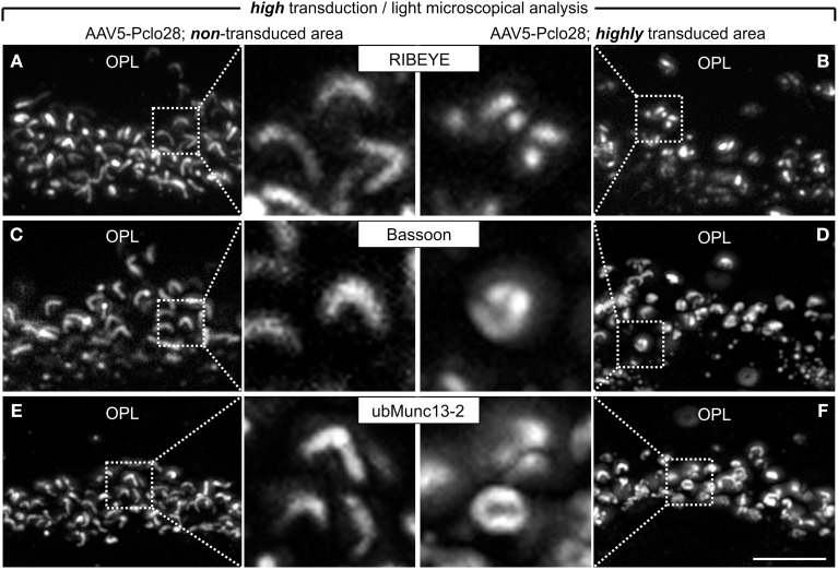
Piccolo is the largest known cytomatrix protein at active zones of chemical synapses. A growing number of studies on conventional chemical synapses assign Piccolo a role in the recruitment and integration of molecules relevant for both endo- and exocytosis of synaptic vesicles, the dynamic assembly of presynaptic F-actin, as well as the proteostasis of presynaptic proteins, yet a direct function in the structural organization of the active zone has not been uncovered in part due to the expression of multiple alternatively spliced isoforms. We recently identified Piccolino, a Piccolo splice variant specifically expressed in sensory ribbon synapses of the eye and ear. Here we down regulated Piccolino in vivo via an adeno-associated virus-based RNA interference approach and explored the impact on the presynaptic structure of mouse photoreceptor ribbon synapses. Detailed immunocytochemical light and electron microscopical analysis of Piccolino knockdown in photoreceptors revealed a hitherto undescribed photoreceptor ribbon synaptic phenotype with striking morphological changes of synaptic ribbon ultrastructure.

Keywords: Aczonin; Piccolino; Piccolo; RIBEYE; active zone; retina; ribbon synapse.


Figures

Figure 1
Figure 1
Targeted delivery of Piccolo shRNA to rod photoreceptors of the mouse retina via subretinal injection of AAV5. (A) Schematic multidomain structure of the conventional and ribbon synaptic Piccolo variants (Piccolo and Piccolino, respectively), and location and sequence of the shRNA (Pclo28) used to down regulate Piccolino. (B) In vivo fluorescence fundus photography showing mouse opsin promoter driven GFP expression in transduced rod photoreceptors 8 weeks post injection (8w p.i.). The asterisk demarcates the approximate site of injection where the transduction rate is highest. (C,D) Images of vertical sections through an AAV5-Pclo28-transduced retina displaying high (C) and low (D) transduction rate, stained with an antibody against GFP (green) and the antibody Pclo44a, labeling both Piccolo and Piccolino (magenta). ONL: outer nuclear layer; OPL: outer plexiform layer; INL: inner nuclear layer; IPL: inner plexiform layer; GCL: ganglion cell layer. Scale bar in (D) for (C,D): 20 μm.
Figure 2
Figure 2
Piccolino knockdown efficiency in rod photoreceptor terminals. (A,B) Staining of GFP (green) and Piccolino (magenta; antibody Pclo44a) in vertical cryostat sections through retinal areas with low (A) and high (B) AAV5-Pclo28-transduction rates 8w p.i. (C,D) 3D reconstructions of the GFP and Piccolino staining of the boxed regions in (A) and (B). (E) Staining of GFP (green) and Piccolino (magenta; antibody Pclo44a) in an AAV5-Pclo28-transduced whole-mounted retina 8w p.i. Projection of a confocal image stack through the outer plexiform layer (OPL). (F) Higher magnification of the boxed region in (E). GFP positive rod spherules are outlined with dotted lines. (G) Quantification of Piccolino fluorescence staining intensity in non-transduced vs. AAV5-Pclo28-transduced rod photoreceptor terminals. Bars show the mean fluorescence intensity ± SD normalized to non-transduced terminals (***p < 0.001; t-test). Scale bar in (A,B): 5 μm, in (E): 10 μm.
Figure 3
Figure 3
Whole-mount preparations of AAV5-Pclo28-transduced retinae and analysis of the Piccolino-knockdown effect on RIBEYE, Bassoon, and ubMunc13-2 in rod photoreceptor terminals. (A-C) Double labeling of GFP (green) with RIBEYE (magenta; A), Bassoon (magenta; B), and ubMunc13-2 (magenta; C) in AAV5-Pclo28-transduced whole-mounted retinae 8w p.i. Projections of confocal image stacks from the OPL. Arrows in (A) point to irregular RIBEYE staining. GFP positive rod spherules are outlined with dotted lines. (D) Quantification of the lateral extension (= active zone length) of RIBEYE, Bassoon, and ubMunc13-2 staining in non-transduced vs. AAV5-Pclo28-transduced rod photoreceptor terminals. Bars show the mean extension in nm (± SD). (E) Percentage of disrupted or punctate RIBEYE, Bassoon, and ubMunc13-2 staining in non-transduced vs. AAV5-Pclo28-transduced rod photoreceptor terminals. OPL: outer plexiform layer. Scale bar in (C) for (A–C): 2 μm.
Figure 4
Figure 4
Ultrastructural analysis of C57BL/6 rod photoreceptor ribbon synapses in retinal areas with low AAV5-Pclo28-transduction rates. (A–C) Representative electron micrographs of non-transduced (A), AAV5-GFP-transduced (B; control), and AAV5-Pclo28-transduced (C) rod photoreceptor terminals from retinal areas with low transduction rate. Pre-embedding immunolabeling of GFP in the transduced photoreceptor terminals (B,C). Arrowheads point to synaptic ribbons, the asterisk demarcates spherical, membrane-associated ribbon material. bc: bipolar cell; hc: horizontal cell; mt: mitochondrion. Scale bar in (C) for (A–C): 0.5 μm.
Figure 5
Figure 5
Vertical sections through retinal areas with high AAV5-Pclo28-transduction rates and analysis of the Piccolino-knockdown effect on RIBEYE, Bassoon, and ubMunc13-2 in rod photoreceptor terminals. (A–F) Staining for RIBEYE (A,B), Bassoon (C,D), and ubMunc13-2 (E,F) in AAV5-Pclo28-transduced retina 8w p.i. For a given staining, images were taken from a non-transduced (A,C,E) and a highly transduced area (B,D,F) of the same retinal slice. GFP fluorescence is not shown. OPL: outer plexiform layer. Scale bar in (F) for (A–F): 5 μm.
Figure 6
Figure 6
Ultrastructural analysis of C57BL/6 rod photoreceptor ribbon synapses in retinal areas with high AAV5-Pclo28-transduction rates. (A) Representative electron micrographs of rod photoreceptor terminals from a highly AAV5-Pclo28-transduced retinal area, processed for best tissue preservation. The asterisks demarcate spherical, membrane-associated ribbon material. (B) Quantification of four different shapes of ribbon profiles—plate-shaped, club-shaped, floating and membrane-attached spheres—in rod photoreceptor terminals from highly AAV5-Pclo28-transduced, non-transduced, and AAV5-GFP-transduced (control) retinal areas. hc: horizontal cell. Scale bar in (A): 0.2 μm.
Figure 7
Figure 7
Ultrastructural analysis of rod photoreceptor ribbon synapses in the AAV5-Pclo28-transduced BALB/c retina. (A) Percentages of the four different shapes of ribbon profiles—plate-shaped, club-shaped, floating and membrane-attached spheres—in non-transduced and AAV5-Pclo28-transduced rod photoreceptor terminals of BALB/c mice after 3 h light and dark adaptation. (B–E) Representative electron micrographs of rod photoreceptor ribbon synapses in non-transduced 3 h light (B) and dark adapted (C), and in AAV5-Pclo28-transduced 3 h light (D) and dark adapted (E) BALB/c mouse retina. Arrowheads point to plate-shaped synaptic ribbons, asterisks demarcate free floating or membrane attached ribbon spheres. (F–I) 3D-reconstructions of representative rod photoreceptor ribbon synapses from non-transduced 3 h light (F) and dark adapted (G), and from AAV5-Pclo28-transduced 3 h light (H) and dark adapted (I) BALB/c mouse retina. Synaptic ribbon material is shown in black, ribbon-tethered vesicles in blue. bc: bipolar cell; hc: horizontal cell. Scale bar in (E) for (B–E) and in (I) for (F–I): 0.5 μm.
Figure 8
Figure 8
Schematic illustration of the effect of the absence of Bassoon and Piccolino on the structure of the rod photoreceptor ribbon synaptic complex. (A) Plate-shaped and anchored ribbon in a wild-type rod photoreceptor synapse. (B) Free floating ribbon in a Bassoon-mutant rod photoreceptor synapse. (C) Membrane attached spherical ribbon in a Piccolino knockdown photoreceptor synapse. Presynaptic proteins and their localization to either the ribbon (green) or the arciform density compartment (red) of the ribbon synaptic complex are depicted in the legend. Bassoon (blue) is the molecular link between the two compartments.

Similar articles

Cited by

References

    1. Adly M. A., Spiwoks-Becker I., Vollrath L. (1999). Ultrastructural changes of photoreceptor synaptic ribbons in relation to time of day and illumination. Invest. Ophthalmol. Vis. Sci. 40, 2165–2172 - PubMed
    1. Altrock W. D., tom Dieck S., Sokolov M., Meyer A. C., Sigler A., Brakebusch C., et al. (2003). Functional inactivation of a fraction of excitatory synapses in mice deficient for the active zone protein bassoon. Neuron 37, 787–800 10.1016/S0896-6273(03)00088-6 - DOI - PubMed
    1. Beltran W. A., Boye S. L., Boye S. E., Chiodo V. A., Lewin A. S., Hauswirth W. W., et al. (2010). rAAV2/5 gene-targeting to rods: dose-dependent efficiency and complications associated with different promoters. Gene Ther. 17, 1162–1174 10.1038/gt.2010.56 - DOI - PMC - PubMed
    1. Cooper B., Hemmerlein M., Ammermüller J., Imig C., Reim K., Lipstein N., et al. (2012). Munc13-independent vesicle priming at mouse photoreceptor ribbon synapses. J. Neurosci. 32, 8040–8052 10.1523/JNEUROSCI.4240-11.2012 - DOI - PMC - PubMed
    1. Dick O., Hack I., Altrock W. D., Garner C. C., Gundelfinger E. D., Brandstätter J. H. (2001). Localization of the presynaptic cytomatrix protein Piccolo at ribbon and conventional synapses in the rat retina: comparison with Bassoon. J. Comp. Neurol. 439, 224–234 10.1002/cne.1344 - DOI - PubMed

LinkOut - more resources