Review
. 2014 Sep 15;6(9):330-43.
doi: 10.4251/wjgo.v6.i9.330.

Radiology of pancreatic neoplasms: An update

Affiliations
Review

Radiology of pancreatic neoplasms: An update

Luis Gijón de la Santa et al. World J Gastrointest Oncol. .

Abstract

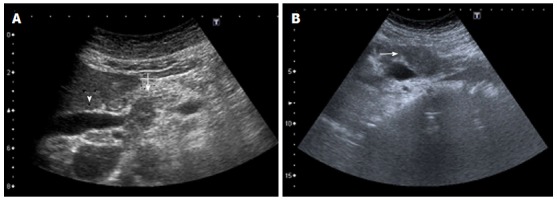
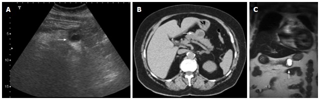
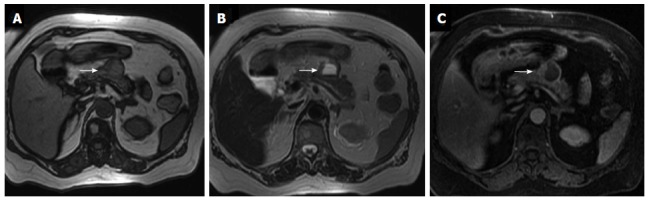
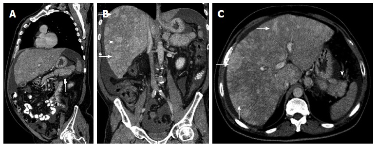
Diagnostic imaging is an important tool to evaluate pancreatic neoplasms. We describe the imaging features of pancreatic malignancies and their benign mimics. Accurate detection and staging are essential for ensuring appropriate selection of patients who will benefit from surgery and for preventing unnecessary surgeries in patients with unresectable disease. Ultrasound, multidetector computed tomography with multiplanar reconstruction and magnetic resonance imaging can help to do a correct diagnosis. Radiologists should be aware of the wide variety of anatomic variants and pathologic conditions that may mimic pancreatic neoplasms. The knowledge of the most important characteristic key findings may facilitate the right diagnosis.

Keywords: Computed tomography; Magnetic resonance imaging; Pancreas; Pancreatic neoplasms; Radiology; Surgery; cancer.


Figures

Figure 1
Figure 1
Multidetector computed tomography image. Multidetector computed tomography shows enlargement of the pancreatic head (arrow), with dilatation and beading of the pancreatic duct (arrowhead) and dilatation of the extra- and intrahepatic bile ducts. A focal calcification can also be visualized. These findings matched with the definite diagnosis of a chronic pancreatitis.
Figure 2
Figure 2
Axial contrast enhanced multidetector computed tomography image. A: Depicts a nodular peripancreatic mass localized between the pancreatic tail (arrowhead) and the splenic hilum (arrow), each well separated by fat planes; B: The sagittal reformatted contrast enhanced multidetector computed tomography image allows a better identification of the surrounding fat planes (arrow and arrowhead) enabling the exclusion of a pancreatic dependency. This mass actually turned out to be a tumoral implant of a gastric neoplasm.
Figure 3
Figure 3
Unresectablility of a pancreatic adenocarcinoma. Contrast enhanced multidetector computed tomography (MDCT) image (A) and coronal reformation image (B) shows dilatation of the distal pancreatic duct caused by a hypodense tumor (arrow) in the pancreatic body. On plain film (C) and coronal reformation image on MDCT (D) of the same patient multiple lung metastases of his pancreatic carcinoma are evident - a definite criteria for unresectability.
Figure 4
Figure 4
Axial contrast enhanced multidetector computed tomography image. Arterial phase imaging allows optimal visualization of the pancreatic neoplasm and peripancreatic arteries: the shown hypodense mass compromises the splenic artery (arrow). Pancreatic adenocarcinoma was proven by biopsy.
Figure 5
Figure 5
Contrast enhanced multidetector computed tomography image. A: In portal venous phase depicts a mass (arrow) in the pancreatic tail with permeability of the splenic vein (arrowhead); B: Focal round focal hypodensities with different sizes, localized in both hepatic lobules, represent metastatic spread to the liver. Pancreatic adenocarcinoma was proven by biopsy.
Figure 6
Figure 6
Axial multidetector computed tomography image. Pancreatic tumor, localized in the pancreatic head (arrow), is hypodense in relation to the pancreatic parenchyma after contrast administration.
Figure 7
Figure 7
Indirect signs of pancreatic neoplasms. Transverse ultrasound image (A) shows a markedly dilated common bile duct, also seen on the coronal reformation image of multidetector computed tomography (B) where the dilated duct terminates abruptly at the level of the pancreatic head (arrow).
Figure 8
Figure 8
Endoscopic retrograde cholangiopancreatography. A short segment of narrowing causing stenosis of the common bile duct was recognized (arrow), without affection of the main pancreatic duct (arrowhead). Pancreatic adenocarcinoma was proven by biopsy.
Figure 9
Figure 9
Axial contrast enhanced multidetector computed tomography image A: Focal hypodense mass in the body of the pancreas (arrow), with upstream dilatation of the main pancreatic duct (arrowhead). Pacreatic adenocarcinoma was histologically proven; B: Image depicts a circumferential soft tissue cuff around the celiac trunk according to vascular invasion (arrow); C: Image shows multiple peritoneal metastases in a patient with a pancreatic tumor (arrow).
Figure 10
Figure 10
Adenocarcinoma has low signal intensity on T1 (A) and T2 (B) weighted magnetic resonance imaging (arrows).
Figure 11
Figure 11
Axial arterial-phase gadolinium-enhanced T1-weighted fat-suppressed gradient-recalled echo magnetic resonance imaging (A) and coronal reformatted (B) show no enhancement of the hypovascular tumor in the pancreatic head (arrow). Pancreatic adenocarcinoma was proven by biopsy.
Figure 12
Figure 12
Use of diffusion-weighted magnetic resonance imaging in the earlier detection of pancreatic tumours. Axial contrast enhanced multidetector computed tomography image (A) and axial arterial-phase gadolinium-enhanced T1-weighted fat-suppressed gradient-recalled echo magnetic resonance image (B) do not depict any abnormality. Axial diffusion-weighted magnetic resonance imaging (C) demonstrates a focal increased signal intensity (arrow) and low apparent diffusion coefficient values in the color coded images (D).
Figure 13
Figure 13
Ultrasound images (A, B) of an ill-defined, heterogeneous hypoechoic mass (arrow) in the pancreas obstructing the common bile duct (arrowhead).
Figure 14
Figure 14
Pancreatic neuroendocrine tumor. Ultrasound images (A), axial unenhanced multidetector computed tomography and coronal magnetic resonance T2-weighted image show a round, heterogeneous mass, localized in the pancreatic body, with variable amounts of cystic-necrotic degeneration (arrows).
Figure 15
Figure 15
Same patient shown in figure 14. Magnetic resonance axial gradient T1 out-of-phase image (A) and T1 fat-suppressed sequence (C) show a hypointense signal in the liquid component of the lesion whereas it reveals a hyperintense signal in the T2-weighted sequence (B) (arrows).
Figure 16
Figure 16
Sagittal multidetector computed tomography image. A: A heterogeneous pancreatic mass (arrow); B, C: Coronal (B) and axial (C) multidetector computed tomography images show multiple hypervascular metastases in the liver (arrows), showing the same enhancement pattern of the primary mass. Neuroendocrine pancreatic tumor and metastases were histologically proven.
Figure 17
Figure 17
Axial contrast enhanced multidetector computed tomography images (A, B) reveal a homogeneously hypodense intraparenchymal fluid collection of the pancreas without any non-liquefied material in it, encapsulated completely by a thin slightly hyperdense layer (arrows). These findings are compatible with a pseudocyst in a patient with a clinical history of pancreatitis.
Figure 18
Figure 18
Axial nonenhanced multidetector computed tomography image. A: A polylobulated cystic lesion with a coarse calcification in its center (arrow), which is the phathognomonic central scar for serous cystadenoma; B-D: Magnetic resonance imaging show a cluster of small cysts (arrows), which are hypointense in T1-weighted images (B) and hyperintense in T2-weighted images (C, D), without visible communication within the cyst or the pancreatic duct. A central signal void is also identifiable.
Figure 19
Figure 19
Ultrasound and multidetector computed tomography images. On ultrasound (A, B) a hypoechogenic multilocular mass with well-definable internal spetations and posterior acoustic enhancement can be seen. Contrast-enhanced multidetector computed tomography images (C-F) show a big round to slightly lobulated mass with an enhancing capsule and different levels of attenuation within the cyst cavities are seen. Some enhancing components are also detectable.
Figure 20
Figure 20
Multidetector computed tomography image. A: Cystic dilatation of the main pancreatic duct and some of its branches in the pancreatic tail. Ductal communication with the tumor cannot be clearly identified; B-D: In contrast-enhanced axial T1 (B) and T2-weighted (C) magnetic resonance images and in magnetic resonance imaging cholangiography (D) ductal communication can be easily detectable.
Figure 21
Figure 21
Oblique reformatted enhanced multidetector computed tomography image reveals a well-defined round mass in the pancreas, slightly hypodense to the pancreatic parenchyma. Pancreatic metastases from melanoma was proven. Note the liver concomitant metastases.

Similar articles

Cited by

References

    1. Lu DS, Reber HA, Krasny RM, Kadell BM, Sayre J. Local staging of pancreatic cancer: criteria for unresectability of major vessels as revealed by pancreatic-phase, thin-section helical CT. AJR Am J Roentgenol. 1997;168:1439–1443. - PubMed
    1. Brennan DD, Zamboni GA, Raptopoulos VD, Kruskal JB. Comprehensive preoperative assessment of pancreatic adenocarcinoma with 64-section volumetric CT. Radiographics. 2007;27:1653–1666. - PubMed
    1. Low G, Panu A, Millo N, Leen E. Multimodality imaging of neoplastic and nonneoplastic solid lesions of the pancreas. Radiographics. 2011;31:993–1015. - PubMed
    1. Ros PR, Mortelé KJ. Imaging features of pancreatic neoplasms. JBR-BTR. 2001;84:239–249. - PubMed
    1. Ross BA, Jeffrey RB, Mindelzun RE. Normal variations in the lateral contour of the head and neck of the pancreas mimicking neoplasm: evaluation with dual-phase helical CT. AJR Am J Roentgenol. 1996;166:799–801. - PubMed

LinkOut - more resources