. 2014 Sep 6;4(5):508-17.
eCollection 2014.

Epigenetic silencing of dual oxidase 1 by promoter hypermethylation in human hepatocellular carcinoma

Affiliations

Epigenetic silencing of dual oxidase 1 by promoter hypermethylation in human hepatocellular carcinoma

Qingxia Ling et al. Am J Cancer Res. .

Abstract

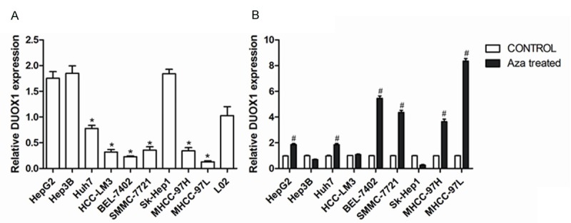
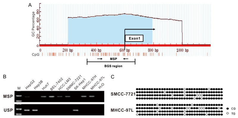
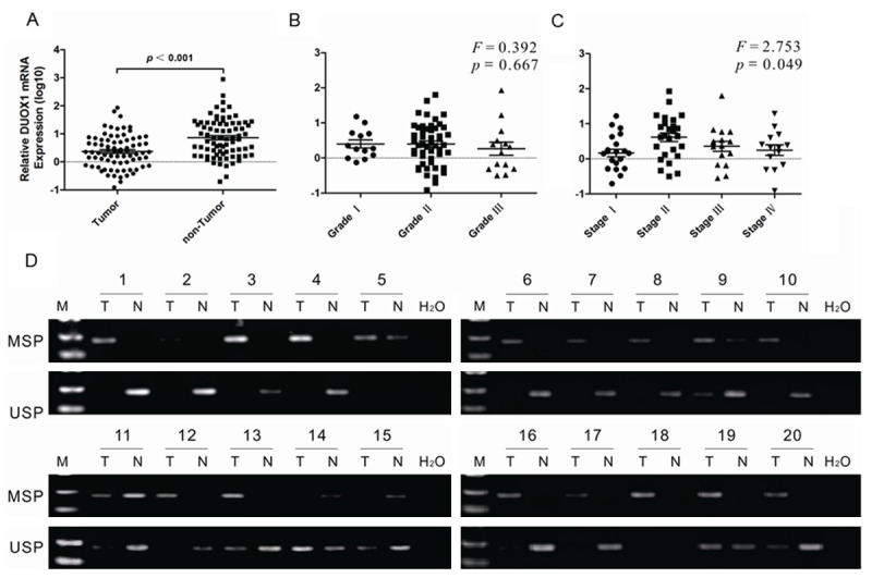
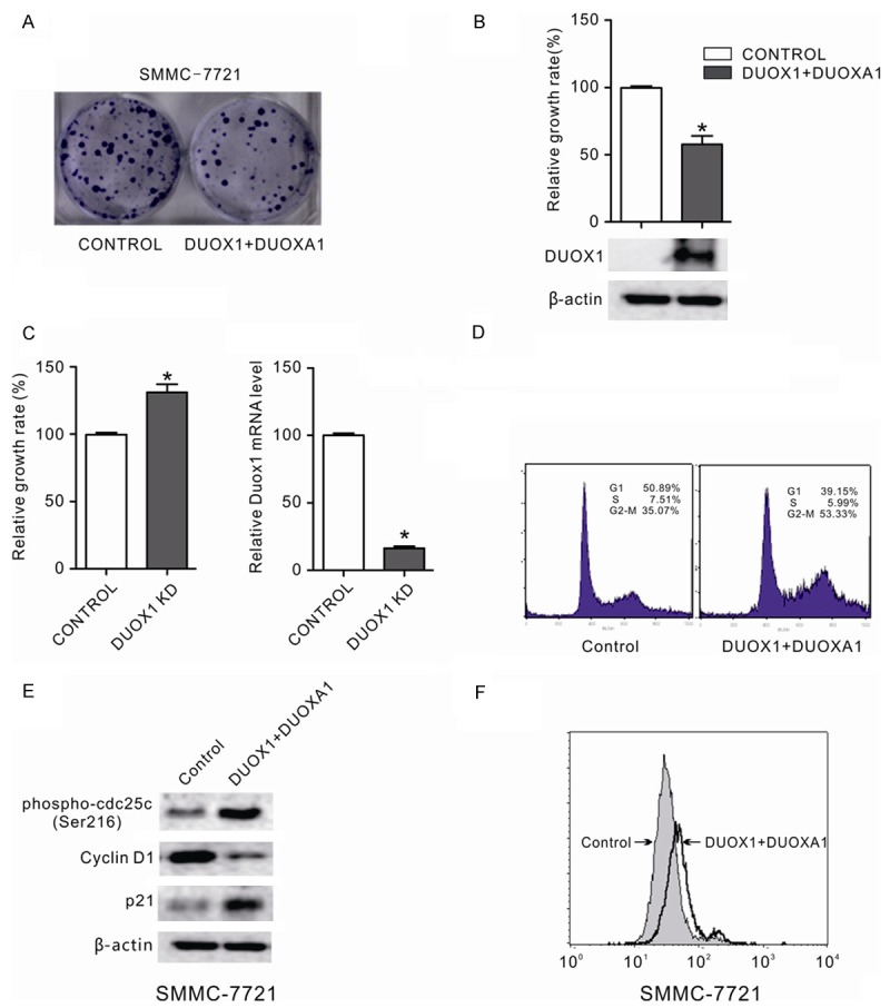
Dual oxidase 1 (DUOX1), which is the main sources for reactive oxygen species (ROS) production in the airway, are frequently silenced in human lung cancer. In poorly differentiated follicular thyroid carcinoma, a high expression of DUOX1 was associated with a reduced risk of death. However, the role of DUOX1 in human hepatocellular carcinoma (HCC) is still not clear. Here, we investigated DUOX1 expression and its promoter methylation status in primary HCC. To date, We found that expression of DUOX1 was decreased significantly in 76.9% (60/78) human hepatocellular carcinoma and 66.7% (6/9) liver cancer cell lines, compared with the paired adjacent non-tumor tissues and immortalized normal cell line. Moreover, which was well correlated with its promoter methylation status. Methylation was further detected in primary HCC, but none or occasionally in paired adjacent non-tumor tissues. Detailed methylation analysis of 35 CpG sites at a 324-bp promoter region by bisulfi te genomic sequencing (BGS) confi rmed its methylation. DUOX1 silencing could be reversed by chemical demethylation treatment with 5-aza-2'-deoxycytidine (5-Aza-dC), indicating direct epigenetic silencing. Restoring DUOX1 expression in lowly expressed cancer cells signifi cantly inhibited cancer cells growth and colony formation ability through the induction of G2/M phase cell cycle arrest and an increase in ROS generation, while knockdown of DUOX1 could markedly promote cancer cells proliferation. In conclusion, we demonstrate that epigenetic silencing of DUOX1 via promoter hypermethylation is common in human liver cancer cells and primary HCC and DUOX1 appears to be a functional tumor suppressor involved in liver carcinogenesis.

Keywords: DUOX1; ROS; hepatocellular carcinoma; methylation.


Figures

Figure 1
Figure 1
Pharmacological demethylation reversed DUOX1 downregulation in human liver cancers. A: DUOX1 expression in human liver cancer cell lines and normal control was determined by real-time PCR. B: Relative DUOX1 expression level in cancer cells before and after 5-Aza-dC treatment was determined by real-time PCR. Data are mean ± SEM from three independent experiments. *P < 0.05, versus L02; #P < 0.05, versus CONTROL.
Figure 2
Figure 2
Methylation of DUOX1 promoter in human liver cancers. (A) Schematic structure of the DUOX1 CGI, with the exon 1 and MSP and BGS region indicated. Each short vertical line represents one CpG site. The position of MSP primers were marked as arrows. The methylation status of the DUOX1 CGI was analyzed by MSP (B) and BGS (C). MSP = Methylation-specific PCR; USP = Unmethylation-specific PCR, the gels have been run under the same experimental conditions. For BGS (C), each circle indicates one CpG site and circles filled in black represent methylated CpG sites. One row of circles represents a single colony.
Figure 3
Figure 3
DUOX1 was downregulated and hypermethylated in primary HCC tissues. The expression of DUOX1 in HCC tissues and adjacent non-tumor tissues was determined by real-time PCR. A: 78 pairs of samples were from liver tissue, including tumor tissue and adjacent non-tumor tissue. B: The expression of DUOX1 mRNA in different Edmondson stage of primary HCC tissues. C: The expression of DUOX1 mRNA in different TNM stage of primary HCC tissues. D: The methylation status of DUOX1 promoter in primary HCC tissues and adjacent non-tumor tissues was detected by MSP. Representative results were shown. “T” indicates tumor tissues and “N” represents adjacent non-tumor tissues. The gels have been run under the same experimental conditions.
Figure 4
Figure 4
DUOX1 suppressed the growth of cancer cells by inducing G2/M phase cell cycle arrest and increasing ROS production. A: The effect of ectopic DUOX1 expression on cancer cell growth was investigated by the monolayer colony formation assay. The scanned colony formation in six-well plates was shown in the panel. B: The growth of cancer cells after DUOX1 overexpression was determined by MTS cell growth assay. Ectopic expression of DUOX1 suppressed the growth of cancer cells. DUOX1 expression level in transfected SMMC-7721 cells was confi rmed by western blot. C: Knockdown of DUOX1 promoted Hep3B cells proliferation. Hep3B cells were transfected with DUOX1 siRNA (100 nM) or NC siRNA (100 nM). After 48h, the cell growth was determined by MTS and knockdown efficiency was confirmed by real-time PCR. D: Effect of Duox1 on the cell cycle in liver cancer cell. The cell cycle distribution of liver cancer cell line SMMC-7721 with and without DUOX1 expression was evaluated by flow cytometry analysis. E: Effect of Duox1 on cell cycle regulators (phospho-cdc25 (Ser216), p21 and cyclin D1) were determined using western blotting. β-actin was used as the internal control. F: Exogenous DUOX1 increased ROS level in liver cancer cells. The effect of ectopic DUOX1 expression on oxidative stress was determined by flow cytometry assay as shown. Stable cells were stained with DCFH-DA and then the redox state of cells was measured by flow cytometry. Data are mean ± SEM from three independent experiments. The gels have been run under the same experimental conditions.

References

    1. Jones PA, Baylin SB. The fundamental role of epigenetic events in cancer. Nat Rev Genet. 2002;3:415–428. - PubMed
    1. Esteller M. CpG island hypermethylation and tumor suppressor genes: a booming present, a brighter future. Oncogene. 2002;21:5427–5440. - PubMed
    1. Baylin SB, Ohm JE. Epigenetic gene silencing in cancer - a mechanism for early oncogenic pathway addiction? Nat Rev Cancer. 2006;6:107–116. - PubMed
    1. Sauer H, Wartenberg M, Hescheler J. Reactive oxygen species as intracellular messengers during cell growth and differentiation. Cell Physiol Biochem. 2001;11:173–186. - PubMed
    1. De Deken X, Wang D, Many MC, Costaqliola S, Libert F, Vassart G, Dumont JE, Miot F. Cloning of two human thyroid cDNAs encoding new members of the NADPH oxidase family. J Biol Chem. 2000;275:23227–23233. - PubMed

LinkOut - more resources