

Incidental findings of pericardial calcification
- PMID: 25232549
- PMCID: PMC4163768
- DOI: 10.12998/wjcc.v2.i9.455
Incidental findings of pericardial calcification
Abstract
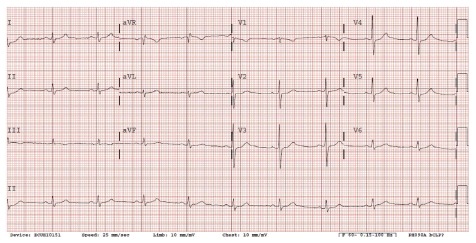
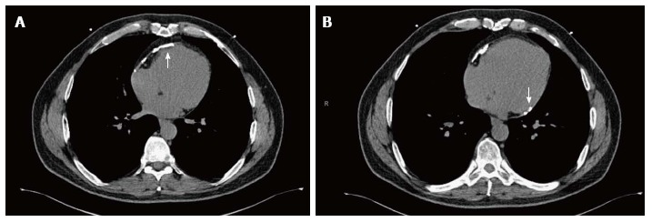
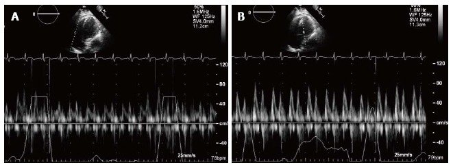
Calcification of the pericardium is a relatively rare finding and often has an uncertain etiology. Incidental findings of pericardial calcification may increase due to widespread application of cardiac computed tomography for the assessment of coronary atherosclerosis in the appropriate clinical setting using coronary artery calcium scoring and/or coronary angiography. Pericardial calcification alone is asymptomatic and is neither necessary nor sufficient for the diagnosis of pericardial constriction. Its presence may suggest of diffused pericardial scarring and consequently, its pathological involvement with pericardial constriction. Calcification of the pericardium must be evaluated with full clinical knowledge to facilitate an accurate diagnosis and an appropriate therapy when required. Our objective is to present a case of asymptomatic pericardial calcification and to discuss the importance of its clinical implications.
Keywords: Cardiac computed tomography; Constrictive pericarditis; Pericardial calcification.
Figures
References
-
- Mathewson FA. Calcification of the pericardium in apparently healthy people; electrocardiographic abnormalities found in tracings from apparently healthy persons with calcification of the pericardium. Circulation. 1955;12:44–51. - PubMed
-
- Isaacs D, Stark P, Nichols C, Antevil J, Shabetai R. Post-traumatic pericardial calcification. J Thorac Imaging. 2003;18:250–253. - PubMed
-
- Sengupta PP, Eleid MF, Khandheria BK. Constrictive pericarditis. Circ J. 2008;72:1555–1562. - PubMed
-
- Ling LH, Oh JK, Breen JF, Schaff HV, Danielson GK, Mahoney DW, Seward JB, Tajik AJ. Calcific constrictive pericarditis: is it still with us? Ann Intern Med. 2000;132:444–450. - PubMed
-
- Ferguson EC, Berkowitz EA. Cardiac and pericardial calcifications on chest radiographs. Clin Radiol. 2010;65:685–694. - PubMed
Publication types
LinkOut - more resources
Full Text Sources
Other Literature Sources
