Review
. 2014 Sep 17;83(6):1284-302.
doi: 10.1016/j.neuron.2014.08.041.

Toward a genetic dissection of cortical circuits in the mouse

Affiliations
Review

Toward a genetic dissection of cortical circuits in the mouse

Z Josh Huang. Neuron. .

Abstract

The mammalian neocortex gives rise to a wide range of mental activities and consists of a constellation of interconnected areas that are built from a set of basic circuit templates. Major obstacles to understanding cortical architecture include the diversity of cell types, their highly recurrent local and global connectivity, dynamic circuit operations, and a convoluted developmental assembly process rooted in the genome. With our increasing knowledge of gene expression and developmental genetic principles, it is now feasible to launch a program of genetic dissection of cortical circuits through systematic targeting of cell types and fate mapping of neural progenitors. Strategic design of even a modest number of mouse driver lines will facilitate efforts to compile a cell type parts list, build a Cortical Cell Atlas, establish experimental access to modern tools, integrate studies across levels, and provide coordinates for tracing developmental trajectory from circuit assembly to functional operation.


Figures

Figure 1
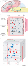
Figure 1. The basic architecture of the neocortical sheet
A). The Swanson model of the cerebral hemisphere highlights the elementary circuit elements characteristic to many cortical areas. It consists of a triple descending projection to the motor system of the brainstem and spinal cord, with feedback to the cortex and basal ganglia via thalamus. This elemental circuit is topographically organized and differentially elaborated in cortical areas that correspond to sensory, motor, emotive, and cognitive function (Adapted and modified from Swanson, 2005). B). Schematic showing the functional organization an unfolded cortical sheet of the right hemisphere and some of its related structures: the cortical sheet (gray) is surrounded by limbic cortices (brown, e.g. cingulate cortex, insula) and their associated nuclei (red, e.g. amygdala). The concept presented here is that the components of behavior are systematically distributed across this regular sheet. Dynamically evolving behaviors are represented schematically as graphical structures composed of ‘nodes’, the regions of active processing, and ‘edges’, which represent the axonal communication channels between active nodes. Multiple behaviors may evolve simultaneously (blue graph), while the red graph represents the various functional relations of the behavior currently being executed. See Douglas & Martin 2012 for more detailed description. (Adapted and modified from Douglas & Martin 2012). C) Simplified schematic representation of the neocortical microcircuitry, including major cell types and synaptic connections. Excitatory neurons and synapses (V-shapes) are in red, inhibitory neurons and synapses (small filled circles) are in blue. Dashed circles depict afferent and efferent extracortical brain regions. Inhibitory synapses onto pyramidal neurons (PC) are displayed according to the target domains: axonal inhibition is provided by chandelier cells (CHC), somatic inhibition by basket cells (BC), and dendritic inhibition by double-bouquet cells (DBC), bipolar cells (BP), neurogliaform cells (NGC), Martinotti cells (MNC) and Cajal-Retzius cells (CRC). PCs projecting to different brain areas reside in different layers: layer 5 PCs project to subcortical regions such as the brainstem (Bs), spinal cord (SC), superior colliculus, basal ganglia (BG) and thalamus (TH). Layer 6 PCs project mainly to the thalamus and claustrum (CL), and PCs in superficial layers project to other cortical targets, such as neighbouring columns and the contralateral cortical hemisphere. SSC, spiny stellate cell; WM, white matter. (Adapted and modified from Markram et al, 2005.)
Figure 2
Figure 2. Cortical areal and laminar patterning
A) Inductive signals (i.e. morphogens) secreted from several patterning centers provide positional information for areal patterning along major tangential axis in the ventricular zone (VZ). R-rostral, C-caudal, M-medial, L-lateral. B) VZ progenitors located at different mediolateral and rostrocaudal coordinates express specific levels of transcription factors, which combinatorially establish a protomap of cortical areas in the VZ. This protomap is later translated, in part through radial glia cells and their neuronal progenies, into a definitive area map in the cortical plate (CP). In addition to cortical intrinsic mechanisms, the subsequent areal differentiation patterns are also regulated by extrinsic influences, such as thalamocortical afferents. C) Schematic of a coronal hemisection of embryonic telencephalon at approximately E13, showing an overview of VZ and CP and the pattern of radial migration of PyNs. The boxed region depicts radial glia fibers linking VZ and CP. D) Schematic depiction of radial units and the generation of PyNs that contributes to laminar patterning through an 'inside-out' sequence of radial migration. Radial glias (RG) in the VZ produce PyNs and also intermediate progenitors (IPs). IPs establish the subventricular zone (SVZ) and act as transit-amplifying cells to increase neuronal production. Later born PyNs migrate along radial glial processes and pass their predecessors to reach their final laminar destinations. Cajal–Retzius (CR) cells primarily migrate into neocortical layer I from non-cortical locations. (Part of B and D are adapted and modified from Greig et al., 2013).
Figure 3
Figure 3. Subpallial generation and tangential migration of cortical GABAergic interneurons
A) A schematic of coronal hemisection through a mid-gestation embryonic mouse brain, showing in different colors the distinct progenitor cell domains of the telencephalon, and highlighting the approximate expression patterns of selected transcription factors that are implicated in regulating telencephalic patterning and differentiation. The dorsal-ventral gradients of Gli3 and Shh signaling contribute to the patterning of progenitor domains along the ventricle wall. AEP, anterior entopeduncular area; DP, dorsal pallium; LGE, lateral ganglionic eminence; LP, lateral pallium; MGE, medial ganglionic eminence; MP, medial pallium; POA, anterior preoptic area; VP, ventral pallium. B) GABAergic neurons are generated from MGE and CGE (not shown) and migrate over long distance to reach piriform cortex (Pcx), neocortex (Ncx), hippocampus (H), and striatum (Str). (A and B are modified from Marin and Rubenstein 2001). C) Schematic showing that migrating GABAergic neurons enter the developing cortex from the marginal zone (MZ) and cortical VZ/SVZ. They then switch to radial migration to reach their proper laminar destination and integrate with PyNs.
Figure 4
Figure 4. Genetic targeting of PyNs and fate mapping of progenitors
A) Different types of PyNs represent inter-areal, inter-hemisphere processing streams and cortio-striatal, cortico-fugal output channels. Several major types are depicted on a schematic sagittal (top left) and coronal (top right) section (adapted from Molyneaux et al., 2007); their laminar location, characteristic dendritic morphology and axon projection are schematized in the bottom panel. B) Several dozen genes (a few representatives are listed here) show laminar restricted expression, but whether these mRNA expression correlate with PyN subtypes are mostly unknown. Combinatorial genetic targeting by intersection (AND) and subtraction (NOT) of expression patterns may identify and capture specific PyN subtypes. C) All PyNs are generated from neural progenitors (e.g. radial glial cells – RGCs, intermediate progenitors-IPC), but the lineage progression from progenitors to PyN subtypes is not well understood. It is unclear whether the same progenitor can generate all subtypes, or there exist fate-restricted progenitors for certain subtypes. D) Genetic fate mapping using temporally controlled and lineage-restricted combinatorial TF drivers may clarify lineage progression and enable tracking the developmental trajectory of PyN subtypes.
Figure 5
Figure 5. Genetic targeting of GABA subtypes and fate mapping of GABA progenitors
A) A generic diagram depicting several subtypes of GABAergic interneurons, with their characteristic cellular and subcellular targets, and molecular markers. The soma location of these interneurons are not accurately represented. ISC, interneuron selective cell; LPC, long projection cell. Other abbreviations are the same as in Figure 1. SOM, somatostatin; AAc, alpha-actinin-2; CR, calretinin; VIP, vasoactive intestinal polypeptide; CCK, cholecystokinin; PV, parvalbumin; NPY, neuropeptide Y; nNOS, neuronal nitric oxide synthase; SPR, substance P receptor. B) Neurochemical markers expressed in cortical GABAergic neurons. No single markers correlate with any individual morpho-physiological GABA subtypes depicted in A); combination of 2 or more markers may significantly increase the correlation. Except for PV, SOM, 5-HT3aR, the size of other shapes represented by a marker does not accurately correlate with the proportion of the marked cell population. C) Strategies for combinatorial targeting of GABAergic cell types. Top panel: intersection of SOM and CR likely target MNC, whereas intersection of SOM and nNOS(I) likely target LPC. Bottom panel: intersection of Ascl1 (TF of a population of IPCs) and PV likely target BSCs in different cortical layers depending the embryonic induction time of the Ascl1 driver. D) Schematic of subpallium progenitor domains in an embryonic brain and their flat sheet view. Spatial axis are: D-dorsal, V-ventral, R-rostral, C-caudal. E) Transcription factors expressed in LGE, MGE, POA. No single TF defines a progenitor subdomain in MGE. Combinatorial targeting with two TFs may capture subdomains of MGE and different progenitor types.
Figure 6
Figure 6. Different types of GABA interneuron types contribute to distinct cortical circuit modules
Although each molecular marker (PV, SOM, VIP, CCK) and the corresponding mouse driver line targets several cell types, a combination with viral approaches begins to enable multiple studies to reveal that interneurons with defined molecular characteristics, connectivity pattern, and physiological properties contribute to distinct circuit operation and behavioral function. Only the key features of cellular and subcellular connectivity are depicted for each cell type. See text for detailed description.
Figure 7
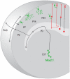
Figure 7. Genetic fate mapping allows tracking the developmental trajectory of chandelier cells
A). ChC identity (green) is specified from Nkx2.1+ progenitors (filled green circles) at the time of neurogenesis. Young ChCs appear to take stereotyped route and schedule in their migration and laminar deployment, before achieving specific innervation at axon initial segment of PyNs (red) which are generated from progenitor along the dorsal ventricle wall (filled red circles).

References

    1. Ackman JB, Crair MC. Role of emergent neural activity in visual map development. Current opinion in neurobiology. 2014;24:166–175. - PMC - PubMed
    1. Adesnik H, Bruns W, Taniguchi H, Huang ZJ, Scanziani M. A neural circuit for spatial summation in visual cortex. Nature. 2012;490:226–231. - PMC - PubMed
    1. Alaynick WA, Jessell TM, Pfaff SL. SnapShot: spinal cord development. Cell. 2011;146:178–178. e171. - PMC - PubMed
    1. Anderson SA, Eisenstat DD, Shi L, Rubenstein JL. Interneuron migration from basal forebrain to neocortex: dependence on Dlx genes. Science. 1997;278:474–476. - PubMed
    1. Angevine JB, Jr., Sidman RL. Autoradiographic study of cell migration during histogenesis of cerebral cortex in the mouse. Nature. 1961;192:766–768. - PubMed

Publication types

LinkOut - more resources