

Fiber-optic system for dual-modality imaging of glucose probes 18F-FDG and 6-NBDG in atherosclerotic plaques
- PMID: 25233472
- PMCID: PMC4169475
- DOI: 10.1371/journal.pone.0108108
Fiber-optic system for dual-modality imaging of glucose probes 18F-FDG and 6-NBDG in atherosclerotic plaques
Abstract
Background: Atherosclerosis is a progressive inflammatory condition that underlies coronary artery disease (CAD)-the leading cause of death in the United States. Thus, the ultimate goal of this research is to advance our understanding of human CAD by improving the characterization of metabolically active vulnerable plaques within the coronary arteries using a novel catheter-based imaging system. The aims of this study include (1) developing a novel fiber-optic imaging system with a scintillator to detect both 18F and fluorescent glucose probes, and (2) validating the system on ex vivo murine plaques.
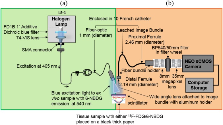
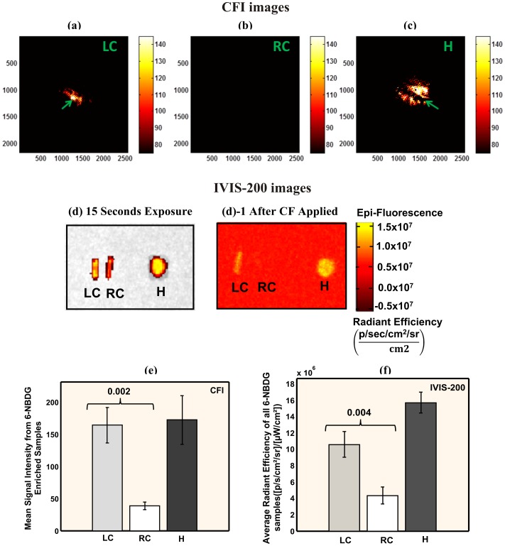
Methods: A novel design implements a flexible fiber-optic catheter consisting of both a radio-luminescence and a fluorescence imaging system to detect radionuclide 18F-fluorodeoxyglucose (18F-FDG) and the fluorescent analog 6-(N-(7-Nitrobenz-2-oxa-1,3-diazol-4-yl)amino)-6-Deoxyglucose (6-NBDG), respectively. Murine macrophage-rich atherosclerotic carotid plaques were imaged ex vivo after intravenous delivery of 18F-FDG or 6-NBDG. Confirmatory optical imaging by IVIS-200 and autoradiography were also performed.
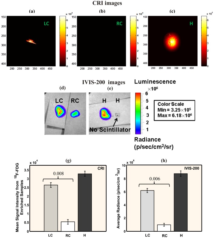
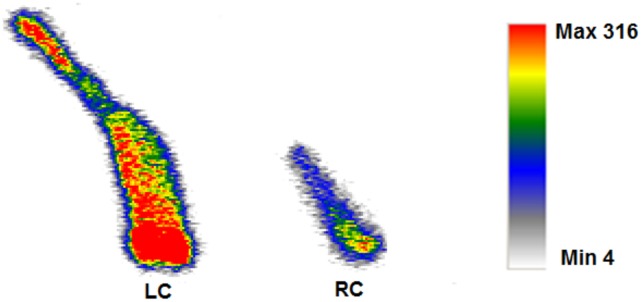
Results: Our fiber-optic imaging system successfully visualized both 18F-FDG and 6-NBDG probes in atherosclerotic plaques. For 18F-FDG, the ligated left carotid arteries (LCs) exhibited 4.9-fold higher radioluminescence signal intensity compared to the non-ligated right carotid arteries (RCs) (2.6 × 10(4) ± 1.4 × 10(3) vs. 5.4 × 10(3) ± 1.3 × 10(3) A.U., P = 0.008). Similarly, for 6-NBDG, the ligated LCs emitted 4.3-fold brighter fluorescent signals than the control RCs (1.6 × 10(2) ± 2.7 × 10(1) vs. 3.8 × 10(1) ± 5.9 A.U., P = 0.002). The higher uptake of both 18F-FDG and 6-NBDG in ligated LCs were confirmed with the IVIS-200 system. Autoradiography further verified the higher uptake of 18F-FDG by the LCs.
Conclusions: This novel fiber-optic imaging system was sensitive to both radionuclide and fluorescent glucose probes taken up by murine atherosclerotic plaques. In addition, 6-NBDG is a promising novel fluorescent probe for detecting macrophage-rich atherosclerotic plaques.
Conflict of interest statement
Figures
Similar articles
-
Scintillating balloon-enabled fiber-optic system for radionuclide imaging of atherosclerotic plaques.J Nucl Med. 2015 May;56(5):771-7. doi: 10.2967/jnumed.114.153239. Epub 2015 Apr 9. J Nucl Med. 2015. PMID: 25858046 Free PMC article.
-
Imaging cellular pharmacokinetics of 18F-FDG and 6-NBDG uptake by inflammatory and stem cells.PLoS One. 2018 Feb 20;13(2):e0192662. doi: 10.1371/journal.pone.0192662. eCollection 2018. PLoS One. 2018. PMID: 29462173 Free PMC article.
-
A Dual-Modality Hybrid Imaging System Harnesses Radioluminescence and Sound to Reveal Molecular Pathology of Atherosclerotic Plaques.Sci Rep. 2018 Jun 12;8(1):8992. doi: 10.1038/s41598-018-26696-8. Sci Rep. 2018. PMID: 29895966 Free PMC article.
-
Nuclear Imaging: Focus on Vascular Probes.Arterioscler Thromb Vasc Biol. 2019 Jul;39(7):1369-1378. doi: 10.1161/ATVBAHA.119.312586. Epub 2019 May 23. Arterioscler Thromb Vasc Biol. 2019. PMID: 31242032 Review.
-
Science to Practice: Does FDG Differentiate Morphologically Unstable from Stable Atherosclerotic Plaque?Radiology. 2017 Apr;283(1):1-3. doi: 10.1148/radiol.2017162495. Radiology. 2017. PMID: 28318446 Review.
Cited by
-
Gamma rays excited radioluminescence tomographic imaging.Biomed Eng Online. 2018 Apr 24;17(1):45. doi: 10.1186/s12938-018-0480-x. Biomed Eng Online. 2018. PMID: 29690883 Free PMC article.
-
Cellular binding and uptake of fluorescent glucose analogs 2-NBDG and 6-NBDG occurs independent of membrane glucose transporters.Biochimie. 2021 Nov;190:1-11. doi: 10.1016/j.biochi.2021.06.017. Epub 2021 Jul 2. Biochimie. 2021. PMID: 34224807 Free PMC article.
-
Scintillating balloon-enabled fiber-optic system for radionuclide imaging of atherosclerotic plaques.J Nucl Med. 2015 May;56(5):771-7. doi: 10.2967/jnumed.114.153239. Epub 2015 Apr 9. J Nucl Med. 2015. PMID: 25858046 Free PMC article.
-
Imaging inflammation and neovascularization in atherosclerosis: clinical and translational molecular and structural imaging targets.Curr Opin Cardiol. 2015 Nov;30(6):671-80. doi: 10.1097/HCO.0000000000000226. Curr Opin Cardiol. 2015. PMID: 26398413 Free PMC article. Review.
References
-
- Pennant M, Davenport C, Bayliss S, Greenheld W, Marshall T, et al. (2010) Community programs for the prevention of cardiovascular disease: a systematic review. Am J Epidemiol 172: 501–516. - PubMed
-
- Waxman S, Ishibashi F, Muller JE (2006) Detection and treatment of vulnerable plaques and vulnerable patients - Novel approaches to prevention of coronary events. Circulation 114: 2390–2411. - PubMed
-
- Yamaguchi Y, Patt BE, Iwanczyk JS, MacDonald LM, Mari C, et al. (2004) Performance of intravascular probe in animal studies. 2003 Ieee Nuclear Science Symposium, Conference Record, Vols 1–5: 2463–2467.
Publication types
MeSH terms
Substances
Grants and funding
LinkOut - more resources
Full Text Sources
Other Literature Sources
Medical
Miscellaneous
