

Optical coherence tomography for the diagnosis of neovascular age-related macular degeneration: a systematic review
- PMID: 25233820
- PMCID: PMC4268457
- DOI: 10.1038/eye.2014.214
Optical coherence tomography for the diagnosis of neovascular age-related macular degeneration: a systematic review
Abstract
The purpose is to study the diagnostic performance of optical coherence tomography (OCT) and alternative diagnostic tests for neovascular age-related macular degeneration (nAMD). Methods employed are as follows:systematic review and meta-analysis;
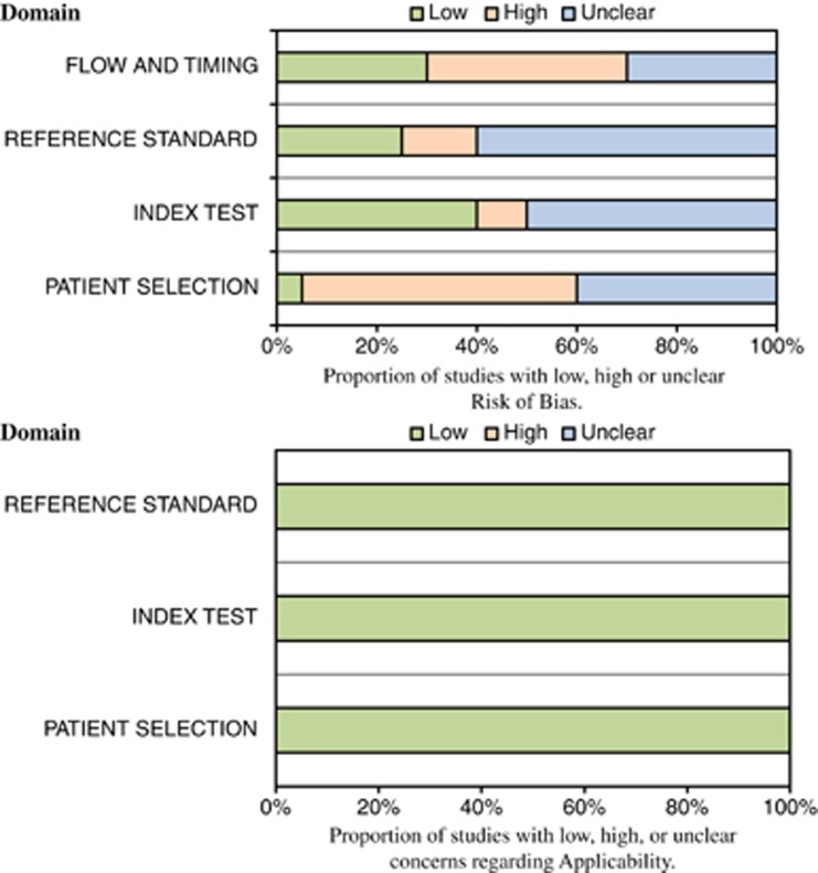
Index test: OCT including time-domain (TD-OCT) and the most recently developed spectral domain (SD-OCT); comparator tests: visual acuity, clinical evaluation (slit lamp), Amsler chart, colour fundus photographs, infra-red reflectance, red-free images/blue reflectance, fundus autofluorescence imaging (FAF), indocyanine green angiography (ICGA), preferential hyperacuity perimetry (PHP), and microperimetry; reference standard: fundus fluorescein angiography. Databases searched included MEDLINE, MEDLINE In Process, EMBASE, Biosis, SCI, the Cochrane Library, DARE, MEDION, and HTA database. Last literature searches: March 2013. Risk of bias assessed using QUADAS-2. Meta-analysis models were fitted using hierarchical summary receiver operating characteristic (HSROC) curves. Twenty-two studies (2 abstracts and 20 articles) enrolling 2124 participants were identified, reporting TD-OCT (12 studies), SD-OCT (1 study), ICGA (8 studies), PHP (3 studies), Amsler grid, colour fundus photography and FAF (1 study each). Most studies were considered to have a high risk of bias in the patient selection (55%, 11/20), and flow and timing (40%, 8/20) domains. In a meta-analysis of TD-OCT studies, sensitivity and specificity (95% CI) were 88% (46-98%) and 78% (64-88%), respectively. There was insufficient information to undertake meta-analysis for other tests. TD-OCT is a sensitive test for detecting nAMD, although specificity was only moderate. Data on SD-OCT are sparse. Diagnosis of nAMD should not rely solely on OCT.
Figures


References
-
- Brown DM, Michels M, Kaiser PK, Heier JS, Sy JP, Ianchulev T, et al. Ranibizumab versus verteporfin photodynamic therapy for neovascular age-related macular degeneration: Two-year results of the ANCHOR study. Ophthalmology. 2009;116:57–65. - PubMed
-
- Martin DF, Maguire MG, Fine SL, Ying GS, Jaffe GJ, Grunwald JE, et al. Comparison of Age-related Macular Degeneration Treatments Trials (CATT) Research Group. Ranibizumab and bevacizumab for treatment of neovascular age-related macular degeneration: two-year results. Ophthalmology. 2012;119:1388–1398. - PubMed
-
- Chakravarthy U, Harding SP, Rogers CA, Downes SM, Lotery AJ, the IVAN Study Investigators et al. Ranibizumab versus bevacizumab to treat neovascular age-related macular degeneration: one-year findings from the IVAN randomized trial. Ophthalmology. 2012;119:1399–1411. - PubMed
-
- Rosenfeld PJ, Brown DM, Heier JS, Boyer DS, Kaiser PK, Chung CY, et al. Ranibizumab for neovascular age-related macular degeneration. N Engl J Med. 2006;355:1419–1431. - PubMed
-
- Macular Photocoagulation Study Group Laser photocoagulation of subfoveal neovascular lesions in age-related macular degeneration. Results of a randomized clinical trial. Arch Ophthalmol. 1991;109:1220–1231. - PubMed
Publication types
MeSH terms
Substances
Grants and funding
LinkOut - more resources
Full Text Sources
Other Literature Sources
