

Cardiac PET perfusion tracers: current status and future directions
- PMID: 25234078
- PMCID: PMC4333146
- DOI: 10.1053/j.semnuclmed.2014.06.011
Cardiac PET perfusion tracers: current status and future directions
Abstract
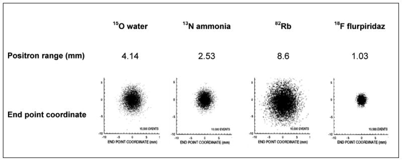
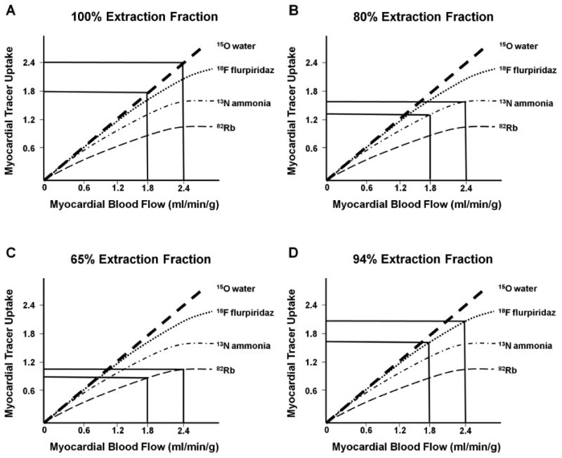
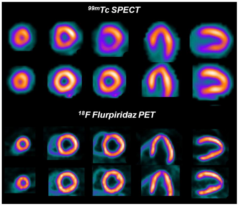
PET myocardial perfusion imaging (MPI) is increasingly being used for noninvasive detection and evaluation of coronary artery disease. However, the widespread use of PET MPI has been limited by the shortcomings of the current PET perfusion tracers. The availability of these tracers is limited by the need for an onsite ((15)O water and (13)N ammonia) or nearby ((13)N ammonia) cyclotron or commitment to costly generators ((82)Rb). Owing to the short half-lives, such as 76 seconds for (82)Rb, 2.06 minutes for (15)O water, and 9.96 minutes for (13)N ammonia, their use in conjunction with treadmill exercise stress testing is either not possible ((82)Rb and (15)O water) or not practical ((13)N ammonia). Furthermore, the long positron range of (82)Rb makes image resolution suboptimal and its low myocardial extraction limits its defect resolution. In recent years, development of an (18)F-labeled PET perfusion tracer has gathered considerable interest. The longer half-life of (18)F (109 minutes) would make the tracer available as a unit dose from regional cyclotrons and allow use in conjunction with treadmill exercise testing. Furthermore, the short positron range of (18)F would result in better image resolution. Flurpiridaz F 18 is by far the most thoroughly studied in animal models and is the only (18)F-based PET MPI radiotracer currently undergoing clinical evaluation. Preclinical and clinical experience with Flurpiridaz F 18 demonstrated a high myocardial extraction fraction, high image and defect resolution, high myocardial uptake, slow myocardial clearance, and high myocardial-to-background contrast that was stable over time-important properties of an ideal PET MPI radiotracer. Preclinical data from other (18)F-labeled myocardial perfusion tracers are encouraging.
Copyright © 2014. Published by Elsevier Inc.
Figures
Comment in
-
Letter from the guest Editor: update in Cardiovascular Nuclear Medicine (Part II).Semin Nucl Med. 2014 Sep;44(5):332. doi: 10.1053/j.semnuclmed.2014.07.002. Semin Nucl Med. 2014. PMID: 25234077 No abstract available.
References
-
- Dilsizian V, Taillefer R. Journey in evolution of nuclear cardiology: will there be another quantum leap with the F-18-labeled myocardial perfusion tracers? JACC Cardiovasc Imaging. 2012;5:1269–1284. - PubMed
-
- Rischpler C, Park MJ, Fung GS, Javadi M, Tsui BM, Higuchi T. Advances in PET myocardial perfusion imaging: F-18 labeled tracers. Ann Nucl Med. 2012;26:1–6. - PubMed
-
- Nekolla SG, Saraste A. Novel F-18-labeled PET myocardial perfusion tracers: bench to bedside. Curr Cardiol Rep. 2011;13:145–150. - PubMed
-
- Bergmann SR, Fox KA, Rand AL, et al. Quantification of regional myocardial blood flow in vivo with H215O. Circulation. 1984;70:724–733. - PubMed
-
- Garcia EV, Galt JR, Faber TL, et al. Principles of nuclear cardiology imaging. In: Dilsizian Vasken, Narula Jagat., editors. Atlas of Nuclear Cardiology. 4th edition. Springer Science; 2013. pp. 1–53. Chapter 1.
Publication types
MeSH terms
Substances
Grants and funding
LinkOut - more resources
Full Text Sources
Other Literature Sources
