

Blockade of excitatory synaptogenesis with proximal dendrites of dentate granule cells following rapamycin treatment in a mouse model of temporal lobe epilepsy
- PMID: 25234294
- PMCID: PMC4252507
- DOI: 10.1002/cne.23681
Blockade of excitatory synaptogenesis with proximal dendrites of dentate granule cells following rapamycin treatment in a mouse model of temporal lobe epilepsy
Abstract
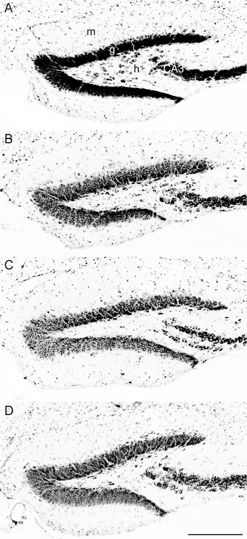
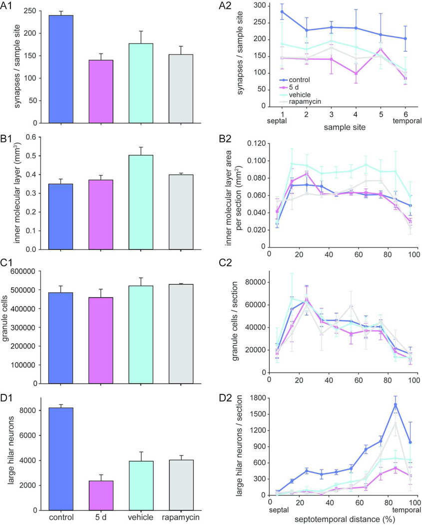
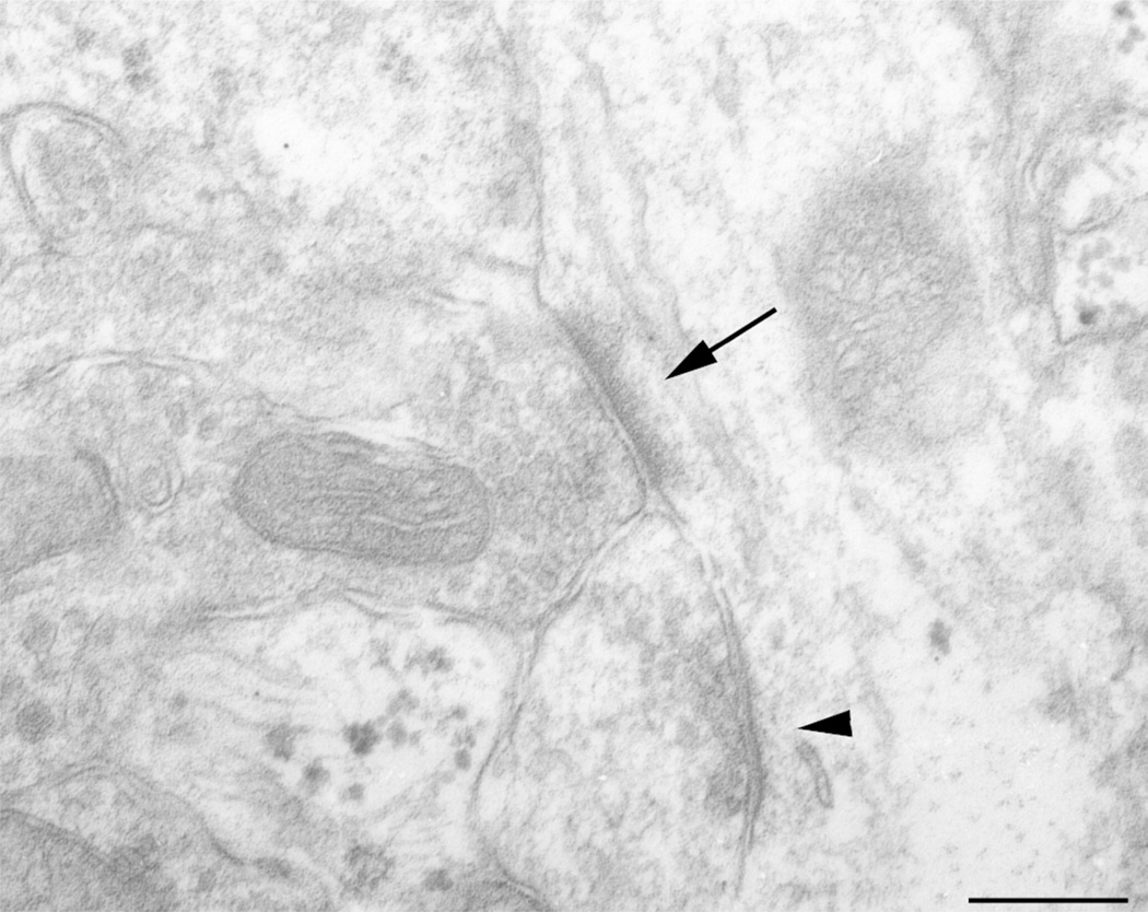
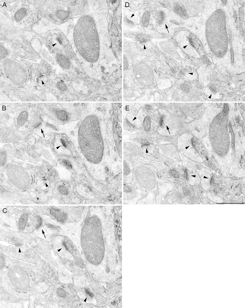
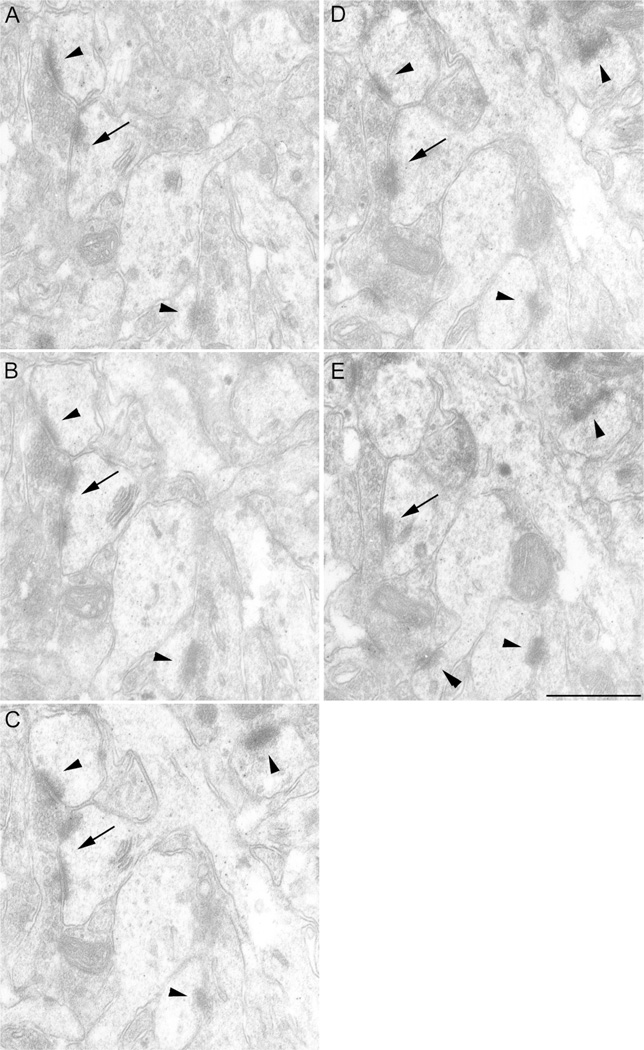
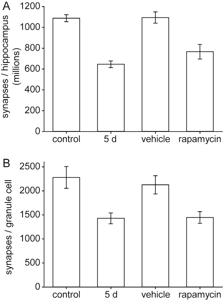
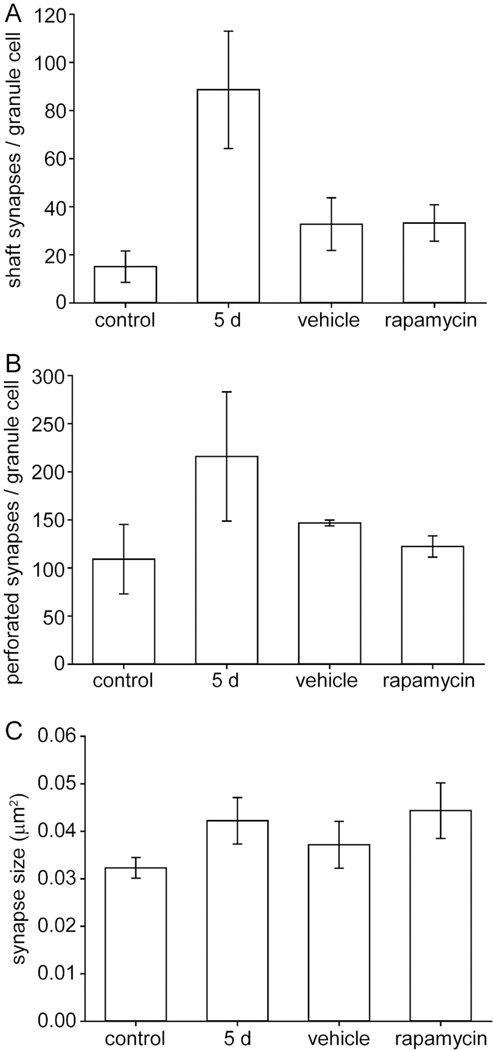
Inhibiting the mammalian target of rapamycin (mTOR) signaling pathway with rapamycin blocks granule cell axon (mossy fiber) sprouting after epileptogenic injuries, including pilocarpine-induced status epilepticus. However, it remains unclear whether axons from other types of neurons sprout into the inner molecular layer and synapse with granule cell dendrites despite rapamycin treatment. If so, other aberrant positive-feedback networks might develop. To test this possibility stereological electron microscopy was used to estimate the numbers of excitatory synapses in the inner molecular layer per hippocampus in pilocarpine-treated control mice, in mice 5 days after pilocarpine-induced status epilepticus, and after status epilepticus and daily treatment beginning 24 hours later with rapamycin or vehicle for 2 months. The optical fractionator method was used to estimate numbers of granule cells in Nissl-stained sections so that numbers of excitatory synapses in the inner molecular layer per granule cell could be calculated. Control mice had an average of 2,280 asymmetric synapses in the inner molecular layer per granule cell, which was reduced to 63% of controls 5 days after status epilepticus, recovered to 93% of controls in vehicle-treated mice 2 months after status epilepticus, but remained at only 63% of controls in rapamycin-treated mice. These findings reveal that rapamycin prevented excitatory axons from synapsing with proximal dendrites of granule cells and raise questions about the recurrent excitation hypothesis of temporal lobe epilepsy.
Keywords: electron microscopy; hippocampus; mossy cell; mossy fiber sprouting; pilocarpine; stereology.
Copyright © 2014 Wiley Periodicals, Inc.
Conflict of interest statement
The authors have no conflicts of interest.
Figures
Similar articles
-
Rapamycin suppresses axon sprouting by somatostatin interneurons in a mouse model of temporal lobe epilepsy.Epilepsia. 2011 Nov;52(11):2057-64. doi: 10.1111/j.1528-1167.2011.03253.x. Epub 2011 Aug 29. Epilepsia. 2011. PMID: 21883182 Free PMC article.
-
Is there a critical period for mossy fiber sprouting in a mouse model of temporal lobe epilepsy?Epilepsia. 2011 Dec;52(12):2326-32. doi: 10.1111/j.1528-1167.2011.03315.x. Epub 2011 Nov 16. Epilepsia. 2011. PMID: 22092282 Free PMC article.
-
Status epilepticus-induced hilar basal dendrites on rodent granule cells contribute to recurrent excitatory circuitry.J Comp Neurol. 2000 Dec 11;428(2):240-53. doi: 10.1002/1096-9861(20001211)428:2<240::aid-cne4>3.0.co;2-q. J Comp Neurol. 2000. PMID: 11064364
-
Synaptic connections of hilar basal dendrites of dentate granule cells in a neonatal hypoxia model of epilepsy.Epilepsia. 2012 Jun;53 Suppl 1:98-108. doi: 10.1111/j.1528-1167.2012.03481.x. Epilepsia. 2012. PMID: 22612814 Review.
-
Neuroplasticity in the damaged dentate gyrus of the epileptic brain.Prog Brain Res. 2002;136:319-28. doi: 10.1016/s0079-6123(02)36027-8. Prog Brain Res. 2002. PMID: 12143392 Review.
Cited by
-
Genetic Inhibition of Plppr5 Aggravates Hypoxic-Ischemie-Induced Cortical Damage and Excitotoxic Phenotype.Front Neurosci. 2022 Mar 24;16:751489. doi: 10.3389/fnins.2022.751489. eCollection 2022. Front Neurosci. 2022. PMID: 35401091 Free PMC article.
-
Combining Constitutively Active Rheb Expression and Chondroitinase Promotes Functional Axonal Regeneration after Cervical Spinal Cord Injury.Mol Ther. 2017 Dec 6;25(12):2715-2726. doi: 10.1016/j.ymthe.2017.08.011. Epub 2017 Aug 19. Mol Ther. 2017. PMID: 28967557 Free PMC article.
-
Editorial: The Developmental Seizure-Induced Hippocampal Mossy Fiber Sprouting: Target for Epilepsy Therapies?Front Neurol. 2019 Nov 13;10:1212. doi: 10.3389/fneur.2019.01212. eCollection 2019. Front Neurol. 2019. PMID: 31849807 Free PMC article. No abstract available.
-
Astrocyte Role in Temporal Lobe Epilepsy and Development of Mossy Fiber Sprouting.Front Cell Neurosci. 2021 Sep 30;15:725693. doi: 10.3389/fncel.2021.725693. eCollection 2021. Front Cell Neurosci. 2021. PMID: 34658792 Free PMC article. Review.
-
Total Number Is Important: Using the Disector Method in Design-Based Stereology to Understand the Structure of the Rodent Brain.Front Neuroanat. 2018 Mar 5;12:16. doi: 10.3389/fnana.2018.00016. eCollection 2018. Front Neuroanat. 2018. PMID: 29556178 Free PMC article. Review.
References
-
- Babb TL, Brown WJ, Pretorious J, Davenport C, Lieb JP, Crandall PH. Temporal lobe volumetric cell densities in temporal lobe epilepsy. Epilepsia. 1984;25:729–740. - PubMed
-
- Babb TL, Kupfer WR, Pretorius JK, Crandall PH, Levesque MF. Synaptic reorganization by mossy fibers in human epileptic fascia dentata. Neuroscience. 1991;42:351–363. - PubMed
-
- Blümcke I, Suter B, Behle K, Kuhn R, Schramm J, Elger CE, Wiestler OD. Loss of hilar mossy cells in Ammon’s horn sclerosis. Epilepsia. 2000;41(suppl 6):S174–S180. - PubMed
-
- Blümcke I, Zuschratter W, Schewe J-C, Suter B, Lie AA, Riederer BM, Meyer B, Schramm J, Elger CE, Wiestler OD. Cellular pathology of hilar neurons in Ammon’s horn sclerosis. J Comp Neurol. 1999;414:437–453. - PubMed
Publication types
MeSH terms
Substances
Grants and funding
LinkOut - more resources
Full Text Sources
Other Literature Sources
Miscellaneous
