. 2015 Feb;61(2):692-702.
doi: 10.1002/hep.27447.

Liver is the major source of elevated serum lipocalin-2 levels after bacterial infection or partial hepatectomy: a critical role for IL-6/STAT3

Affiliations

Liver is the major source of elevated serum lipocalin-2 levels after bacterial infection or partial hepatectomy: a critical role for IL-6/STAT3

Ming-Jiang Xu et al. Hepatology. 2015 Feb.

Abstract

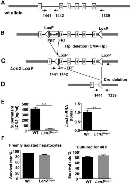
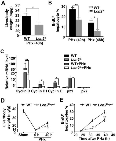
Lipocalin-2 (LCN2) was originally isolated from human neutrophils and termed neutrophil gelatinase-associated lipocalin (NGAL). However, the functions of LCN2 and the cell types that are primarily responsible for LCN2 production remain unclear. To address these issues, hepatocyte-specific Lcn2 knockout (Lcn2(Hep-/-)) mice were generated and subjected to bacterial infection (with Klesbsiella pneumoniae or Escherichia coli) or partial hepatectomy (PHx). Studies of Lcn2(Hep-/-) mice revealed that hepatocytes contributed to 25% of the low basal serum level of LCN2 protein (∼ 62 ng/mL) but were responsible for more than 90% of the highly elevated serum LCN2 protein level (∼ 6,000 ng/mL) postinfection and more than 60% post-PHx (∼ 700 ng/mL). Interestingly, both Lcn2(Hep-/-) and global Lcn2 knockout (Lcn2(-/-)) mice demonstrated comparable increases in susceptibility to infection with K. pneumoniae or E. coli. These mice also had increased enteric bacterial translocation from the gut to the mesenteric lymph nodes and exhibited reduced liver regeneration after PHx. Treatment with interleukin (IL)-6 stimulated hepatocytes to produce LCN2 in vitro and in vivo. Hepatocyte-specific ablation of the IL-6 receptor or Stat3, a major downstream effector of IL-6, markedly abrogated LCN2 elevation in vivo. Furthermore, chromatin immunoprecipitation (ChIP) assay revealed that STAT3 was recruited to the promoter region of the Lcn2 gene upon STAT3 activation by IL-6.

Conclusion: Hepatocytes are the major cell type responsible for LCN2 production after bacterial infection or PHx, and this response is dependent on IL-6 activation of the STAT3 signaling pathway. Thus, hepatocyte-derived LCN2 plays an important role in inhibiting bacterial infection and promoting liver regeneration.


Figures

Figure 1
Figure 1. Targeted disruption of the mouse Lcn2 gene
(A) Structures of the WT type Lcn2 gene and positions of the PCR primers used to analyze gene expression. (B) Schematic drawing showing the positions of the flippase recognition target (FRT)-flanked NeoR in intron 1 and the loxP site preceding the Neo cassette in intron 1 as well as the loxP site downstream of exon 6. (C) Following the selection of neomycin-resistant ES cells (129/SvPas), these cells were introduced into C57BL/6 blastocysts. ES cell electroporation and blastocyst injection were performed. The Neo cassette was removed by crossing these mice with CMV-Flip mice. (D) Several steps were used to cross the Lcn2-floxed mice with albumin-Cre+ mice, generating hepatocyte-specific Lcn2 knockout mice (albumin Cre+ Lcn2flox/flox, Lcn2Hep−/−), in which exons 2 to 6 of the Lcn2 gene have been deleted in hepatocytes. (E) Hepatocytes were isolated from WT and Lcn2Hep−/− mice and cultured for 24 hours. LCN2 protein levels in supernatants were measured; and Lcn2 mRNA expression was measured by real-time PCR. (G) Freshly isolated hepatocytes from WT and Lcn2Hep−/− mice were subjected to trypan blue staining. Hepatocytes survival rates (%) were calculated. In addition, WT and Lcn2Hep−/− hepatocytes were cultured for 48 h and hepatocytes survival rates were assessed by LDH release assay. Values represent the means ± SEM (n=4). **P<0.01, ***P<0.001.
Figure 2
Figure 2. Lcn2Hep−/− mice present lower levels of serum and hepatic LCN2 than WT mice following bacterial infection or PHx
(A, B) WT and Lcn2Hep−/− mice were infected with K. pneumoniae or E. coli, and serum were collected to measure LCN2 protein levels. Various organs were collected 24 hours post-infection and subjected to real-time PCR analyses for Lcn2 mRNA. (C) WT and Lcn2Hep−/− mice were subjected to PHx. Serum LCN2 protein levels were measured using an ELISA kit, and hepatic Lcn2 mRNA was measured with real-time PCR analysis. Values represent the means ± SEM (n=6-10). *P<0.05, **P<0.01, ***P<0.001.
Figure 3
Figure 3. Lcn2Hep−/− and Lcn2−/− mice demonstrate greater mortality and organ damage post-bacterial infection than WT mice
WT, Lcn2−/−, and Lcn2Hep−/− mice were infected with K. pneumoniae or E. coli. (A) The survival rate was measured (P<0.05 WT vs Lcn2Hep−/− or Lcn2−/−). (B) Blood bacterial counts were determined 24 hours post-infection. (C-D) Representative histology images of lung and liver tissues 24 hours post-infection. (E-F) Serum ALT and AST levels were measured 24 hours post-infection. Values represent the means ± SEM (n=6-10), *P<0.05, **P<0.01.
Figure 4
Figure 4. PHx-induced liver regeneration is suppressed in Lcn2Hep−/− and Lcn2−/− mice
(A, B) WT and Lcn2−/− mice were subjected to PHx. Liver/body weight ratios and the percentages of BrdU+ hepatocytes were determined 40 hours or 48 hours post-PHx. (C) Real-time PCR analyses of liver tissues from sham or PHx mice 40 hours post-PHx. (D, E) WT and Lcn2Hep−/− mice were subjected to PHx. Liver/body weight ratios and percentages of BrdU+ hepatocytes were determined post-PHx at the indicated time points. Values represent the means ± SEM (n=5-11), *P<0.05, **P<0.01 in comparison with corresponding WT groups.
Figure 5
Figure 5. Lcn2Hep−/− and Lcn2−/− mice exhibit increased bacterial translocation in MLNs post-PHx compared to WT mice
WT and Lcn2−/− mice (A, B) or WT and Lcn2Hep−/− mice (C, D) were subjected to PHx without bacterial infection. MLN/body weight ratios were recorded, and bacterial loads in MLNs were measured to determine bacterial transolation. Values represent the means ± SEM (n=3-14), *P<0.05, ***P<0.001.
Figure 6
Figure 6. Hepatic IL-6R mediates hepatic LCN2 production during bacterial infection or after PHx
(A) Mice were infected with K. pneumoniae, and serum IL-6 and IL-22 protein levels were determined. (B) WT and IL-6RHep−/− mice were infected with K. pneumoniae, and serum LCN2 protein levels and blood bacterial counts were determined. (C, D) WT, IL-6−/−, and IL-6RHep−/− mice were subjected to PHx without bacterial infection, and serum LCN2 protein levels (panel C), MLN/body weight ratios and bacterial translocation in MLNs were determined 24 hours post-surgery (panel D). Values represent the means ± SEM (n=6-8), *P<0.05, **P<0.01, ***P<0.001
Figure 7
Figure 7. STAT3 plays an important role in promoting LCN2 production in hepatocytes after bacterial infection or PHx
(A) Hepatocytes were isolated from WT and Stat3Hep−/− mice and treated with IL-6, and LCN2 protein levels in the supernatant were determined. (B, C) WT and Stat3Hep−/− mice were injected with IL-6, and serum protein levels (panel B) and hepatic mRNA levels (panel C) for LCN2 were determined. (D) The STAT3 binding site is identified in the Lcn2 promoter region as shown in the left panel. A representative ChIP assay blot is shown in the right panel. Socs3 and G6Pase were used as positive and negative controls, respectively. (E) WT and Stat3Hep−/− mice were infected with K. pneumoniae, and serum LCN2 protein levels, hepatic Lcn2 mRNA levels, and blood bacterial counts were determined 24 hours post-infection. (F) WT and Stat3Hep−/− mice were subjected to PHx, and serum LCN2 protein levels were determined 24 hours post-surgery. Values represent the means ± SEM (n=6-8), *P<0.05; **P<0.01; ***P<0.001.
Fig. 8
Fig. 8. A model depicting the critical role of hepatocytes in the production of LCN2 protein post-bacterial infection or PHx
Hepatocytes contribute to 25% of the basal levels of LCN2 protein (approximately 62 ng/ml) but are primarily responsible for the production of the highly elevated serum LCN2 protein levels following bacterial infection or PHx. This production is mainly dependent on the activation of the hepatic STAT3 signaling pathway activated by IL-6. Thus, hepatocyte-derived LCN2 proteins play an important role in controlling bacterial infection and promoting liver regeneration.

Comment in

References

    1. Gao B, Jeong WI, Tian Z. Liver: An organ with predominant innate immunity. Hepatology. 2008;47:729–36. - PubMed
    1. Cowland JB, Borregaard N. Molecular characterization and pattern of tissue expression of the gene for neutrophil gelatinase-associated lipocalin from humans. Genomics. 1997;45:17–23. - PubMed
    1. Wu H, Santoni-Rugiu E, Ralfkiaer E, et al. Lipocalin 2 is protective against E. coli pneumonia. Respir Res. 2010;11:96. - PMC - PubMed
    1. Kjeldsen L, Johnsen AH, Sengelov H, et al. Isolation and primary structure of NGAL, a novel protein associated with human neutrophil gelatinase. J Biol Chem. 1993;268:10425–32. - PubMed
    1. Chakraborty S, Kaur S, Guha S, et al. The multifaceted roles of neutrophil gelatinase associated lipocalin (NGAL) in inflammation and cancer. Biochim Biophys Acta. 2012;1826:129–69. - PMC - PubMed

Publication types