. 2014 Apr;135(4):1941-9.
doi: 10.1121/1.4868389.

Adaptation of distortion product otoacoustic emissions predicts susceptibility to acoustic over-exposure in alert rabbits

Affiliations

Adaptation of distortion product otoacoustic emissions predicts susceptibility to acoustic over-exposure in alert rabbits

Anne E Luebke et al. J Acoust Soc Am. 2014 Apr.

Abstract

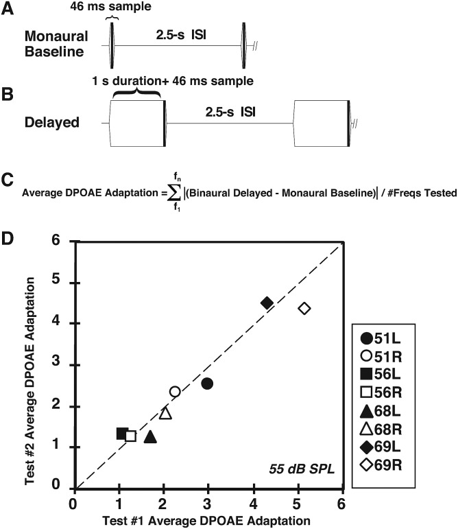
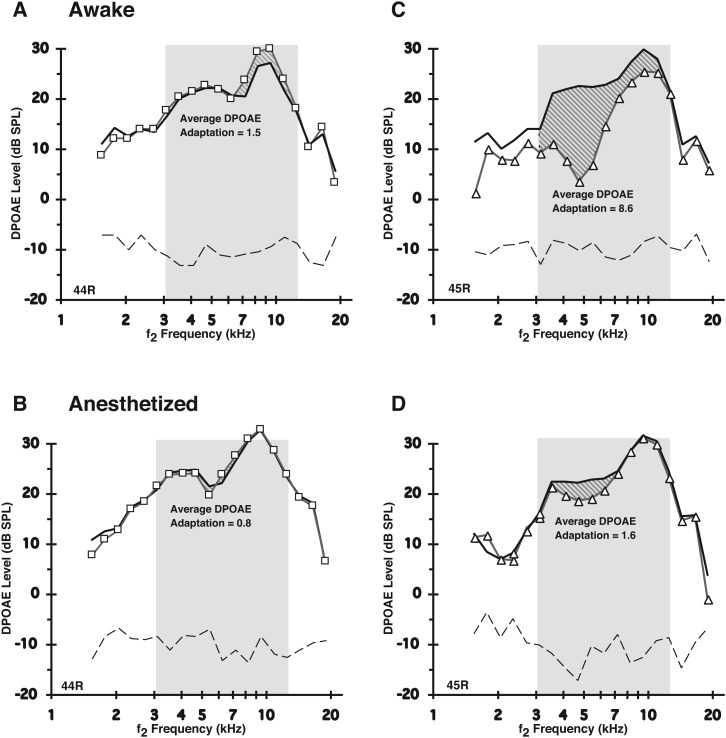
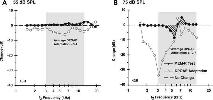
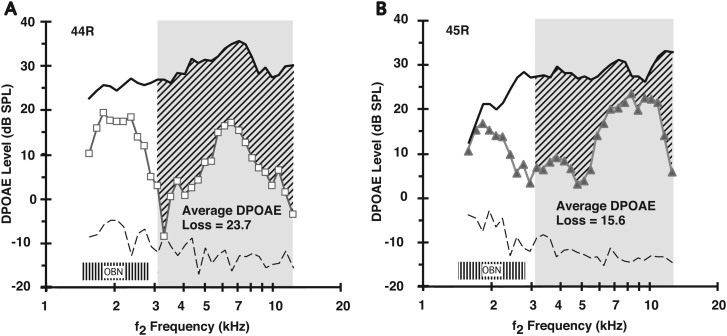
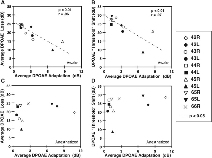
A noninvasive test was developed in rabbits based on fast adaptation measures for 2f1-f2 distortion-product otoacoustic emissions (DPOAEs). The goal was to evaluate the effective reflex activation, i.e., "functional strength," of both the descending medial olivocochlear efferent reflex (MOC-R) and the middle-ear muscle reflex (MEM-R) through sound activation. Classically, it is assumed that both reflexes contribute toward protecting the inner ear from cochlear damage caused by noise exposure. The DP-gram method described here evaluated the MOC-R effect on DPOAE levels over a two-octave (oct) frequency range. To estimate the related activation of the middle-ear muscles (MEMs), the MEM-R was measured by monitoring the level of the f1-primary tone throughout its duration. Following baseline measures, rabbits were subjected to noise over-exposure. A main finding was that the measured adaptive activity was highly variable between rabbits but less so between the ears of the same animal. Also, together, the MOC-R and MEM-R tests showed that, on average, DPOAE adaptation consisted of a combined contribution from both systems. Despite this shared involvement, the amount of DPOAE adaptation measured for a particular animal's ear predicted that ear's subsequent susceptibility to the noise over-exposure for alert but not for deeply anesthetized rabbits.


Figures

FIG. 1.
FIG. 1.
Determination of the average DPOAE adaptation index. (A) Schematic representation of the monaural DPOAE-adaptation measurement protocol. Essentially, two condensed DP-grams (f2 = 3–12.7 kHz) were obtained consisting of the monaural baseline measure and either the delayed-monaural or binaural measure. In the baseline condition, DPOAEs were routinely elicited monaurally from the test ear and their magnitudes measured at 46 ms after the ramped onset of the f1 and f2 primary tones. (B) Schematic showing the delayed DPOAE-adaptation condition in which DPOAEs were measured either monaurally or binaurally at about 1046 ms (1 s + 46 ms) after the presentation of the continuous f1 and f2 primary tones. For both the baseline and delayed-monaural or binaural conditions, a 2.5-ISI was interspersed between each pair of primary-tone presentations. (C) Equation showing the computation of the average DPOAE-adaptation index. That is, the absolute value of the difference between the monaural-baseline DPOAE level and the DPOAE level for either the delayed-monaural or binaural conditions was determined for each f2 frequency tested. This difference was then summed across the test frequencies and divided by the number of frequencies tested (n = 11) over a selected f2 range to yield the average DPOAE-adaptation index. (D) Scatterplot illustrating repeatability of determining the average DPOAE adaptation Index within each rabbit and the correspondence between right and left ears of an individual rabbit. The dotted diagonal line at 45° represents the condition in which the amount of the average DPOAE change from f2 = 3.1–12.3 kHz was identical between the right and left ears of the same rabbit.
FIG. 2.
FIG. 2.
Anesthesia suppressed the magnitude of the average DPOAE-adaptation index measured in alert rabbits. Shaded regions show, for two ears from different awake rabbits (A = right ear of rabbit 44 R; C = right ear of rabbit 45 R), the absolute DPOAE levels elicited by 55-dB SPL primaries for the baseline monaural (thick linewithout symbols) condition vs the delayed binaural condition (line with symbols), for f2 frequencies tested over the selected 2-oct interval designated by the slanted-line region in each plot. (B and D) These show counterpart responses for the same rabbits illustrated in (A) and (C), respectively, while under deep anesthesia. Note only the slight variability in baseline DPOAE levels between the two states of consciousness in contrast to the decreased adaptation index, i.e., for A (8.6) vs B (1.6) for rabbit 45 R, and for C (1.5) vs D (0.8) for rabbit 44 R. Also, note that anesthesia tended to make the average DPOAE-adaptation indices more similar between different rabbit subjects (see also numeric data in Table I).
FIG. 3.
FIG. 3.
DP-gram difference plots showing that the average DPOAE adaptation in the alert rabbit elicited by either 55 -(A) or 75-dB SPL (B) primaries was likely composed of both a presumed olivocochlear efferent reflex (MOC-R, open circles) plus a middle-ear muscle reflex (MEM-R, filled circles) component. The presumed “MEM-R” contribution to DPOAE adaptation measured with an f1 primary-tone constancy test (see text) increased with the higher 75-dB SPL sound pressure levels, yet was still detectable at 55 dB SPL, which is below the reported acoustic threshold for the MEM response in alert rabbits (see text). The bold dashed line at “0” on the ordinate represents a “no change” outcome in which the level of f1 stayed at a constant level between the monaural baseline and, in this case, the delayed-binaural condition.
FIG. 4.
FIG. 4.
Average amount of noise-induced loss in DPOAE levels was variable in different rabbits. The average DPOAE loss was plotted as a function of the f2 frequency to compute a DPOAE-loss index for each rabbit, here shown for both 44 R (A: open squares) and 45 R (B: closed triangles). Rabbit 44 R, which exhibited an average DPOAE-adaptation index of 1.5 [see Fig. 2(A)], showed an appreciable OBN-induced DPOAE loss at 23.7 dB. In contrast, rabbit 45 R, which displayed a more hardy average DPOAE-adaptation index of 8.6 [Fig. 2(C)], showed a lesser noise-induced average loss of 15.6 dB. The shaded “OBN” box around 2 kHz represents the 2-kHz OBN exposure.
FIG. 5.
FIG. 5.
Relationship of the average DPOAE-adaptation index to the average DPOAE-loss value that estimated the amount of adverse effects on emissions produced by the OBN exposure. (A) For all rabbits, the average DPOAE-adaptation index computed while awake predicted the amount of noise-induced decrement in DPOAE levels in that there was an inverse correlation between the adaptive-index value and the value representing the average DPOAE loss. (B) A similar outcome occurred when measuring the OBN-induced DPOAE “threshold” shift. That is, the average DPOAE-adaptation index measured for the awake rabbit was inversely related to the amount of “threshold” shift, with larger indices (e.g., rabbit 45 L = filled triangle) associated with small threshold shifts, and smaller indices (e.g., rabbit 44 R = open square) associated with large threshold shifts. (C) In anesthetized rabbits, the average DPOAE-adaptation index did not predict the amount of noise-induced loss in DPOAE levels. (D) Similarly, DPOAE-adaptation indices determined while the animal was anesthetized, did not predict the amount of DPOAE threshold shift. Note that statistical correlations were based on the mean value for the left and right ears of individual rabbits. Symbols missing in these plots represent instances in which the overlay of data points prevented data for individual ears to be distinguished.

Similar articles

Cited by

References

    1. Astl, J., Popelar, J., Kvasnak, E., and Syka, J. (1996). “Comparison of response properties of neurons in the inferior colliculus of guinea pigs under different anesthetics,” Audiology 35, 335–34510.3109/00206099609071954 - DOI - PubMed
    1. Backus, B. C. , and Guinan, J. J. , Jr. (2007). “Measurement of the distribution of medial olivocochlear acoustic reflex strengths across normal-hearing individuals via otoacoustic emissions,” J. Assoc. Res. Otolaryngol. 8, 484–49610.1007/s10162-007-0100-0 - DOI - PMC - PubMed
    1. Chambers, A. R. , Hancock, K. E. , Maison, S. F. , Liberman, M. C. , and Polley, D. B. (2012). “Sound-evoked olivocochlear activation in unanesthetized mice,” J. Assoc. Res. Otolaryngol. 13, 209–21710.1007/s10162-011-0306-z - DOI - PMC - PubMed
    1. Engdahl, B., and Kemp, D. T. (1996). “The effect of noise exposure on the details of distortion product otoacoustic emissions in humans,” J. Acoust. Soc. Am. 99, 1573–158710.1121/1.414733 - DOI - PubMed
    1. Guinan, J. J. , Jr., Backus, B. C. , Lilaonitkul, W., and Aharonson, V. (2003). “Medial olivocochlear efferent reflex in humans: Otoacoustic emission (OAE) measurement issues and the advantages of stimulus frequency OAEs,” J. Assoc. Res. Otolaryngol. 4, 521–54010.1007/s10162-002-3037-3 - DOI - PMC - PubMed

Publication types