Case Reports
. 2014 Sep 18:8:311.
doi: 10.1186/1752-1947-8-311.

Gossypiboma 19 years after laminectomy mimicking a malignant spinal tumour: a case report

Affiliations
Case Reports

Gossypiboma 19 years after laminectomy mimicking a malignant spinal tumour: a case report

Takashi Kobayashi et al. J Med Case Rep. .

Abstract

Introduction: Gossypiboma is rare and mostly asymptomatic in chronic cases. It can be confused with other soft tissue masses.

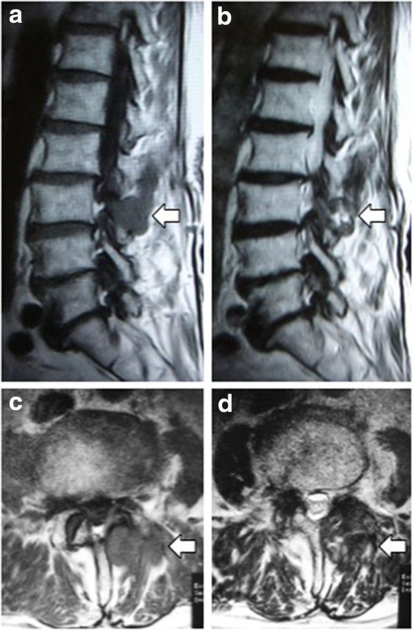
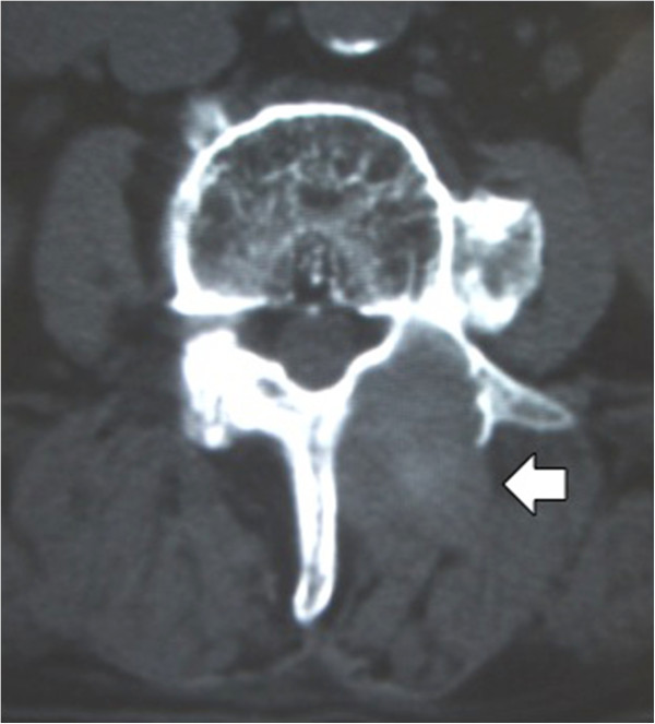
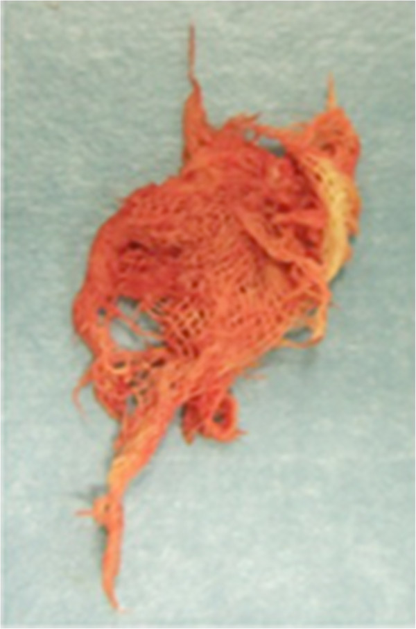
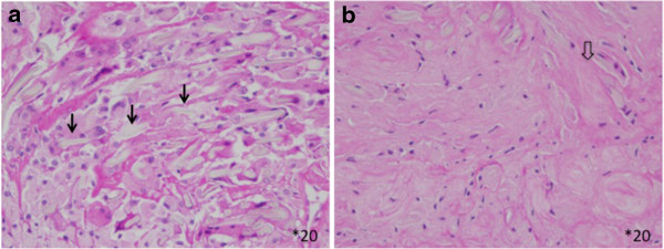
Case presentation: Our patient was an 87-year-old Japanese man with a history of surgery for a lumbar lesion causing lumbar canal stenosis 19 years earlier. Computed tomography showed a soft tissue mass with osteolysis and periosteal thickening of the vertebral lamina. On magnetic resonance imaging, the mass showed heterogeneous signal intensity on T2-weighted imaging, suggesting a malignancy. At the time of biopsy, small pieces of retained surgical sponge were collected. Surgical treatment was performed to excise the soft tissue tumour.

Conclusions: Gossypiboma should be included in the differential diagnosis of soft tissue masses in the paraspinal region in patients with a history of previous spinal surgery.


Figures

Figure 1
Figure 1
Magnetic resonance imaging sagittal and axial images. A soft tissue mass (open arrow) at the L3 to 4 level is hypointense compared with the spinal cord on the T1-weighted image (a, c), and heterogeneous signal intensity is seen on the T2-weighted image (b, d).
Figure 2
Figure 2
Computed tomography axial image. A mass (open arrow) is seen posterior to the lamina with osteolytic change.
Figure 3
Figure 3
Macroscopic appearance of the retained sponge material at biopsy.
Figure 4
Figure 4
Histological findings. A fresh foreign body reaction around the gauze fibre (arrow, a) and fibrin deposition (open arrow), which indicates an old foreign body reaction (b) are seen (hematoxylin and eosin ×20).

Similar articles

Cited by

References

    1. Aydogan M, Mirzanli C, Ganiyusufoglu K, Tezer M, Ozturk I. A 13-year-old textiloma (gossypiboma) after discectomy for lumbar disc herniation: a case report and review of the literature. Spine J. 2007;7:618–621. doi: 10.1016/j.spinee.2006.08.004. - DOI - PubMed
    1. Atabey C, Turgut M, Ilica AT. Retained surgical sponge in differential diagnosis of paraspinal soft-tissue mass after posterior spinal surgery: report of eight cases. Neurol India. 2009;57:320–323. doi: 10.4103/0028-3886.53289. - DOI - PubMed
    1. Hakan T, Aydoseli A, Demir K, Aker F. Clinical, pathological and radiological features of paraspinal textiloma: report of two cases and review of the literature. Neurol Neurochir Pol. 2009;43:475–478. - PubMed
    1. Is M, Karatas A, Akgul M, Yildirim U, Gezen F. A retained surgical sponge (gossypiboma) mimicking a paraspinal abscess. Br J Neurosurg. 2007;21:307–308. doi: 10.1080/02688690701367701. - DOI - PubMed
    1. Kim HS, Chung T-S, Suh SH, Kim SY. MR imaging findings of paravertebral gossypiboma. AJNR Am J Neuroradiol. 2007;28:709–713. - PMC - PubMed

Publication types