

Axial T1ρ MRI as a diagnostic imaging modality to quantify proteoglycan concentration in degenerative disc disease
- PMID: 25236594
- PMCID: PMC5603078
- DOI: 10.1007/s00586-014-3582-6
Axial T1ρ MRI as a diagnostic imaging modality to quantify proteoglycan concentration in degenerative disc disease
Abstract
Purpose: The aim of the study was to investigate if axial T1ρ MR images had similar accuracy as established sagittal T1ρ MRI for the assessment of proteoglycan concentration and content in intervertebral degenerated discs (IDDs).
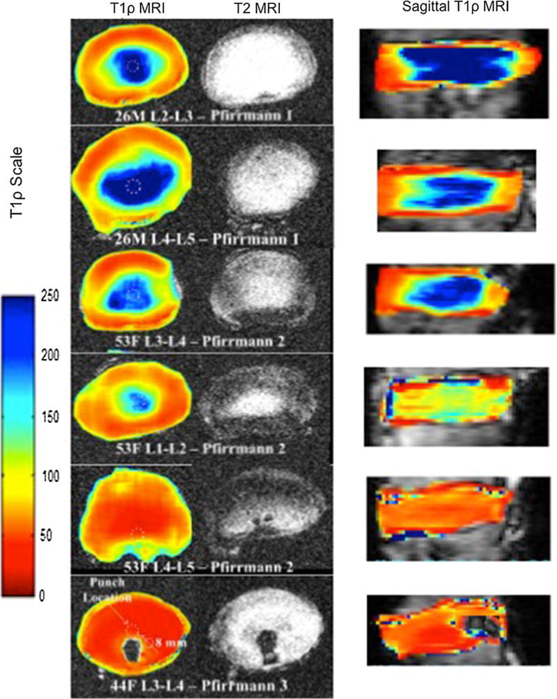
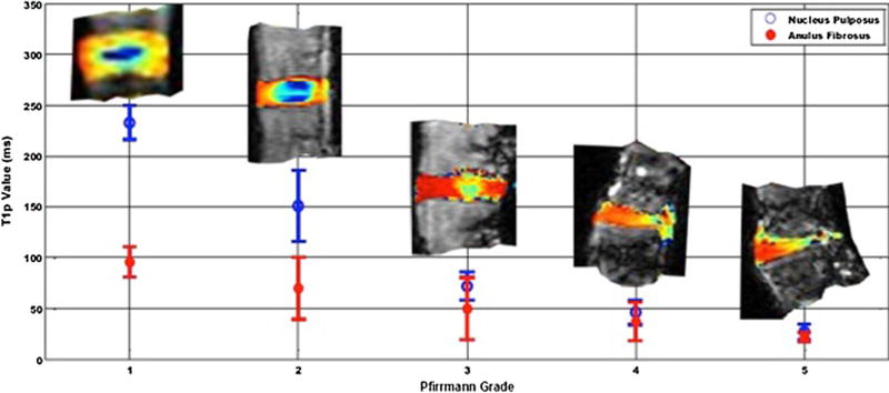
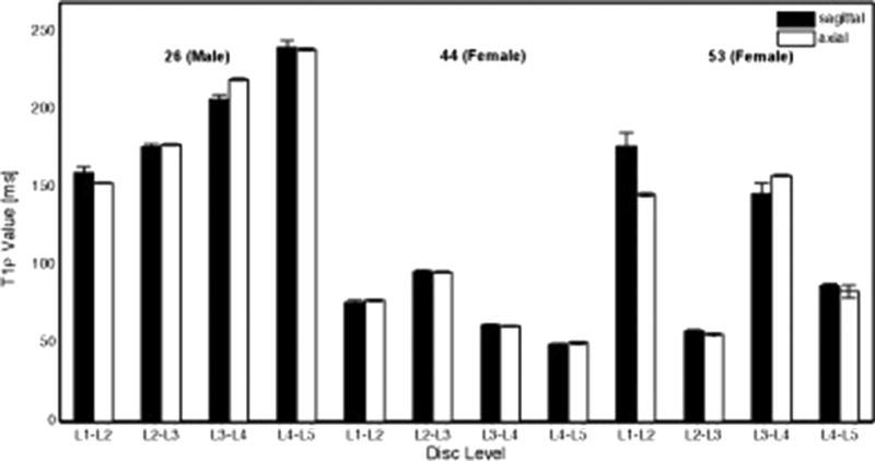
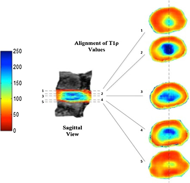
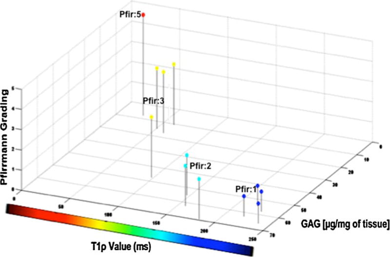
Methods: T1ρ and T2-weighted MR images of 12 intervertebral discs (IVDs) from 3 harvested human lumbar spines (levels L1-L2 to L5-S1) were grouped across their degenerative grade (Pfirrmann scores) and analyzed using a 3T MRI scanner in the axial and sagittal views. Post-processing of axial T1ρ-weighted images was performed using a Wiener filter. Median axial T1ρ values for traced regions of interest (ROIs) on color maps were compared against ROIs in the corresponding location in the sagittal plane of each disc. Assessment of sulfated glycosaminoglycans (GAGs) content was also performed.
Results: Comparison of post Wiener filtered mid-axial T1ρ values in the NP with corresponding mid-sagittal values revealed no statistical difference (P > 0.05). Higher axial T1ρ and biochemically measured GAGs content corresponded to a lower Pfirrmann grading of the IVDs. A strong association between the T1ρ values and the GAG contents was observed (r = 0.85, P = 0.0002).
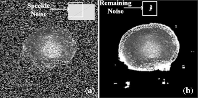
Conclusions: The axial T1ρ methodology was validated against sagittal T1ρ providing an augmented spatial representation of IVD and can facilitate localization of focal degeneration within IVDs. T1ρ values provided a better granularity assessment of degenerative disc disease as it correlated with proteoglycan concentration. Thus, Wiener filtering is an effective tool for removing noise from T1ρ-weighted axial MR images.
Keywords: Axial T1ρ magnetic resonance imaging (MRI); Intervertebral disc (IVD); Pfirrmann grade; Proteoglycan; Wiener filtering.
Figures
Similar articles
-
Glycosaminoglycan Chemical Exchange Saturation Transfer of Lumbar Intervertebral Discs in Healthy Volunteers.Spine (Phila Pa 1976). 2016 Jan;41(2):146-52. doi: 10.1097/BRS.0000000000001144. Spine (Phila Pa 1976). 2016. PMID: 26583472
-
Assessment of human disc degeneration and proteoglycan content using T1rho-weighted magnetic resonance imaging.Spine (Phila Pa 1976). 2006 May 15;31(11):1253-7. doi: 10.1097/01.brs.0000217708.54880.51. Spine (Phila Pa 1976). 2006. PMID: 16688040 Free PMC article.
-
T1ρ magnetic resonance imaging quantification of early lumbar intervertebral disc degeneration in healthy young adults.Spine (Phila Pa 1976). 2012 Jun 15;37(14):1224-30. doi: 10.1097/BRS.0b013e31824b2450. Spine (Phila Pa 1976). 2012. PMID: 22281486 Free PMC article.
-
Imaging diagnosis for intervertebral disc.JOR Spine. 2019 Sep 29;3(1):e1066. doi: 10.1002/jsp2.1066. eCollection 2020 Mar. JOR Spine. 2019. PMID: 32211585 Free PMC article. Review.
-
T1ρ magnetic resonance: basic physics principles and applications in knee and intervertebral disc imaging.Quant Imaging Med Surg. 2015 Dec;5(6):858-85. doi: 10.3978/j.issn.2223-4292.2015.12.06. Quant Imaging Med Surg. 2015. PMID: 26807369 Free PMC article. Review.
Cited by
-
Imaging of the spine: Where do we stand?World J Radiol. 2019 Apr 28;11(4):55-61. doi: 10.4329/wjr.v11.i4.55. World J Radiol. 2019. PMID: 31110605 Free PMC article.
-
Critical aspects and challenges for intervertebral disc repair and regeneration-Harnessing advances in tissue engineering.JOR Spine. 2018 Sep;1(3):e1029. doi: 10.1002/jsp2.1029. Epub 2018 Jul 30. JOR Spine. 2018. PMID: 30895276 Free PMC article. Review.
-
Quantitative MRI in early intervertebral disc degeneration: T1rho correlates better than T2 and ADC with biomechanics, histology and matrix content.PLoS One. 2018 Jan 30;13(1):e0191442. doi: 10.1371/journal.pone.0191442. eCollection 2018. PLoS One. 2018. PMID: 29381716 Free PMC article.
-
Senotherapeutic drugs for human intervertebral disc degeneration and low back pain.Elife. 2020 Aug 21;9:e54693. doi: 10.7554/eLife.54693. Elife. 2020. PMID: 32821059 Free PMC article.
-
Multimodal quantitative magnetic resonance imaging for lumbar intervertebral disc degeneration.Exp Ther Med. 2017 Sep;14(3):2078-2084. doi: 10.3892/etm.2017.4786. Epub 2017 Jul 12. Exp Ther Med. 2017. PMID: 28962127 Free PMC article.
References
-
- Beattie PF, Meyers SP. Magnetic resonance imaging in low back pain: general principles and clinical issues. Phys Ther. 1998;78(7):738–753. - PubMed
-
- Antoniou J, Steffen T, Nelson F, Winterbottom N, Hollander AP, Poole RA, Aebi M, Alini M. The human lumbar intervertebral disc: evidence for changes in the biosynthesis and denaturation of the extracellular matrix with growth, maturation, ageing, and degeneration. J Clin Investig. 1996;98(4):996–1003. - PMC - PubMed
-
- Keshari KR, Zektzer AS, Swanson MG, Majumdar S, Lotz JC, Kurhanewicz J. Characterization of intervertebral disc degeneration by high-resolution magic angle spinning (HR-MAS) spectroscopy. Magn Reson Med Off J Soc Magn Reson Med Soc Magn Reson Med. 2005;53(3):519–527. - PubMed
Publication types
MeSH terms
Substances
Grants and funding
LinkOut - more resources
Full Text Sources
Other Literature Sources
Medical
Miscellaneous
