. 2014 Dec:77:57-63.
doi: 10.1016/j.freeradbiomed.2014.08.016. Epub 2014 Sep 16.

Mechanism of oxidative inactivation of human presequence protease by hydrogen peroxide

Affiliations

Mechanism of oxidative inactivation of human presequence protease by hydrogen peroxide

Jue Chen et al. Free Radic Biol Med. 2014 Dec.

Abstract

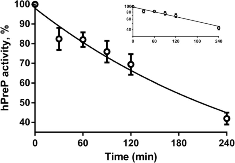
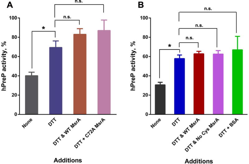
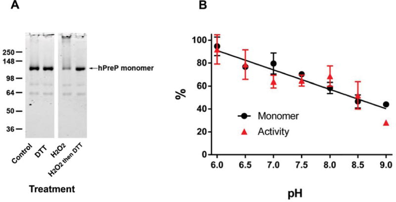
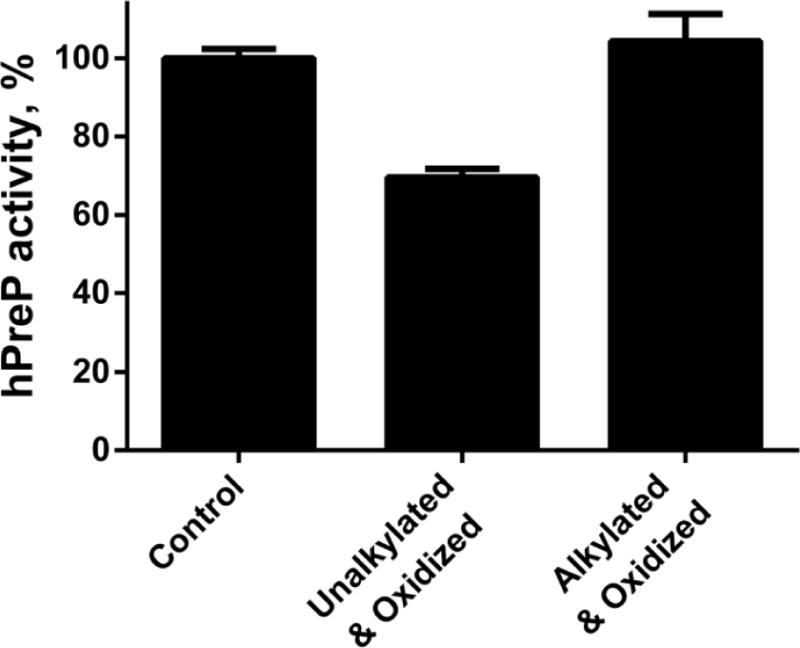
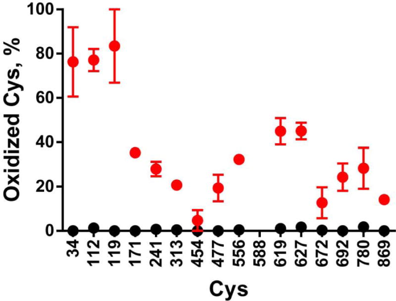
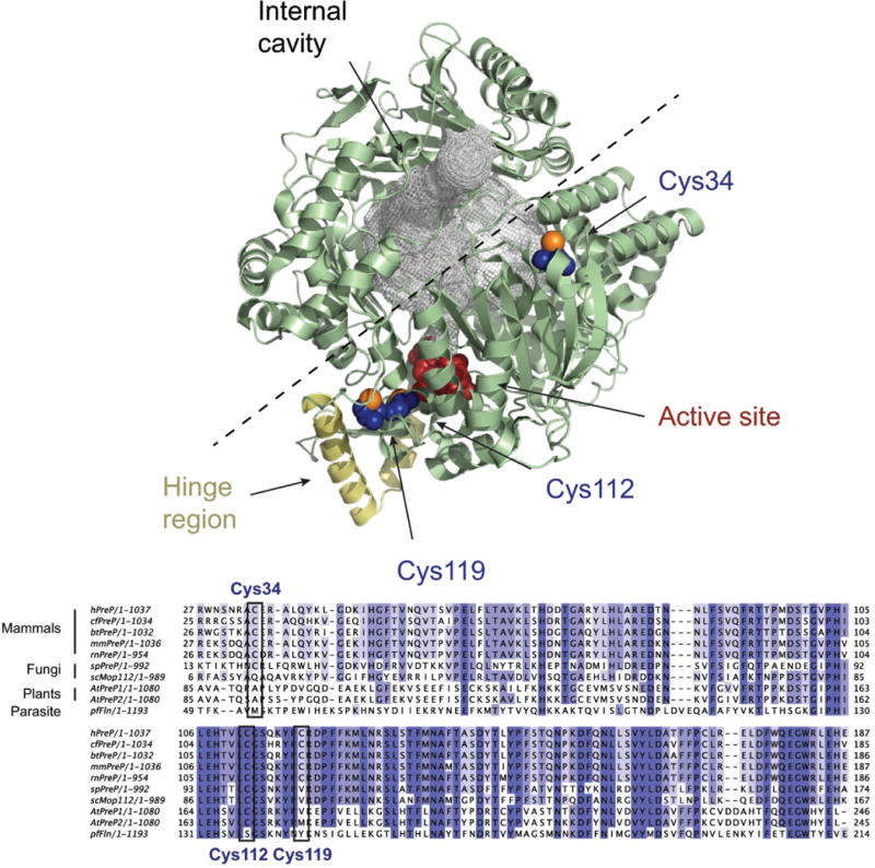
The mitochondrial presequence protease (PreP) is a member of the pitrilysin class of metalloproteases. It degrades the mitochondrial targeting presequences of mitochondria-localized proteins as well as unstructured peptides such as amyloid-β peptide. The specific activity of PreP is reduced in Alzheimer patients and animal models of Alzheimer disease. The loss of activity can be mimicked in vitro by exposure to oxidizing conditions, and indirect evidence suggested that inactivation was due to methionine oxidation. We performed peptide mapping analyses to elucidate the mechanism of inactivation. None of the 24 methionine residues in recombinant human PreP was oxidized. We present evidence that inactivation is due to oxidation of cysteine residues and consequent oligomerization through intermolecular disulfide bonds. The most susceptible cysteine residues to oxidation are Cys34, Cys112, and Cys119. Most, but not all, of the activity loss is restored by the reducing agent dithiothreitol. These findings elucidate a redox mechanism for regulation of PreP and also provide a rational basis for therapeutic intervention in conditions characterized by excessive oxidation of PreP.

Keywords: Cysteine oxidation; Free radicals; Methionine sulfoxide; Peptide degradation; Presequence protease; Protein oxidation.


Figures

Fig. 1
Fig. 1
Kinetics of inactivation of hPreP by 500 μM hydrogen peroxide at pH 8.2. The plotted points are the mean and SEM for 3 experiments performed on separate days. The insert plots the same data as a semilogarithmic plot and demonstrates that inactivation is first order. The semilogarithmic plot was fit by linear regression and drawn as a solid line in both plots. Log(hPreP activity) = 1.99 – 0.00141(min), R2=0.86. The time to half-inactivation is 3.4 h.
Fig. 2
Fig. 2
Recovery of hPreP activity by reduction. Four h oxidized hPreP, 2.4 μM, was incubated for 3 h at 37 °C with the indicated additions and then assayed for activity. The activity of unoxidized hPreP was set to 100%. When added, DTT was 10 mM and methionine sulfoxide reductase A was 1 μM. Panels A and B are results from two separate preparations of hPreP. The results are plotted as the mean and SEM with n=4 in panel A and n=3 in panel B. Comparisons were assessed by Student’s t test with the Bonferroni correction for multiple comparisons. Pairs whose p values are less than 0.05 are marked with an asterisk, and those whose p values were ≥ 0.05 are marked n.s. (not significant).
Fig. 3
Fig. 3
Oxidation of hPreP induces oligomerization whose extent matches the loss of activity. (A) Non-reducing SDS-PAGE gel of hPreP subjected to varying treatments. 1 μg hPreP was loaded in each lane. Oligomerization was induced by hydrogen peroxide and reversed by subsequent incubation with DTT. (B) Oligomerization and inactivation are quantitatively linked. hPreP was oxidized in 50 mM HEPES at varying pH. Regression lines fit separately to oligomerization and inactivation did not differ from each other (p=0.62) and thus a single regression line is shown.
Fig. 4
Fig. 4
Alkylation of accessible cysteines prevents hydrogen peroxide-mediated inactivation of hPreP. Control and alkylated hPreP were incubated with 500 μM hydrogen peroxide at pH 7.4 for 4 h and then assayed for hPreP activity. Each bar plots the mean and SEM of 3 independent experiments.
Fig. 5
Fig. 5
Quantitation of cysteine oxidation. hPreP was oxidized by 500 μM hydrogen peroxide for 4 h in HEPES buffer at pH 8.2. The oxidation state of each cysteine was determined by peptide mapping as described in Experimental Procedures. Black circles plot the control not exposed to hydrogen peroxide, and red circles plot the hPreP incubated with hydrogen peroxide. The points are the mean and SEM from 3 independent experiments.
Fig. 6
Fig. 6
Crystal structure of hPreP. Upper panel. Overview of the hPreP structure [27] (4L3T, pale green) with the residues composing the active site shown in red and the hinge region colored in yellow. The internal cavity in hPreP is filled with a gray mesh, and the dashed line shows the proposed path of opening. The oxidation-susceptible residues Cys34, Cys112, and Cys119 are shown in blue, except for the sulfur atoms which are orange. The figure was prepared using PyMOL [30]. Lower panel. Alignment of PreP sequences from Homo sapiens (hPreP), Canis familiaris (cfPreP), Bos taurus (btPreP), Mus musculus (mmPreP), Rattus norvergicus (rnPreP), Schizosaccharomyces pombe (spPreP), S. cerevisiae (scMop112), A. thaliana (AtPreP1 and AtPreP2) and Plasmodium falciparum (pfFln). The alignment was generated with Clustal Omega [31] and is restricted to the region around Cys34 to Cys119.

References

    1. Mzhavia N, Berman YL, Qian Y, Yan L, Devi LA. Cloning, expression, and characterization of human metalloprotease 1: a novel member of the pitrilysin family of metalloendoproteases. DNA and cell biology. 1999;18:369–380. - PubMed
    1. Glaser E, Alikhani N. The organellar peptidasome, PreP: A journey from Arabidopsis to Alzheimer’s disease. Biochimica et Biophysica Acta (BBA) – Bioenergetics. 2010;1797:1076–1080. - PubMed
    1. Falkevall A, Alikhani N, Bhushan S, Pavlov PF, Busch K, Johnson KA, Eneqvist T, Tjernberg L, Ankarcrona M, Glaser E. Degradation of the Amyloid β-Protein by the Novel Mitochondrial Peptidasome, PreP. Journal of Biological Chemistry. 2006;281:29096–29104. - PubMed
    1. Pinho CM, Teixeira PF, Glaser E. Mitochondrial import and degradation of amyloid-beta peptide. Biochim Biophys Acta. 2014 - PubMed
    1. Johnson KA, Bhushan S, Stahl A, Hallberg BM, Frohn A, Glaser E, Eneqvist T. The closed structure of presequence protease PreP forms a unique 10 000 A3 chamber for proteolysis. EMBO J. 2006;25:1977–1986. - PMC - PubMed

Publication types

LinkOut - more resources