. 2014 Sep 4:8:267.
doi: 10.3389/fncel.2014.00267. eCollection 2014.

Differential contribution of TRPM4 and TRPM5 nonselective cation channels to the slow afterdepolarization in mouse prefrontal cortex neurons

Affiliations

Differential contribution of TRPM4 and TRPM5 nonselective cation channels to the slow afterdepolarization in mouse prefrontal cortex neurons

Ya-Ting Lei et al. Front Cell Neurosci. .

Abstract

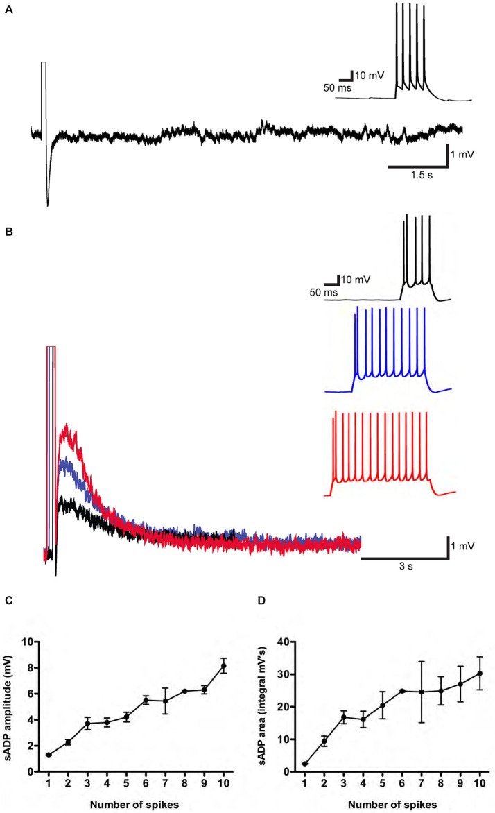
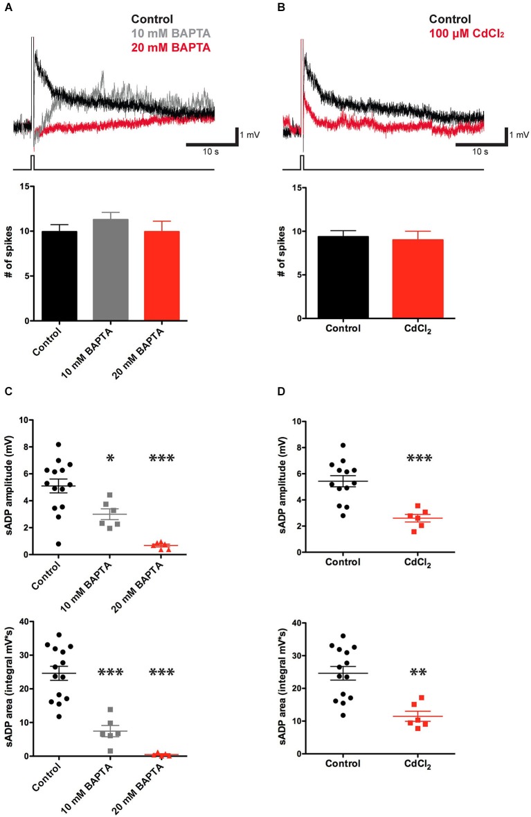
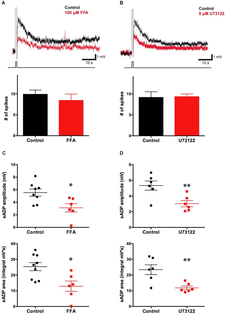
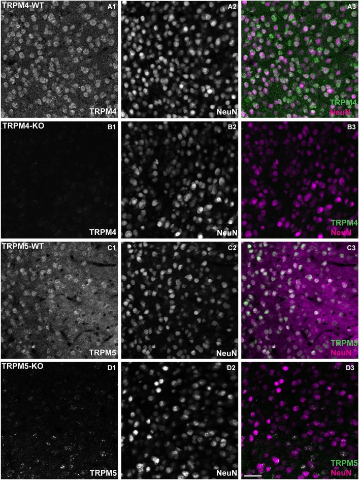
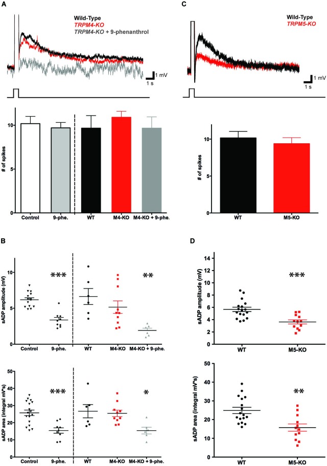
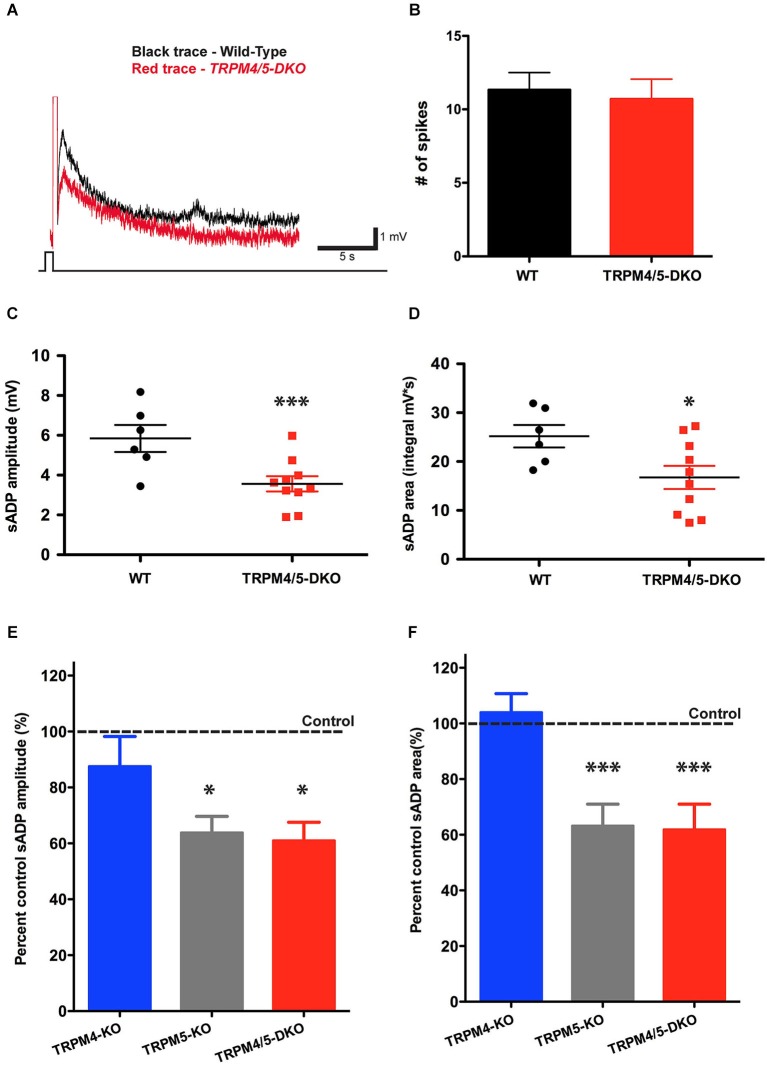
In certain neurons from different brain regions, a brief burst of action potentials can activate a slow afterdepolarization (sADP) in the presence of muscarinic acetylcholine receptor agonists. The sADP, if suprathreshold, can contribute to persistent non-accommodating firing in some of these neurons. Previous studies have characterized a Ca(2+)-activated non-selective cation (CAN) current (ICAN ) that is thought to underlie the sADP. ICAN depends on muscarinic receptor stimulation and exhibits a dependence on neuronal activity, membrane depolarization and Ca(2+)-influx similar to that observed for the sADP. Despite the widespread occurrence of sADPs in neurons throughout the brain, the molecular identity of the ion channels underlying these events, as well as ICAN , remains uncertain. Here we used a combination of genetic, pharmacological and electrophysiological approaches to characterize the molecular mechanisms underlying the muscarinic receptor-dependent sADP in layer 5 pyramidal neurons of mouse prefrontal cortex. First, we confirmed that in the presence of the cholinergic agonist carbachol a brief burst of action potentials triggers a prominent sADP in these neurons. Second, we confirmed that this sADP requires activation of a PLC signaling cascade and intracellular calcium signaling. Third, we obtained direct evidence that the transient receptor potential (TRP) melastatin 5 channel (TRPM5), which is thought to function as a CAN channel in non-neural cells, contributes importantly to the sADP in the layer 5 neurons. In contrast, the closely related TRPM4 channel may play only a minor role in the sADP.

Keywords: Ca2+-activated non-selective cation (CAN) current; muscarinic receptors; persistent firing; slow afterdepolarization (sADP); transient receptor potential melastatin 5 channel (TRPM5).


Figures

Figure 1
Figure 1
A muscarinic receptor-dependent slow afterdepolarization (sADP) is observed in PFC layer 5 pyramidal neurons. (A) In the absence of carbachol (CCh). Representative traces showing an AHP following a burst of action potentials induced by a 200 pA depolarizing current step. Inset shows action potential firing during the current step. Initial resting potential was −66 mV. (B) In the presence of 10 μM CCh. Representative traces show sADPs following a burst of action potentials in response to 200 pA depolarizing current steps of increasing duration (black trace: 100 ms; blue trace: 300 ms; red trace: 400 ms). Insets show action potential firing during the depolarization. Initial Vm was −65 mV. (C) and (D) Input-output relations plotting sADP amplitude and area as a function of the number of action potentials elicited by increasing amounts of depolarizing charge injection in the presence of CCh. n = 4–8 for each point.
Figure 2
Figure 2
CCh-induced sADP requires intracellular Ca2+. (A) Applying BAPTA in the patch pipette solution decreased the sADP in presence of 10 μM CCh. Inset: Superimposed traces showing the sADP following action potentials induced by a depolarizing current step (bottom trace) in CCh under control conditions (black trace) or with 10 mM (gray trace) or 20 mM (red trace) BAPTA in the patch pipette solution. Initial Vm was −66 mV. Bar graph, number of action potentials during the depolarizing step (200 pA for 500 ms) was not significantly different among groups (n = 7–16, P = 0.31 and 1 respectively). (B) Bath application of Cd2+ reduced the sADP. Inset: sADPs induced by a depolarizing current step (in CCh) under control conditions (black trace) or in the presence of 100 μM Cd2+ (red trace). Bar graph, number of spikes during the depolarization was not affected by Cd2+ (n = 6–16, P = 0.77). (C) BAPTA significantly reduced peak amplitude (n = 6–14, * P < 0.05, *** P < 0.0001) and area (n = 6–14, *** P < 0.0001) of sADP triggered by a 200 pA depolarizing current step for 500 ms. (D) Cd2+ decreased sADP amplitude (n = 6–13, *** P < 0.001) and area (n = 6–14, ** P < 0.01).
Figure 3
Figure 3
The sADP is sensitive to a blocker of non-specific cationic currents and requires the PLC pathway. (A) The CAN current blocking agent FFA decreased the sADP induced by a depolarizing current step (in 10 μM CCh). Inset: sADP under control conditions (black trace) and in presence of FFA (10 μM; red trace). Initial Vm was −66 mV. Bar graph, number of spikes during the depolarization (200 pA for 500 ms) was not affected by FFA (n = 7–10, P = 0.42). (B) The PLC blocker U73122 decreased the CCh-induced sADP. Inset: Superimposed traces showing sADP induced by a depolarizing current step (bottom trace) in CCh under control conditions (black trace) or with 5 μM U73122 in bath (red trace). Initial Vm was −65 mV. Number of spikes during the depolarization (200 pA for 500 ms) was not affected by U73122 (n = 6, P = 0.91). (C) FFA decreased the sADP amplitude (n = 6–8, * P < 0.05) and area (n = 6–9, * indicates P < 0.05). (D) The PLC blocker U73122 decreased the sADP amplitude (n = 6, ** indicates P < 0.01) and area (n = 6, ** indicates P < 0.01).
Figure 4
Figure 4
TRPM4 and TRPM5 expression in coronal slices of adult medial PFC (mPFC). Immunofluorescence using antibodies directed against TRPM4, TRPM5 and Neuronal Nuclei (NeuN) performed in coronal slices of mPFC. TRPM4 (magenta) and NeuN (green) double/labeled cells were observed in medial prefrontal cortex (A1–A3) from TRPM4+/– mice, whereas no TRPM4 immunostaining was observed in sections from TRPM4−/− (TRPM4-KO) mice (B1–B3). TRPM5+/– heterozygous mice showed TRPM5 immunoreactivity (C1–C3), whereas no TRPM5 immunostaining was observed in sections from TRPM5−/− (TRPM5-KO) mice (D1–D3). Staining with TRPM4 and TRPM5 antibody produced predominantly cytosolic labeling and staining of NeuN was primarily localized in the nucleus of the neurons. Scale bars: 50 μm.
Figure 5
Figure 5
TRPM5 but not TRPM4 contributes to the CCh-induced sADP. (A) TRPM4 deletion did not significantly alter the sADP. Inset: superimposed representative traces show sADP induced by a depolarizing current step (200 pA, 500 ms) in CCh for wild-type littermate control mice (black trace), Trpm4−/− mice (red trace) and Trpm4−/− mice in the presence of 9-phenanthrol (dark gray trace). Initial Vm was −66 mV. Bar graph, there was no statistically significant difference in numbers of spikes in wild-type mice under control conditions vs. in the presence of 9-phenanthrol (n = 10–22, P = 0.71). There were no significant differences in number of spikes among wild-type (WT) mice or Trpm4−/− mice (M4-KO) under control conditions or in presence of 9-phenanthrol (M4-KO + 9-phe.); n = 6–11, P = 0.39 and P = 1 respectively. (B) Genetic deletion of TRPM4 did not significantly reduce sADP amplitude (top graph; n = 6–10, P = 0.31) or sADP area (bottom graph; n = 6–10, P = 0.74). However, application of 9-phenanthrol in both control and Trpm4−/− mice decreased significantly peak sADP amplitude (wild-type control vs. 9-phenanthrol, n = 9–17, *** P < 0.001; wild-type control vs. KO with 9-phenanthrol, n = 6, ** P < 0.01) and sADP area (in control vs. with 9-phenanthrol, n = 9–17, *** P < 0.001; in WT vs. KO with 9-phenanthrol, n = 6, * P < 0.05) by a depolarizing step current (200 pA for 500 ms). (C) TRPM5 deletion reduced the sADP. Inset: sADP induced by depolarizing current (200 pA, 500 ms) in CCh in wild-type littermate control mice (black trace) and Trpm5−/− KO mice (red trace). Initial Vm was −65 mV. Bar graph, TRPM5 deletion (M5-KO) had no effect on number of spikes during the depolarizing step (n = 13–18, P = 0.54). (D) Deletion of TRPM5 significantly decreased sADP amplitude (top graph; n = 12–17, *** P < 0.001) and area (bottom graph; n = 12–17, ** P < 0.01).
Figure 6
Figure 6
TRPM4 does not contribute to the residual sADP in TRPM5 KO mice. (A) Superimposed representative traces show the sADP induced by a depolarizing current step (200 pA, 500 ms) in CCh for wild-type mice (black trace) and Trpm4/5 double knockout (DKO) mice (red trace). Scale bars, 1 mV and 5 s. Initial Vm was −66 mV. (B) There is no difference in spike number during current injection between populations of mice (n = 6–10, P = 0.75). (C) Trpm4/5 -DKO mice generated sADPs with smaller amplitude (n = 6–10, ** indicates P < 0.01) (C) and area (n = 6–10, * indicates P < 0.05) (D) compared to wild-type littermate controls. (E,F) Bar graphs showing sADP amplitude (E) and area (F) in three different genotypes expressed as percentage of sADP in wild-type littermate controls. Normalized sADP amplitude and sADP area show significant differences between Trpm4−/− KO vs. Trpm5−/− KO and between Trpm4−/− KO vs. Trpm4/5 -DKO mice (one-way ANOVA; n = 10–12, * indicates P < 0.05 and *** indicates P < 0.0001, respectively).

Similar articles

Cited by

References

    1. Anagnostaras S. G., Murphy G. G., Hamilton S. E., Mitchell S. L., Rahnama N. P., Nathanson N. M., et al. (2003). Selective cognitive dysfunction in acetylcholine M1 muscarinic receptor mutant mice. Nat. Neurosci. 6, 51–58 10.1038/nn992 - DOI - PubMed
    1. Araneda R., Andrade R. (1991). 5-Hydroxytryptamine2 and 5-hydroxytryptamine 1A receptors mediate opposing responses on membrane excitability in rat association cortex. Neuroscience 40, 399–412 10.1016/0306-4522(91)90128-b - DOI - PubMed
    1. Barbet G., Demion M., Moura I. C., Serafini N., Leger T., Vrtovsnik F., et al. (2008). The calcium-activated nonselective cation channel TRPM4 is essential for the migration but not the maturation of dendritic cells. Nat. Immunol. 9, 1148–1156 10.1038/ni.1648 - DOI - PMC - PubMed
    1. Clapham D. E. (2003). TRP channels as cellular sensors. Nature 426, 517–524 10.1038/nature02196 - DOI - PubMed
    1. Constanti A., Bagetta G., Libri V. (1993). Persistent muscarinic excitation in guinea-pig olfactory cortex neurons: involvement of a slow post-stimulus afterdepolarizing current. Neuroscience 56, 887–904 10.1016/0306-4522(93)90135-3 - DOI - PubMed