

Sonoporation: Gene transfer using ultrasound
- PMID: 25237622
- PMCID: PMC4145571
- DOI: 10.5662/wjm.v3.i4.39
Sonoporation: Gene transfer using ultrasound
Abstract
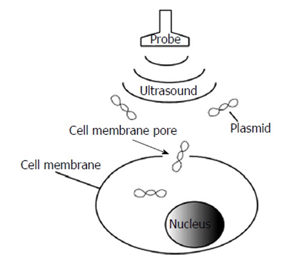
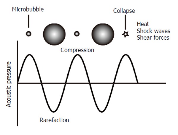
Genes can be transferred using viral or non-viral vectors. Non-viral methods that use plasmid DNA and short interference RNA (siRNA) have advantages, such as low immunogenicity and low likelihood of genomic integration in the host, when compared to viral methods. Non-viral methods have potential merit, but their gene transfer efficiency is not satisfactory. Therefore, new methods should be developed. Low-frequency ultrasound irradiation causes mechanical perturbation of the cell membrane, allowing the uptake of large molecules in the vicinity of the cavitation bubbles. The collapse of these bubbles generates small transient holes in the cell membrane and induces transient membrane permeabilization. This formation of small pores in the cell membrane using ultrasound allows the transfer of DNA/RNA into the cell. This phenomenon is known as sonoporation and is a gene delivery method that shows great promise as a potential new approach in gene therapy. Microbubbles lower the threshold of cavity formation. Complexes of therapeutic genes and microbubbles improve the transfer efficiency of genes. Diagnostic ultrasound is potentially a suitable sonoporator because it allows the real-time monitoring of irradiated fields.
Keywords: Cavity; Contrast agent; Diagnostic ultrasound; Gene therapy; Microbubbles.
Figures
References
-
- Miura K, Okada Y, Aoi T, Okada A, Takahashi K, Okita K, Nakagawa M, Koyanagi M, Tanabe K, Ohnuki M, et al. Variation in the safety of induced pluripotent stem cell lines. Nat Biotechnol. 2009;27:743–745. - PubMed
-
- Raper SE, Chirmule N, Lee FS, Wivel NA, Bagg A, Gao GP, Wilson JM, Batshaw ML. Fatal systemic inflammatory response syndrome in a ornithine transcarbamylase deficient patient following adenoviral gene transfer. Mol Genet Metab. 2003;80:148–158. - PubMed
Publication types
LinkOut - more resources
Full Text Sources
Other Literature Sources
