

Mediterranean Diet and Magnetic Resonance Imaging-Assessed Brain Atrophy in Cognitively Normal Individuals at Risk for Alzheimer's Disease
- PMID: 25237654
- PMCID: PMC4165397
Mediterranean Diet and Magnetic Resonance Imaging-Assessed Brain Atrophy in Cognitively Normal Individuals at Risk for Alzheimer's Disease
Abstract
Objectives: Epidemiological evidence linking diet, one of the most important modifiable environmental factors, and risk of Alzheimer's disease (AD) is rapidly increasing. Several studies have shown that higher adherence to a Mediterranean diet (MeDi) is associated with reduced risk of AD. This study examines the associations between high vs. lower adherence to a MeDi and structural MRI-based brain atrophy in key regions for AD in cognitively normal (NL) individuals with and without risk factors for AD.
Design: Cross-sectional study.
Setting: Manhattan (broader area).
Participants: Fifty-two NL individuals (age 54+12 y, 70% women) with complete dietary information and cross-sectional, 3D T1-weighted MRI scans were examined.
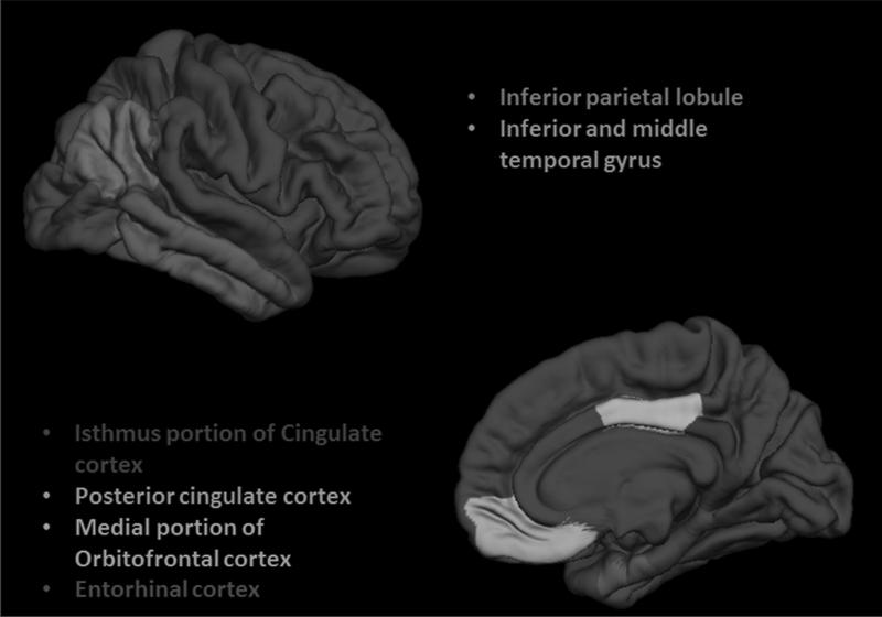
Measurements: Subjects were dichotomized into those showing higher vs. lower adherences to the MeDi using published protocols. Estimates of cortical thickness for entorhinal cortex (EC), inferior parietal lobe, middle temporal gyrus, orbitofrontal cortex (OFC) and posterior cingulate cortex (PCC) were obtained by use of automated segmentation tools (FreeSurfer). Multivariate general linear models and linear regressions assessed the associations of MeDi with MRI measures.
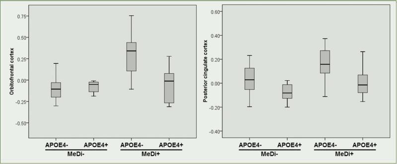
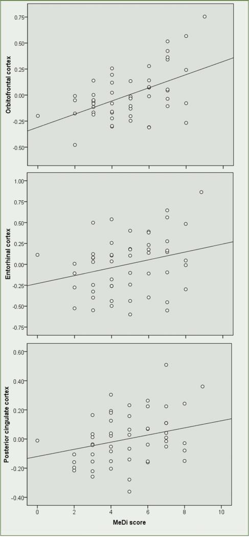
Results: Of the 52 participants, 20 (39%) showed higher MeDi adherence (MeDi+) and 32 (61%) showed lower adherence (MeDi-). Groups were comparable for clinical, neuropsychological measures, presence of a family history of AD (FH), and frequency of Apolipoprotein E (APOE) ε4 genotype. With and without controlling for age and total intracranial volume, MeDi+ subjects showed greater thickness of AD-vulnerable ROIs as compared to MeDi- subjects (Wilk's Lambda p=0.026). Group differences were most pronounced in OFC (p=0.001), EC (p=0.03) and PCC (p=0.04) of the left hemisphere. Adjusting for gender, education, FH, APOE status, BMI, insulin resistance scores and presence of hypertension did not attenuate the relationship.
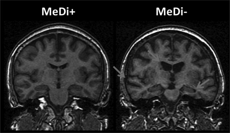
Conclusion: NL individuals showing lower adherence to the MeDi had cortical thinning in the same brain regions as clinical AD patients compared to those showing higher adherence. These data indicate that the MeDi may have a protective effect against tissue loss, and suggest that dietary interventions may play a role in the prevention of AD.
Keywords: Alzheimer's disease; Mediterranean diet; brain imaging; diet; early detection; magnetic resonance imaging (MRI).
Figures
Similar articles
-
Lifestyle and vascular risk effects on MRI-based biomarkers of Alzheimer's disease: a cross-sectional study of middle-aged adults from the broader New York City area.BMJ Open. 2018 Mar 23;8(3):e019362. doi: 10.1136/bmjopen-2017-019362. BMJ Open. 2018. PMID: 29574441 Free PMC article.
-
Physical Activity, Mediterranean Diet and Biomarkers-Assessed Risk of Alzheimer's: A Multi-Modality Brain Imaging Study.Adv J Mol Imaging. 2014 Oct;4(4):43-57. doi: 10.4236/ami.2014.44006. Adv J Mol Imaging. 2014. PMID: 25599008 Free PMC article.
-
Nutrient patterns and brain biomarkers of Alzheimer's disease in cognitively normal individuals.J Nutr Health Aging. 2015 Apr;19(4):413-23. doi: 10.1007/s12603-014-0534-0. J Nutr Health Aging. 2015. PMID: 25809805 Free PMC article.
-
Spermidine intake is associated with cortical thickness and hippocampal volume in older adults.Neuroimage. 2020 Nov 1;221:117132. doi: 10.1016/j.neuroimage.2020.117132. Epub 2020 Jul 3. Neuroimage. 2020. PMID: 32629145
-
The relationship between gender differences in dietary habits, neuroinflammation, and Alzheimer's disease.Front Aging Neurosci. 2024 Apr 17;16:1395825. doi: 10.3389/fnagi.2024.1395825. eCollection 2024. Front Aging Neurosci. 2024. PMID: 38694261 Free PMC article. Review.
Cited by
-
Impact of the Level of Adherence to Mediterranean Diet on the Parameters of Metabolic Syndrome: A Systematic Review and Meta-Analysis of Observational Studies.Nutrients. 2021 Apr 30;13(5):1514. doi: 10.3390/nu13051514. Nutrients. 2021. PMID: 33946280 Free PMC article.
-
Sex and Gender Driven Modifiers of Alzheimer's: The Role for Estrogenic Control Across Age, Race, Medical, and Lifestyle Risks.Front Aging Neurosci. 2019 Nov 15;11:315. doi: 10.3389/fnagi.2019.00315. eCollection 2019. Front Aging Neurosci. 2019. PMID: 31803046 Free PMC article. Review.
-
Omega-3 Fatty Acids, Cognition, and Brain Volume in Older Adults.Brain Sci. 2023 Sep 2;13(9):1278. doi: 10.3390/brainsci13091278. Brain Sci. 2023. PMID: 37759879 Free PMC article.
-
Lifestyle and vascular risk effects on MRI-based biomarkers of Alzheimer's disease: a cross-sectional study of middle-aged adults from the broader New York City area.BMJ Open. 2018 Mar 23;8(3):e019362. doi: 10.1136/bmjopen-2017-019362. BMJ Open. 2018. PMID: 29574441 Free PMC article.
-
Mediterranean Diet, Its Components, and Amyloid Imaging Biomarkers.J Alzheimers Dis. 2018;64(1):281-290. doi: 10.3233/JAD-171121. J Alzheimers Dis. 2018. PMID: 29889074 Free PMC article.
References
-
- Kalmijn S, Launer LJ, Ott A, Witteman JC, Hofman A, Breteler MM. Dietary fat intake and the risk of incident dementia in the Rotterdam Study. Ann Neurol. 1997;42:776–782. - PubMed
Grants and funding
LinkOut - more resources
Full Text Sources
Miscellaneous