. 2014 Sep 19;9(9):e108006.
doi: 10.1371/journal.pone.0108006. eCollection 2014.

Promoting cell proliferation using water dispersible germanium nanowires

Affiliations

Promoting cell proliferation using water dispersible germanium nanowires

Michael Bezuidenhout et al. PLoS One. .

Abstract

Group IV Nanowires have strong potential for several biomedical applications. However, to date their use remains limited because many are synthesised using heavy metal seeds and functionalised using organic ligands to make the materials water dispersible. This can result in unpredicted toxic side effects for mammalian cells cultured on the wires. Here, we describe an approach to make seedless and ligand free Germanium nanowires water dispersible using glutamic acid, a natural occurring amino acid that alleviates the environmental and health hazards associated with traditional functionalisation materials. We analysed the treated material extensively using Transmission electron microscopy (TEM), High resolution-TEM, and scanning electron microscope (SEM). Using a series of state of the art biochemical and morphological assays, together with a series of complimentary and synergistic cellular and molecular approaches, we show that the water dispersible germanium nanowires are non-toxic and are biocompatible. We monitored the behaviour of the cells growing on the treated germanium nanowires using a real time impedance based platform (xCELLigence) which revealed that the treated germanium nanowires promote cell adhesion and cell proliferation which we believe is as a result of the presence of an etched surface giving rise to a collagen like structure and an oxide layer. Furthermore this study is the first to evaluate the associated effect of Germanium nanowires on mammalian cells. Our studies highlight the potential use of water dispersible Germanium Nanowires in biological platforms that encourage anchorage-dependent cell growth.


Conflict of interest statement

Competing Interests: The authors have declared that no competing interests exist.

Figures

Figure 1
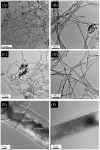
Figure 1. TEM image comparing the surface of pristine unseeded Germanium nanowires and water dispersible Germanium nanowires (WDW) taken on an Electron Microscope JEOL JEM 2100F.
(A, B) the high aspect ratio of the pristine Germanium nanowires (GeNW) which span for micrometres. (C, D) the complex surface morphology of water dispersible Germanium nanowires (WDW) after treatment with Glutamic acid, the wires still maintain their aspect ratio span for micrometres. (E) The HRTEM of WDW after 3 hours at 21°C in treatment solution, in comparison to the HRTEM of the pristine wires (F).
Figure 2
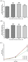
Figure 2. Water dispersible Germanium nanowires promote the proliferation of MCF-7 cells.
(A) MCF-7 cells were seeded at 27,000 cells/well, before MTT were carried out over 24 hr as described in the Materials and Methods. Data analysis to determine the level of significance was performed using Welch's t test to determine the level of significance between treatments and control, results were considered to be significant with P<0.05. The results for the above indicate a high degree of significance ***P≤0.001. (B) The normalised viability of MCF-7 cells seeded at 10,000 per well (N>3), was measured after 24 hours of exposure to wires at varying concentrations and measured by WST-1 test. Data analysis was performed using Welch's t test to determine the level of significance.(C) MCF-7 cells were seeded at 10,000 cells/well on increasing concentrations of WDW's for 4 days. Organic cell growth was determined by trypan blue exclusion (n = 5 for each time point and n = 3 for each experiment).
Figure 3
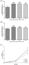
Figure 3. Water dispersible Germanium nanowires promote the proliferation of L929 cells.
(A) L929 cells were seeded at 27,000 cells/well, before MTT were carried out over 24 hr as described in the Materials and Methods. Data analysis to determine the level of significance was performed using Welch's t test to determine the level of significance between treatments and control, results were considered to be significant with P<0.05. The results for the above indicate a high degree of significance ***P≤0.001. (B) The normalised viability of L929 cells seeded at 10,000 per well (N>3), was measured after 24 hours of exposure to wires at varying concentrations and measured by WST-1 test. Data analysis was performed using Welch's t test to determine the level of significance. (C) L929 cells were seeded at 10,000 cells/well on increasing concentrations of WDW's for 4 days. Organic cell growth was determined by trypan blue exclusion (n = 5 for each time point and n = 3 for each experiment).
Figure 4
Figure 4. Confocal microscopy to study the morphological features of MCF-7 cells exposed to WDWs.
(A) MCF-7 control cells cultured on a 10 µg collagen glass cover slip and stained with phosphorylated FAK (pFAK397), Hoechst and Phalloidin TRICI under a 63X oil immersion lens using a Zeiss LSM 710. (B): MCF-7 cells cultured on a 10 µg collagen glass cover slip exposed to 4 µM of Germanium nanowires for 24 hours and stained with phosphorylated FAK (pFAK397), Hoechst and Phalloidin TRICI under a 63X oil immersion lens using a Zeiss LSM 710. (C) Dispersible Germanium Nanowires were added to cultures of MCF-7 cells for 24Hr. Lysates were prepared and the lysates ran on 12% SDS PAGE gels before probing with antibodies against Actin (Santa Cruz Biological) pERK (Cell signalling technology phosphor-p44/42 (ERK1/2)), RACK1 (BD Transduction Laboratories) and FAK (Santa Cruz Biotechnology FAK(C-20):sc558). Results were presented as a Histogram of relative FAK expression, RACK1 expression and pERK expression.
Figure 5
Figure 5. Confocal microscopy to study the morphological features of the L929 cells exposed to WDWs.
(A) L929 cells cultured on a 10 µg collagen glass cover slip and stained with phosphorylated FAK (pFAK397), Hoechst and Phalloidin TRICI under a 63X oil immersion lens using a Zeiss LSM 710. (B): L929 cells cultured on a 10 µg collagen glass cover slip exposed to 4µM of Germanium nanowires for 24 hours and stained with phosphorylated FAK (pFAK397), Hoechst and Phalloidin TRICI under a 63X oil immersion lens using a Zeiss LSM 710. (C) Dispersible Germanium Nanowires were added to cultures of MCF-7 cells for 24 Hr. Lysates were prepared and the lysates ran on 12% SDS PAGE gels before probing with antibodies against Actin (Santa Cruz Biological) pERK (Cell signalling technology phosphor-p44/42 (ERK1/2)), RACK1 (BD Transduction Laboratories) and FAK (Santa Cruz Biotechnology FAK(C-20):sc558). Results were presented as a Histogram of relative FAK expression, RACK1 expression and pERK expression.
Figure 6
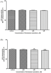
Figure 6. Measuring LDH release into the serum free media when cells are exposed to wires for 1hr.
(A): The MCF-7 results at varying concentrations after 1hr, seeded at 27,000 cells/ well, N>3. (B): The L929 results at varying concentrations after 1hr, seeded at 10,000 cells/ well, N>3. Data analysis was performed using one way Anova and Student-Newman-Keuls post hoc test to determine the level of significance between treatments and control, results were considered to be significant with P<0.05. The results for the L929 cells above (B) indicate a significant decrease in the LDH release into the media in which 4 µM displays a mean of 97.68±0.5237 N = 12 **P≤0.01 and 7 µM displays a mean of 94.83±0.3497 N = 12 *P≤0.5.
Figure 7
Figure 7. Using live cell impedance based platforms to monitor cell behaviour.
xCELLigence (ACEA) platforms were used to monitor cells cultured under the same conditions as those performed in the MTT, WST and LDH assays. All experiments were carried out using E-plates following the ACEA manual protocol where N = 3. (A) Shows MCF-7 cells seeded at 27,000 cells, with and without the presence of WDW (with appropriate controls and blanks). The data indicates that there is an increase in the cell index number a measure of increased impedance across the surface of the plate. (C) Shows L929 seeded at 10,000 cells with and without the presence of WDW (with appropriate controls and blanks). The data indicates that there is an increase in the cell index number a measure of increased impedance across the surface of the plate. (B), (D). MCF-7 cells and L929 cells grown on separate E plates together with increasing concentrations of WDW's to show how the Germanium nanowires themselves have no effect on the impedance as measured by the electrodes.

Similar articles

Cited by

References

    1. Tian B, Cohen-Karni T, Qing Q, Duan X, Xie P, et al. (2010) Three-Dimensional, Flexible Nanoscale Field-Effect Transistors as Localized Bioprobes. Science 329: 830–834. - PMC - PubMed
    1. Chan CK, Zhang XF, Cui Y (2008) High Capacity Li Ion Battery Anodes Using Ge Nanowires. Nano Letters 8: 307–309. - PubMed
    1. Huang J-S, Hsiao C-Y, Syu S-J, Chao J-J, Lin C-F (2009) Well-aligned single-crystalline silicon nanowire hybrid solar cells on glass. Solar Energy Materials and Solar Cells 93: 621–624.
    1. Jiang K, Fan D, Belabassi Y, Akkaraju G, Montchamp J-L, et al. (2009) Medicinal Surface Modification of Silicon Nanowires: Impact on Calcification and Stromal Cell Proliferation. ACS Applied Materials & Interfaces 1: 266–269. - PMC - PubMed
    1. Canham LT, Newey JP, Reeves CL, Houlton MR, Loni A, et al. (1996) The effects of DC electric currents on the in-vitro calcification of bioactive silicon wafers. Advanced Materials 8: 847–849.

Publication types

LinkOut - more resources