

Three-dimensional organotypic culture: experimental models of mammalian biology and disease
- PMID: 25237826
- PMCID: PMC4352326
- DOI: 10.1038/nrm3873
Three-dimensional organotypic culture: experimental models of mammalian biology and disease
Abstract
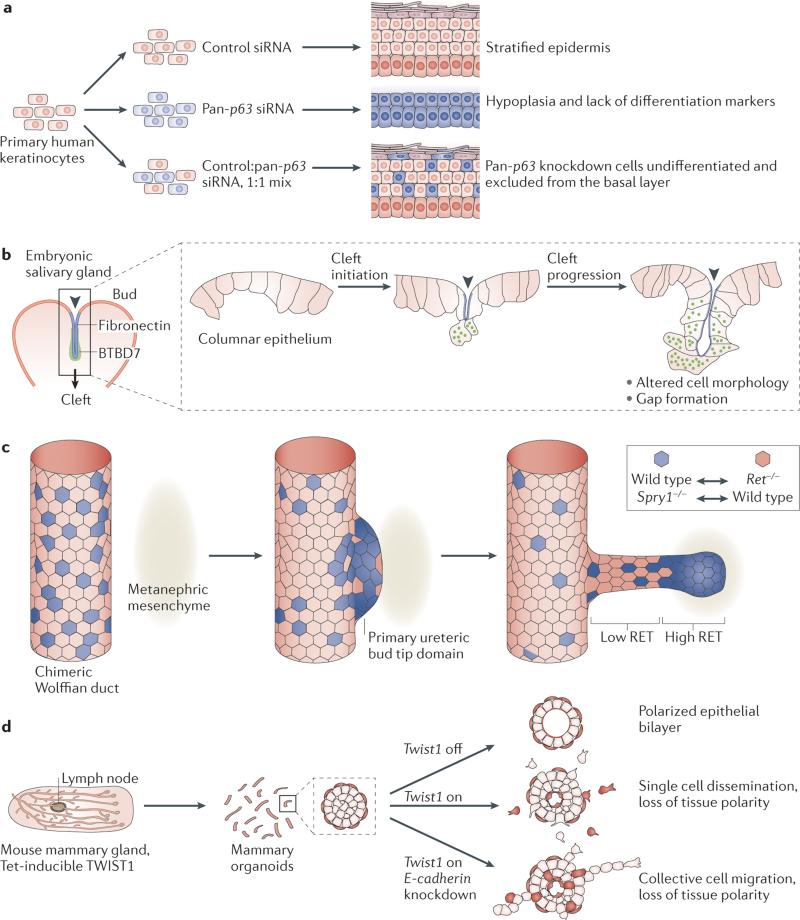
Mammalian organs are challenging to study as they are fairly inaccessible to experimental manipulation and optical observation. Recent advances in three-dimensional (3D) culture techniques, coupled with the ability to independently manipulate genetic and microenvironmental factors, have enabled the real-time study of mammalian tissues. These systems have been used to visualize the cellular basis of epithelial morphogenesis, to test the roles of specific genes in regulating cell behaviours within epithelial tissues and to elucidate the contribution of microenvironmental factors to normal and disease processes. Collectively, these novel models can be used to answer fundamental biological questions and generate replacement human tissues, and they enable testing of novel therapeutic approaches, often using patient-derived cells.
Figures


References
-
- Bichat X. General Anatomy, Applied to Physiology and Medicine. (Richardson and Lord. 1822
-
- Virchow R. De Witt RM, editor. Cellular Pathology, as Based upon Physiological and Pathological Histology. Twenty Lectures Delivered in the Pathological Institute of Berlin During the Months of February, March and April, 1858. 1860
-
- Sobotta J, Huber GC, De Witt LMB. Atlas and Epitome of Human Histology and Microscopic Anatomy. W. B. Saunders & company; 1903.
-
- Harrison RG, Greenman MJ, Mall FP, Jackson CM. Observations on the living developing nerve fiber. Anat. Rec. 1907;1:116–128.
Publication types
MeSH terms
Grants and funding
LinkOut - more resources
Full Text Sources
Other Literature Sources
Medical
