. 2014 Sep 19;9(9):e107961.
doi: 10.1371/journal.pone.0107961. eCollection 2014.

Arsenite-activated JNK signaling enhances CPEB4-Vinexin interaction to facilitate stress granule assembly and cell survival

Affiliations

Arsenite-activated JNK signaling enhances CPEB4-Vinexin interaction to facilitate stress granule assembly and cell survival

Yu-Wei Chang et al. PLoS One. .

Abstract

Stress granules (SGs) are compartmentalized messenger ribonucleoprotein particles (mRNPs) where translationally repressed mRNAs are stored when cells encounter environmental stress. Cytoplasmic polyadenylation element-binding protein (CPEB)4 is a sequence-specific RNA-binding protein and translational regulator. In keeping with the results obtained from the study of other RNA-binding proteins, we found CPEB4 localized in SGs in various arsenite-treated cells. In this study, we identified that Vinexin, a CPEB4-interacting protein, is a novel component of SGs. Vinexin is a SH3-domain-containing adaptor protein and affects cell migration through its association with Vinculin to localize at focal adhesions (FAs). Unexpectedly, Vinexin is translocated from FAs to SGs under arsenite-induced stress. The recruitment of Vinexin to SGs depends on its interaction with CPEB4 and influences SG formation and cell survival. Arsenite-activated c-Jun N-terminal kinase (JNK) signaling enhances the association between CPEB4 and Vinexin, which consequently facilitates SG localization of Vinexin. Taken together, this study uncovers a novel interaction between a translational regulator and an adaptor protein to influence SG assembly and cell survival.


Conflict of interest statement

Competing Interests: The authors have declared that no competing interests exist.

Figures

Figure 1
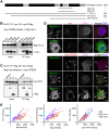
Figure 1. Vinexin interacts with CPEB4 and localizes at SGs in CPEB4-overexpressing cells.
(A) Molecular architectures of mouse Vinexin (mVinexin), showing the sorbin homology (SoHo) and three Src-homology (SH3) motifs. The α, β and γ isoforms vary at the N-terminal end. Using the CPEB4 N-terminus as the bait, a yeast two hybrid (Y2H) screen identified two clones corresponding to the a.a. 482–610 and 483–633 of mVinexin α. The full-length mVinexin β was also shown for positive (+) interaction with CPEB4. N/A: not applicable. (B) Co-immunoprecipitation (Co-IP) assay. The 293T cell lysates containing flag-Vinexin β along with myc-tagged CPEB2, 3 or 4 were precipitated with myc IgG and immunoblotted with flag and myc antibodies. IB: immunoblotting, IP: immunoprecipitation. (C) Similarly, the 293T cell lysates containing myc-CPEB4 (myc-CP4) without (vector) or with flag-Vxn α, β or γ, were precipitated with flag IgG and immunoblotted with flag and myc antibodies. (D) Subcellular distribution of flag-Vxn α and β forms in COS-7 cells in the absence or presence of myc-CPEB4 expression. Arrow heads and arrows denote focal adhesion (FA) and stress granule (SG) structures, respectively. TIA-1 immunostained signal detected by the AlexaFluor 647-conjugated secondary antibody was pseudo-colored in magenta and used as a SG marker. The signal intensities of TIA-1, myc-CPEB4, flag-Vxn α and β in one hundred SGs randomly selected from ten transfected cells were pairwisely compared and plotted in (E). Scale: 10 µm.
Figure 2
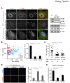
Figure 2. Redistribution of Vinexin from FAs to SGs in arsenite-stressed cells.
(A) U2OS cells were treated without (control) or with arsenite ± cycloheximide (CHX) for 30 min prior to immunostaining of Vinexin, Vinculin (FA marker) and TIA-1 (SG marker). Arrow heads and arrows indicate FAs and SGs, respectively. One hundred FAs were randomly selected from ten cell images in each group to quantify the fluorescence intensities of Vinexin and Vinculin. Similarly, a hundred SGs from ten arsenite-treated cells were analyzed for the signals of Vinexin and TIA-1. The top scatter plot shows the reduction of Vinexin signal in FAs after the addition of arsenite. (B) Distribution of Vinexin and CPEB4 in arsenite or heat shock (42°C, 20 min)-treated HeLa cells. Arrows denote colocalization of CPEB4 and Vinexin in SGs. (C) Live imaging of EGFP-Vinexin β and RFP-TIA-1 distribution in HeLa cells treated with arsenite (see Figure S3 for the entire cell images). Arrow heads and arrows indicate FAs and SGs, respectively. Scale: 10 µm.
Figure 3
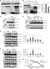
Figure 3. SG localization of Vinexin depends on CPEBs.
(A) The wild-type (wt) and CPEB2/CPEB4 double-knockout (CP2/CP4 dKO) MEFs were treated with arsenite and then fixed for immunodetection of Vinexin, CPEB4 and TIA-1. Arrows indicate TIA-1-positive SGs. Scale: 10 µm. The western blots show the expression of indicated proteins in the wt and dKO MEFs. (B) One hundred SGs were randomly selected in ten cell images taken from arsenite-treated wt or dKO MEFs to quantify the signal intensities of Vinexin and TIA-1. The analyzed results show the Vinexin signal is significantly decreased in TIA-1-containing SGs in the dKO cells. (C) For each cell, the number of TIA-1-positive SGs were analyzed and displayed in the dot plot. The average SG number per cell (mean ± s.e.m.) and the number of analyzed cells are listed at the bottom (see Figure S6A for the analyses using CP2KO and CP4 KO MEFs). The percent of (D) apoptotic and (E) survived wt and dKO cells at 4 h and 10 h after arsenite exposure was analyzed by TUNEL and PrestoBlue assays, respectively. The data was presented as mean ± s.e.m. from three independent experiments. One and two asterisks denote *P<0.05 and **P<0.01 (Student’s t-test).
Figure 4
Figure 4. Vinexin binds to CPEB4 and localizes in SGs through its first two SH3 motifs.
(A) The domain organization of Vinexin β and various truncated mutants. (B) Co-IP assay. The 293T cells were co-transfected with myc-CPEB4 plasmid along with various EGFP-tagged Vinexin β constructs. The cell lysates were immunoprecipitated with GFP antibody, and immunoblotted with myc and GFP antibodies. (C) The 293T cells expressing myc-CPEB4 and varied SH3-deleted flag-Vinexin β mutants were pulled down with myc IgG and immunodetected with myc and flag antibodies. IP: immunoprecipitation, IB: immunoblotting. (D) SG distribution of wt and Vinexin mutants in COS-7 cells overexpressing myc-CPEB4. If the signal of Vinexin in CPEB4 and TIA-1-positive SGs higher than that in the cytosol was considered as SG-localized (dark grey bar), and the rest was grouped as diffuse in cytosol (light grey bar). One hundred transfected cells per group were analyzed.
Figure 5
Figure 5. Vinexin promotes SG formation and assembly.
HeLa cells infected with lentiviruses expressing without (siCtrl) or with Vinexin shRNA (siVxn) were puromycin-selected, treated with ± arsenite and then harvested at different time points for (A) western blotting or (B) TIA-1 immunostaining to detect SGs. The representative images of siCtrl and siVxn cells during (30 min) and recovering from (180 min) arsenite-induced stress are shown. (C) The number of TIA-1-positive SGs in siCtrl and siVxn cells were analyzed and expressed as the mean ± s.e.m. Approximately 1000 cells in each group collected from three independent experiments were analyzed using the MetaMorph software. The significant difference between siCtrl and siVxn groups at each time point was analyzed with Student’s t-test. (D) The percent of survived siCtrl and siVxn cells after arsenite treatment at the indicated time was determined by PrestoBlue viability assay and presented as mean ± s.e.m. from three independent experiments. (E) To test wt or mutant Vinexin in rescuing SG-assembly defect in siVxn cells, the transfected cells were analyzed by western blotting to examine the expression of indicated proteins or TIA-1 immunostaining to score the SG numbers in ∼1000 transfected cells of each group collected from three independent experiments. One and two asterisks denote *P<0.05 and **P<0.01 (Student’s t-test).
Figure 6
Figure 6. JNK signaling promotes CPEB4-Vinexin interaction in arsenite-treated cells.
The 293T cells expressing EGFP or EGFP-Vinexin β were treated with ± arsenite and then harvested for immunoprecipitation using the GFP antibody. The precipitated substances were immunoblotted with (A) CPEB4 and GFP antibodies or (B) Vinculin and GFP antibodies. (C) The 293T cells expressing myc-CPEB4 (CP4) and flag-Vinexin β (Vxn β) were treated with ± arsenite for the denoted times and then precipitated with the flag IgG and immunoblotted using myc and flag antibodies. (D) (E) Similarly to (C) except the cells were pre-treated with various kinase inhibitors, PD98059 (PD), SB2035800 (SB) or SP600125 (SP) for 30 min, followed by arsenite stimulation and co-IP to monitor CPEB4-Vinexin interaction. (F) The 293T cells expressing EGFP-Vinexin β, myc-CPEB4 along with the flag-tagged wt or dominant negative (DN)-JNK1 were harvested at the indicated time after arsenite treatment for co-IP assay. The pull-down substances by the GFP antibody were used for western blotting. For (E, F), the results from three independent experiments were quantified and displayed as mean ± s.e.m. (G) The number of TIA-1-positive SGs in approximately 1000 cells treated with ± SP600125 were analyzed using the MetaMorph software and expressed as the mean ± s.e.m. One and two asterisks denote *P<0.05 and **P<0.01 (Student’s t-test).
Figure 7
Figure 7. The proline-rich domains (PRDs) of CPEB4 bind to Vinexin in SGs.
(A) The domain organization of CPEB4, showing the N-terminal four PRDs and the C-terminal RNA-binding domain composed of two RNA recognition motifs (RRM) and zinc fingers (Zif). The various CPEB4 mutants were illustrated. (B) The 293T lysates containing flag-Vinexin β along with myc-tagged wt or mutant (mut) CPEB4 were precipitated by myc IgG, followed by immunoblotting with myc and flag antibodies. (C) FRET analysis. The plasmids encoding the FRET donor EGFP-Vxn β and acceptor RFP-CPEB4 (CP4) or mutants (CP4mut7 and ΔPRD) were co-transfected to COS-7 cells. The formaldehyde-fixed samples were used for FRET analysis to detect CPEB4-Vineixn interaction in SGs. The example images and the line graph show that the fluorescent signal of EGFP-Vxn β increases after photobleaching the acceptor RFP-CP4 in the selected SG (red circle). In contrast, no increasing change in EGFP signal if RFP-CP4mut7 was used as the acceptor. (D) The changes in fluorescence intensity of EGFP-Vxn β right before and after photobleaching RFP were calculated for FRET efficiency. The FRET interaction between EGFP-Vxn β and RFP-CP4 wt, mut7 or ΔPRD with ± arsenite was determined. Similarly, the FRET interaction of EGFP-Vxn β and RFP-CP4 was also measured in the presence of JNK inhibitor, SP600125 (SP). All of the data were expressed as the mean ± s.e.m. n: the number of SGs in each group (one SG per cell was analyzed by FRET acceptor bleaching). The significant difference between wt and CPEB4 mutants as well as between mock and arsenite (Ars) ± SP treatments was analyzed with Student’s t-test. One and two asterisks denote *P<0.05 and **P<0.01. (E) Schematic model of arsenite-induced redistribution of CPEB4 and Vinexin to SGs. Arsenite stress activates JNK signaling, which somehow promotes the association of CPEB4 and Vinexin to recruit Vinexin in SGs. Once translocated to SGs, Vinexin plays an active role in facilitating SG assembly, most likely recruiting additional factors (e.g., RTKN: Rhotekin) through its third SH3 motif. Meanwhile, the dissociation of Vinexin from cytoskeletal proteins, such as Vinculin, weakens focal adhesions and promotes Vinexin translocation from FAs to SGs. ECM, extracellular matrix.

Similar articles

Cited by

References

    1. Anderson P, Kedersha N (2009) RNA granules: post-transcriptional and epigenetic modulators of gene expression. Nat Rev Mol Cell Biol 10: 430–436. - PubMed
    1. Buchan JR, Parker R (2009) Eukaryotic stress granules: the ins and outs of translation. Mol Cell 36: 932–941. - PMC - PubMed
    1. Higashi S, Kabuta T, Nagai Y, Tsuchiya Y, Akiyama H, et al. (2013) TDP-43 associates with stalled ribosomes and contributes to cell survival during cellular stress. J Neurochem 126: 288–300. - PubMed
    1. Kim B, Cooke HJ, Rhee K (2012) DAZL is essential for stress granule formation implicated in germ cell survival upon heat stress. Development 139: 568–578. - PubMed
    1. Takahashi M, Higuchi M, Matsuki H, Yoshita M, Ohsawa T, et al. (2013) Stress granules inhibit apoptosis by reducing reactive oxygen species production. Mol Cell Biol 33: 815–829. - PMC - PubMed

Publication types