. 2014 Sep 18;41(3):427-439.
doi: 10.1016/j.immuni.2014.08.012.

Transforming growth factor β-mediated suppression of antitumor T cells requires FoxP1 transcription factor expression

Affiliations

Transforming growth factor β-mediated suppression of antitumor T cells requires FoxP1 transcription factor expression

Tom L Stephen et al. Immunity. .

Abstract

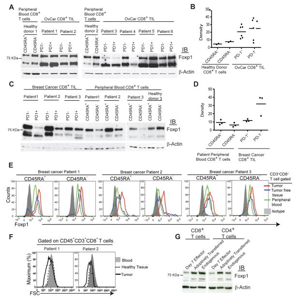
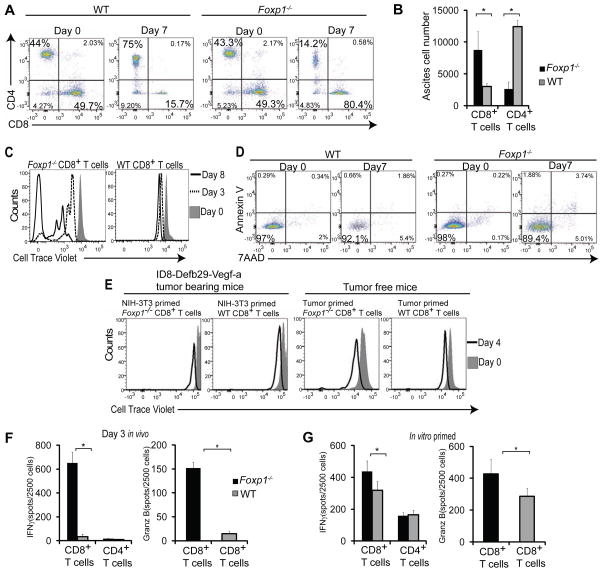
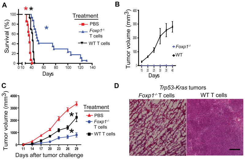
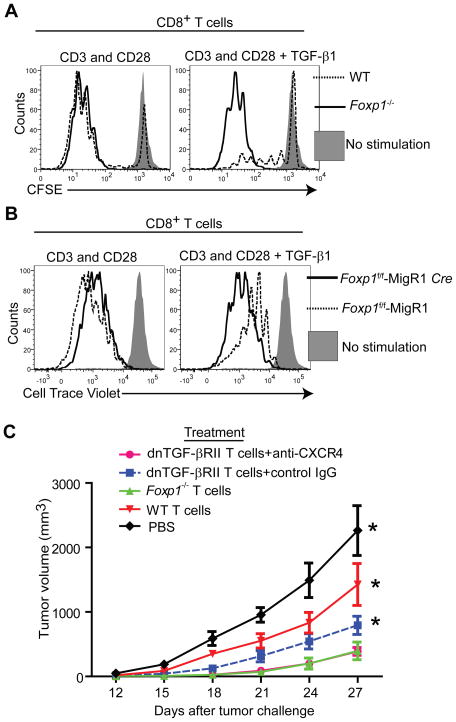
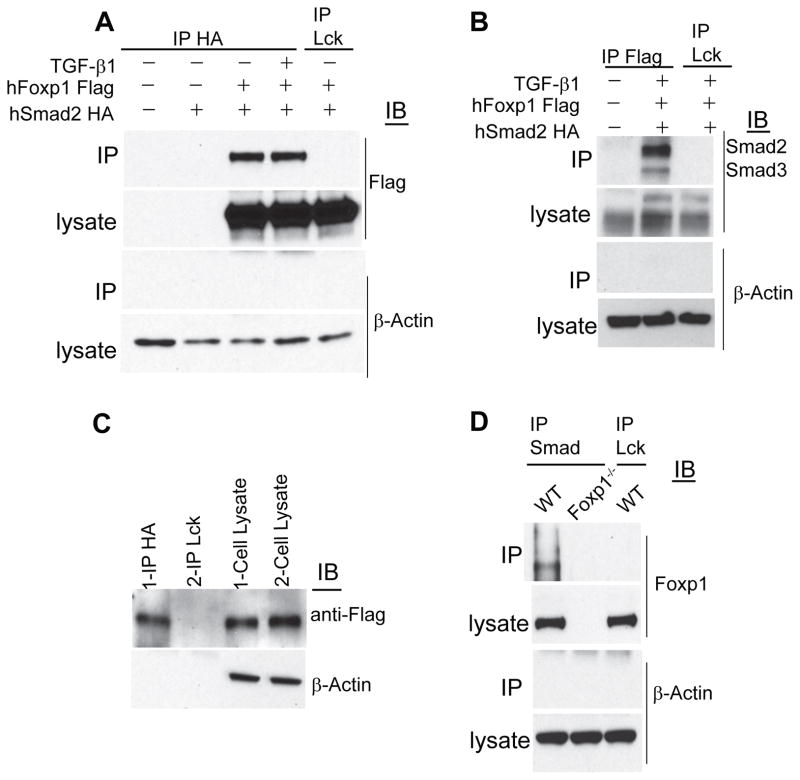
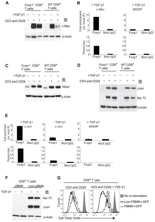
Tumor-reactive T cells become unresponsive in advanced tumors. Here we have characterized a common mechanism of T cell unresponsiveness in cancer driven by the upregulation of the transcription factor Forkhead box protein P1 (Foxp1), which prevents CD8⁺ T cells from proliferating and upregulating Granzyme-B and interferon-γ in response to tumor antigens. Accordingly, Foxp1-deficient lymphocytes induced rejection of incurable tumors and promoted protection against tumor rechallenge. Mechanistically, Foxp1 interacted with the transcription factors Smad2 and Smad3 in preactivated CD8⁺ T cells in response to microenvironmental transforming growth factor-β (TGF-β), and was essential for its suppressive activity. Therefore, Smad2 and Smad3-mediated c-Myc repression requires Foxp1 expression in T cells. Furthermore, Foxp1 directly mediated TGF-β-induced c-Jun transcriptional repression, which abrogated T cell activity. Our results unveil a fundamental mechanism of T cell unresponsiveness different from anergy or exhaustion, driven by TGF-β signaling on tumor-associated lymphocytes undergoing Foxp1-dependent transcriptional regulation.


Conflict of interest statement

The authors declare no conflict of interest.

Figures

Figure 1
Figure 1. CD8+ T cells up-regulate Foxp1 in the TME
(A) Foxp1 expression in PD-1 and PD-1+ CD8+ T cells flow cytometry-sorted from six freshly dissociated stage III and IV human ovarian carcinoma specimens (samples 4 and 5, primary tumors; samples 1, 2, 3, and 6, metastatic masses). CD45RA+ (resting or naïve) and CD45RA (activated) CD8+ T cells from the peripheral blood of two healthy donors were sorted and analyzed in parallel in two independent experiments. (B) Normalization of Foxp1 protein expression in PD-1 and PD-1+ ovarian tumor-infiltrating CD8+ T cells with β-actin. (C) Foxp1 expression in PD-1 and PD-1+ CD8+ T cells flow cytometry-sorted from three freshly dissociated breast cancer specimens, as well as matching peripheral blood from the same patients and a healthy donor. (D) Densitometric normalization performed as in (B). (E) Intracellular staining for Foxp1 in CD3+CD8+ gated CD45RA+ and CD45RA T cells from human breast tumors, tumor free tissues and peripheral blood from same patients. (F) Comparative FSC analysis of CD8+ T cells contained in a freshly dissociated advanced ovarian carcinoma and matching peripheral blood. (G) CD45.2+ naive T cell splenocytes were primed in vitro with bone marrow-derived DCs (BMDCs) pulsed with double irradiated (UV+gamma) ID8-Defb29-Vegf-a tumor cells for 7 days (day 7 effectors) and administered into the peritoneal cavity of congenic ID8-Defb29-Vegf-a tumor-bearing mice at day 23 after tumor challenge. Three days later, transferred (CD45.2+) and endogenous (CD45.1+) CD8+ and CD4+ T cells were flow cytometry-sorted from tumor ascites and analyzed by western blot. Representative of two independent experiments. IB, immunoblotting.
Figure 2
Figure 2. Foxp1 expression regulates anti-tumor effector functions and proliferation of CD8+ T cells in the TME
(A) Flow cytometric analysis of CD45.2+ Foxp1-deficient vs. wild-type T cells identically primed against tumor antigens as in Figure 1G. (B) Absolute cell number of these lymphocytes recovered from peritoneal wash 7 days after adoptive transfer into day 24 ID8-Defb29-Vegf-a tumor-bearing congenic mice. Representative of four independent experiments (p<0.05, Student’s t-test). (C) In vivo proliferation of tumor-reactive Foxp1-deficient vs. wild-type CD8+ T cells. Lymphocytes primed for 7 d against tumor antigens were labeled with cell trace violet, adoptively transferred into orthotopic advanced ovarian cancer-bearing congenic mice, and recovered from peritoneal wash 3 and 8 days later. Representative of three experiments. (D) Annexin V and 7AAD staining of tumor antigen-primed Foxp1-deficient and wild-type T cells recovered from tumor ascites at the indicated days after adoptive transfer in three independent experiments. (E) Proliferation of Foxp1−/− and WT T cells either primed with BMDCs pulsed with irradiated and UV-treated NIH-3T3 cells followed by transfer into day 24 ID8-Defb29-Vegf-a tumor-bearing congenic mice (left), or primed with tumor antigen and transferred into the peritoneal cavity of tumor free congenic mice (right). Representative of three experiments. (F) ELISPOT analysis of identically primed T cells, sorted from tumor ascites 3 d after adoptive transfer into advanced ID8-Defb29-Vegf tumor-bearing congenic mice, and re-stimulated with PMA and Ionomycin for 4 h. Representative of three independent experiments (G) IFN-γ and Granzyme B ELISPOT analysis of T cells primed against tumor antigens as above for 7 d. (p<0.05, Student’s t-test).
Figure 3
Figure 3. Foxp1 expression impairs the protective function of tumor-reactive T cells
(A) On day 24 after ID8-Defb29-Vegf-a tumor challenge, 47 different CD45.1+ mice received 106 tumor antigen-primed (day 7) T cells from Foxp1-deficient (n=16) or wild-type (n=15) CD45.2+ mice. Sixteen additional control tumor-bearing mice were treated with PBS. Data pooled from three independent experiments. P<0.0001, Mantel-Cox test. (B) Four tumor-bearing mice surviving >60 d. after treatment with Foxp1−/− T cells and six control age-matched wild-type mice were re-challenged with 2 × 10⁶ ID8-Defb29-Vegf-a tumor cells, administered into the axillary flank. Tumor growth was monitored in three independent experiments. P<0.003, Student’s t-test. (C) Naïve T cell splenocytes from Foxp1-deficient or wild-type mice were primed for 7 d with BMDCs pulsed with double (γ- plus UV- irradiated) MPKAS cells. Tumor-reactive T cells were delivered into tumors formed from this cell line (2 × 10⁶ cells) in congenic mice (n=9 receiving Foxp1−/− and n=9 receiving wild-type T cells), at days 8 and 14 after flank tumor challenge. Eight additional flank tumor-bearing mice received PBS. Pooled from 3 independent experiments. P<0.005 between tumor volume of Foxp1-deficient T cells and either wild-type T cell or PBS treatment groups (Mann-Whitney). (D) Massive necrosis induced by intratumoral administration of Foxp1−/−, but not wild-type tumor-reactive T cells in C57BL/6 Trp53-Kras mice challenged with s.c. adenovirus-Cre to induce flank sarcomas. Palpable tumors were injected 3–4 times with 106 tumor antigen-primed Foxp1-deficient vs. wild-type T cells, at 5–6 d intervals. Representative of three independent experiments. Scale bar 200 μM.
Figure 4
Figure 4. Foxp1 is required for TGF-β-induced suppression of CD8+ T cells
(A) Proliferation analysis of cell trace violet-labeled Foxp1-deficient and wild-type T cells, stimulated for 5 days with CD3 and CD28 beads, in the presence or the absence of 5 ng/mL of TGF-β1. Cells were stained for CD8, Annexin V and 7AAD. Representative of four independent experiments. (B) T cells from the spleen and lymph nodes of Foxp1f/f were transduced with Cre-recombinase expressing MigR1-GFP retroviruses or the empty vector. GFP+ (excised) CD8+ T cells were flow cytometry-sorted after 48 hours, cell trace violet-labeled and CD3 and CD28-stimulated for 4–5 days, in the presence or the absence of TGF-β1 (5 ng/mL). Representative of three independent experiments. (C) Growth kinetics of MPKAS sarcomas (n=6/group) intratumorally treated at days 7 and 10 with 2×10⁶ tumor antigen-primed (6 d.) T cells that were dnTGF-βRII (pre-incubated for 30 min with 5 μg/ml of anti-mouse CXCR4 or control IgG; both from R&D), Foxp1−/−, or WT. Additional controls received PBS.
Figure 5
Figure 5. Foxp1 deficiency does not affect TGF-β-induced nuclear translocation of Smad signaling molecules in CD8+ T cells
(A) Western blot analysis of TGF-βRII expression in Foxp1-deficient and wild-type CD8+ T cells under various stimulation conditions. (B) Expressions of Smad2 (upper band), Smad3 (lower band) and phosphorylated Smad2 (p-Smad2) in Foxp1-deficient and wild-type CD8+ T cells at rest or CD3 and CD28-activated for 24 hours, in the presence or the absence of TGF-β1 (5 ng/mL). (C) Expression of p-Smad3 (Ser423 and Ser425) in Foxp1-deficient and wild-type CD8+ T cells stimulated in vitro with CD3 and CD28-beads for 24 h, followed by TGF-β1 (5ng/ml) treatment for 30 minutes. (D–E) Confocal microscopy analysis of resting and CD3 and CD28-stimulated (24 h) Foxp1-deficient and wild-type CD8+ T cells (+/−TGF-β1; 5 ng/ml). Representative of three independent experiments. Scale bar 10 μM.
Figure 6
Figure 6. Foxp1 interacts with the TGF-β-induced Smad repressor complex
(A) HeLa cells were transiently transfected with HA-tagged human Smad2 (Smad2-HA) and Flag-tagged human Foxp1.1 (Foxp1-Flag), treated or not with 5 ng/ml of TGF-β1 for 12 hours, cross-linked and lysed. HA was immunoprecipitated (IP) from the extracted proteins, followed by immunoblotting for Flag. HA IP from un-transfected, Smad2-HA only transfected HeLa cell lysates, and irrelevant (α-Lck) IgG IP from Smad2-HA and Foxp1-Flag transfected cell lysates were used as IP controls. Representative of four independent experiments. (B) Reverse IP with Flag and immunoblotting for Smad2 and Smad3 from transiently transfected HeLa cells described above. Representative of two independent experiments. (C) Primary human CD8+ T cells were electroporated with Smad2-HA and Foxp1-Flag. After 6–12 hours, cells were CD3 and CD28-stimulated for 24 h (+/− 5 ng/ml TGF-β1), cross-linked, and lysed. IP was carried out with anti-HA antibody and irrelevant IgG followed by immunoblotting for Flag. (D) Endogenous Smad2 and Smad3 was IPed from the whole cell lysates of wild-type and Foxp1-deficient T cells CD3 and CD28-activated in the presence of TGF-β1 (5 ng/ml). IP with irrelevant IgG was simultaneously performed. IPed proteins were immunoblotted and probed with anti-Foxp1 antibodies. Representative of three independent experiments. IP, Immunoprecipitation.
Figure 7
Figure 7. Foxp1 is required for TGF-β-induced repression of c-Myc and c-Jun in CD8+ T cells
(A) Foxp1−/− and WT CD8+ T cells were CD3 and CD28-stimulated (+/−TGF-β1, 5 ng/ml) for 24 h. Proteins were isolated and immunoblotted for c-Myc. (B) Foxp1 binding to the c-Myc promoter. Chromatin was immunoprecipitated with anti-Foxp1 or control IgG from negatively immunopurified mouse CD8+ T cells activated for 24 hours (+TGF-β1; 5 ng/ml). Enrichment of the c-Myc promoter sequence in chromatin IPed with anti-Foxp1 Abs vs. irrelevant IgG (top) and percent of input samples before immunoprecipitation (2.5% gel input values; bottom) were quantified by Real-Time Q-PCR. The mouse MISIIR gene promoter (GenBank#AF092445) was used as an additional negative control. Representative of two independent experiments. (C–D); Western blot analysis of Foxp1−/− and WT stimulated with CD3 and CD28 (+/−TGF-β1, 5 ng/ml) for 24 h for Nfat 2 (C) or for Jun and phospho-(serine 73)-c-Jun (D). Blots were stripped and reprobed with β-actin as an endogenous normalization control. Representative of three independent experiments. (E) ChIP PCR of mouse CD8+ T cells activated for 24 hours (+/−TGF-β1; 5 ng/ml) for c-Jun promoter sequence as described above. Representative of two independent experiments. (F) CD45.2+ wild-type mouse T cells were transduced with murine c-Jun, and congenic CD45.1+ T cells were infected with the empty pBMN-I-GFP retroviral vector. GFP+CD8+ cells were flow cytometry-sorted based upon CD45.1 and CD45.2 after 48 hours and CD3 and CD28-re-stimulated for 24 hours (+/−TGF-β1; 5 ng/ml). Proteins isolated from cell lysates were immunoblotted for total and Ser73 phosphorylated c-Jun. Representative of two independent experiments. (G) Positively c-Jun-transduced CD45.2+ T cells and mocked-transduced CD45.1+ congenic T cells were flow cytometry-sorted based on GPF expression, pooled at 1:1 ratio, rested for one day, and labeled with cell trace violet. Proliferation in response to CD3 and CD28-stimulation for 3–5 days, in the presence or the absence of TGF-β1 (5 ng/mL), was quantified by flow cytometry. Gated on CD8+ T cells. Annexin V and 7ADD staining were performed to discard dead and apoptotic cells. Representative of three independent experiments.

Comment in

Similar articles

Cited by

References

    1. Ahmadzadeh M, Rosenberg SA. TGF-beta 1 attenuates the acquisition and expression of effector function by tumor antigen-specific human memory CD8 T cells. J Immunol. 2005;174:5215–5223. - PMC - PubMed
    1. Baitsch L, Fuertes-Marraco SA, Legat A, Meyer C, Speiser DE. The three main stumbling blocks for anticancer T cells. Trends Immunol 2012 - PubMed
    1. Chen L, Glover JN, Hogan PG, Rao A, Harrison SC. Structure of the DNA-binding domains from NFAT, Fos and Jun bound specifically to DNA. Nature. 1998;392:42–48. - PubMed
    1. Chen ML, Pittet MJ, Gorelik L, Flavell RA, Weissleder R, von Boehmer H, Khazaie K. Regulatory T cells suppress tumor-specific CD8 T cell cytotoxicity through TGF-beta signals in vivo. Proc Natl Acad Sci U S A. 2005;102:419–424. - PMC - PubMed
    1. Crespo J, Sun H, Welling TH, Tian Z, Zou W. T cell anergy, exhaustion, senescence, and stemness in the tumor microenvironment. Curr Opin Immunol. 2013;25:214–221. - PMC - PubMed

Publication types

MeSH terms