. 2014 Oct 30;5(20):9811-22.
doi: 10.18632/oncotarget.2443.

Combined VEGF and CXCR4 antagonism targets the GBM stem cell population and synergistically improves survival in an intracranial mouse model of glioblastoma

Affiliations

Combined VEGF and CXCR4 antagonism targets the GBM stem cell population and synergistically improves survival in an intracranial mouse model of glioblastoma

Amy Barone et al. Oncotarget. .

Abstract

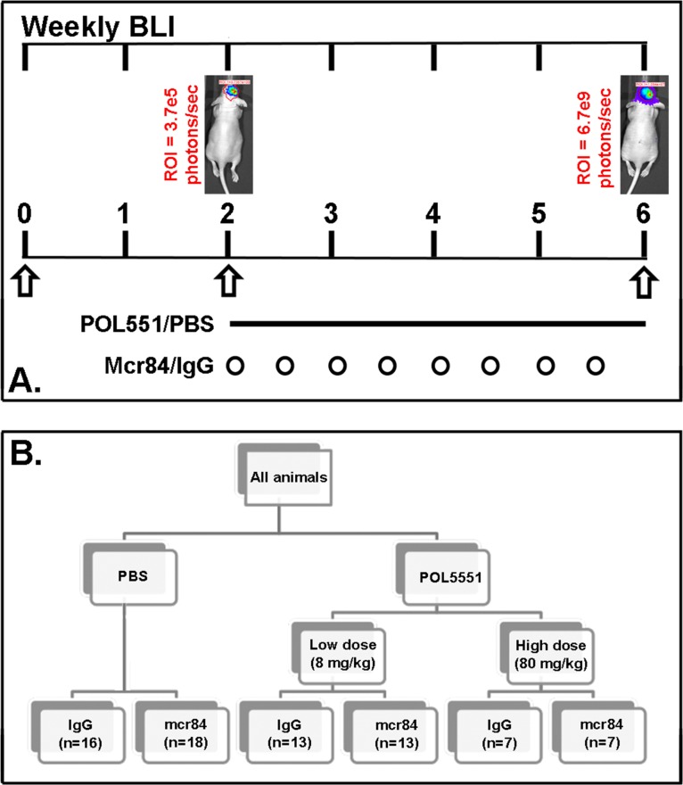
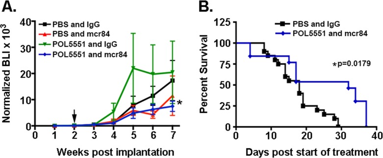
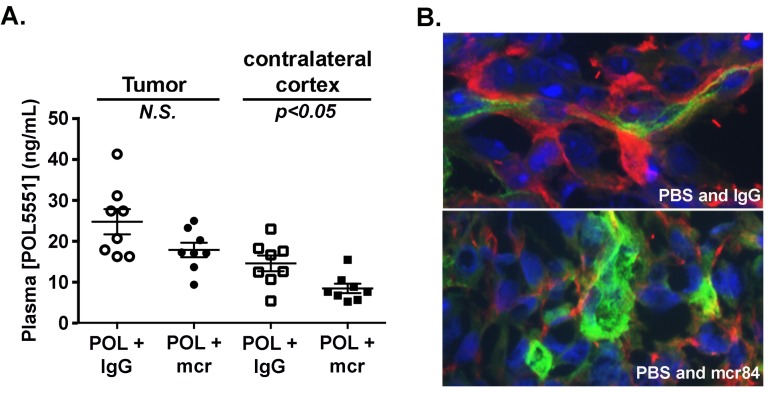
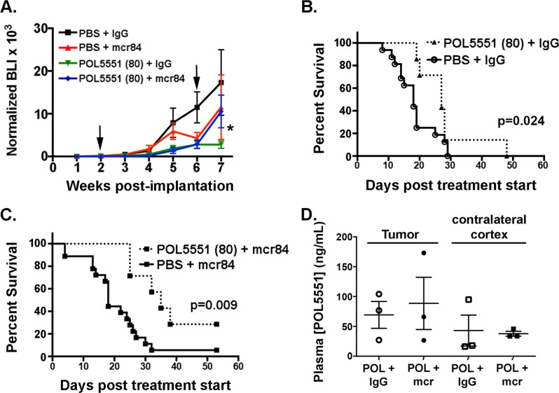
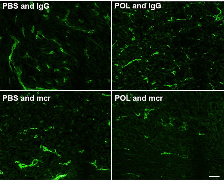
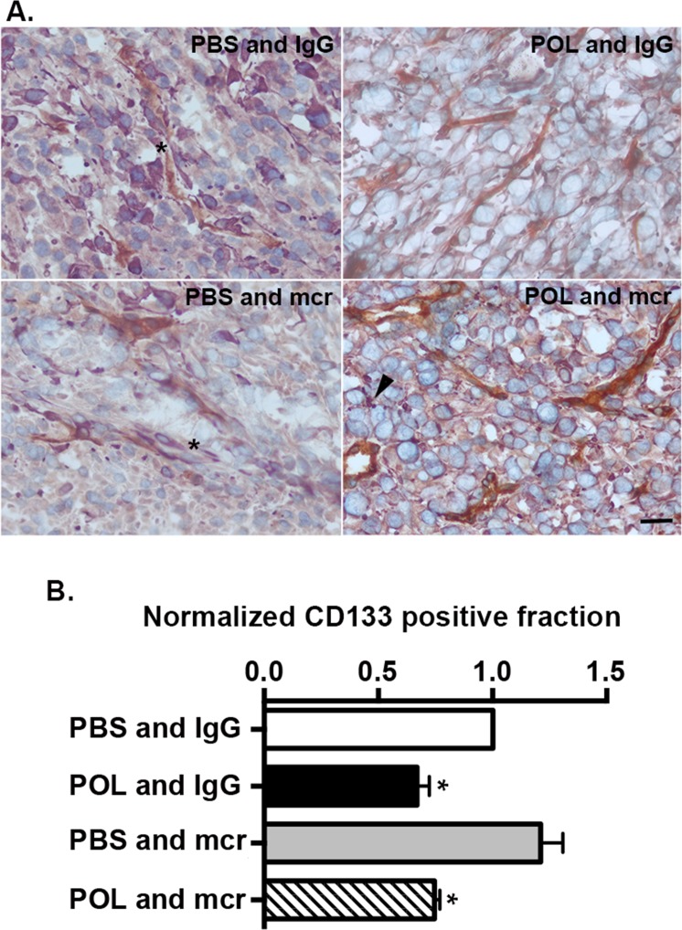
Glioblastoma recurrence involves the persistence of a subpopulation of cells with enhanced tumor-initiating capacity (TIC) that reside within the perivascular space, or niche (PVN). Anti-angiogenic therapies may prevent the formation of new PVN but have not prevented recurrence in clinical trials, suggesting they cannot abrogate TIC activity. We hypothesized that combining anti-angiogenic therapy with blockade of PVN function would have superior anti-tumor activity. We tested this hypothesis in an established intracranial xenograft model of GBM using a monoclonal antibody specific for murine and human VEGF (mcr84) and a Protein Epitope Mimetic (PEM) CXCR4 antagonist, POL5551. When doses of POL5551 were increased to overcome an mcr84-induced improvement in vascular barrier function, combinatorial therapy significantly inhibited intracranial tumor growth and improved survival. Anti-tumor activity was associated with significant changes in tumor cell proliferation and apoptosis, and a reduction in the numbers of perivascular cells expressing the TIC marker nestin. A direct effect on TICs was demonstrated for POL5551, but not mcr84, in three primary patient-derived GBM isolates. These findings indicate that targeting the structure and function of the PVN has superior anti-tumor effect and provide a strong rationale for clinical evaluation of POL5551 and Avastin in patients with GBM.


Conflict of interest statement

Conflicts of Interest

BR, GD, EC, MPB and KD are all employed by Polyphor and own shares in the company. RAB is a consultant for, has equity interest in, and is a recipient of a sponsored research grant from Peregrine Pharmaceuticals and Affitech AS. R. A. Brekken is also an author of a patent on technology that was used to develop the antibody r84 by Peregrine Pharmaceuticals and Affitech. Peregrine and Affitech did not participate in the planning, execution, or interpretation of the experiments.

Figures

Figure 1
Figure 1. Treatment scheme
(A.) Engraftment of intracranial tumors was confirmed by serial BLI over the two week post implantation period. (B) A subcutaneous osmotic pump delivered either PBS or POL5551 (low dose or high dose) continuously over 28 days. Mice received either mcr84 or vehicle IgG antibody (10 mg/kg i.p. twice weekly for 4 weeks).
Figure 2
Figure 2. Combined mcr84 and LD-POL5551 blocks brain tumor growth and increases survival in vivo
(A) Tumor growth was measured by weekly BLI. Shown are the mean and SEM for weekly BLI measurements for each treatment group (n=13-18 mice per group) normalized to fold over initial BLI. Arrows indicate the start and end of treatment. *p=0.0137 for the effect of treatment (weeks 2-6) on tumor growth (BLI). (B) LD-POL5551 in combination with mcr84 (n=13) increased median survival in comparison to vehicle controls (n=16) (p=0.0179). There were no significant improvements in survival for either of the monotherapy groups.
Figure 3
Figure 3. VEGF blockade with mcr84 enhances vascular barrier functions
(A) POL5551 concentrations in tumor tissue and non-tumor tissue from the contralateral cortex of xenograft bearing mice were determined by LC-MS/MS. POL5551 concentration was higher in tumor tissue versus non-tumor tissue. In both the tumor and non-tumor tissue, POL5551 concentration was decreased in the presence of mcr84. Bars represent the mean±SEM values. (B) mcr84 treatment markedly reduced the amount of albumin (red) detectable within the perivascular space of tumor sections compared to mice treated with vehicle control. Vascular endothelial cells were identified by endoglin staining (green) and nuclei are counterstained in blue with DAPI.
Figure 4
Figure 4. HD-POL5551 blocks brain tumor growth and increases survival in vivo
(A) Tumor growth was measured by weekly bioluminescence imaging. Shown are the mean and SEM for weekly BLI measurements each treatment group (n=7-18 mice per group). Arrows indicate the start and end of treatment. *p=0.0129 for the effect of treatment (weeks 2-6) on tumor growth (BLI). (B,C) HD-POL5551 alone (n=7) and in combination with mcr84 (n=7) increased mean survival in comparison to vehicle controls (n=16) (p=0.014 and p=0.009 respectively). (D) POL5551 concentrations in tumor tissue and non-tumor tissue from the contralateral cortex of xenograft bearing mice were determined by LC-MS/MS. POL5551 concentration was higher in tumor tissue versus non-tumor tissue. Bars represent the mean±SEM values. There was no significant decrease in tumor levels upon combination with mcr84.
Figure 5
Figure 5. Treatment with mcr84 reduces endoglin positivity
Tumor tissue from each of the treatment groups was evaluated for endoglin positive endothelial cells (green). mcr84 alone or in combination with HD-POL5551 reduced the amount of endoglin positivity. HD-POL5551 had no effect on endoglin positivity. Scale bar equals 20 microns.
Figure 6
Figure 6. HD-POL5551 reduces the GBM stem cell fraction in vivo and in vitro
(A) Tumor tissue from each of the treatment groups was evaluated for endoglin (brown) and nestin (purple) expression. Nuclei were counterstained with methyl green. In the PBS and IgG and PBS and mcr84 groups, numerous nestin positive cells can be seen within the perivascular space (asterisks). In the HD-POL5551 and IgG and the HD-POL5551 and mcr84 groups, there are few to no nestin positive cells within the perivascular space. Nestin positivity is evident as punctate staining (arrowhead) rather than the usual diffuse cytoplasmic staining. Scale bar equals 20 microns. (B) CD133 fraction was measured by MACS in three different low passage isolates from GBM patients. Presented are the means and SEM of these measurements. * Indicates P<0.05 as determined by two-tailed t-test.

References

    1. CBTRUS CBTRUS Statistical Report: Primary Brain and Central Nervous System Tumors Diagnosed in the United States in 2004-2006. 2010. - PMC - PubMed
    1. Stewart LA. Chemotherapy in adult high-grade glioma: a systematic review and meta-analysis of individual patient data from 12 randomised trials. Lancet. 2002;359(9311):1011–1018. - PubMed
    1. Stupp R, Hegi ME, Mason WP, van den Bent MJ, Taphoorn MJ, Janzer RC, Ludwin SK, Allgeier A, Fisher B, Belanger K, Hau P, Brandes AA, Gijtenbeek J, Marosi C, Vecht CJ, Mokhtari K, et al. Effects of radiotherapy with concomitant and adjuvant temozolomide versus radiotherapy alone on survival in glioblastoma in a randomised phase III study: 5-year analysis of the EORTC-NCIC trial. The Lancet Oncology. 2009;10(5):459–466. - PubMed
    1. Zhou BB, Zhang H, Damelin M, Geles KG, Grindley JC, Dirks PB. Tumour-initiating cells: challenges and opportunities for anticancer drug discovery. Nature reviews Drug discovery. 2009;8(10):806–823. - PubMed
    1. Singh SK, Hawkins C, Clarke ID, Squire JA, Bayani J, Hide T, Henkelman RM, Cusimano MD, Dirks PB. Identification of human brain tumour initiating cells. Nature. 2004;432(7015):396–401. - PubMed

Publication types

MeSH terms