. 2014 Sep 19;9(9):e107913.
doi: 10.1371/journal.pone.0107913. eCollection 2014.

HBx inhibits CYP2E1 gene expression via downregulating HNF4α in human hepatoma cells

Affiliations

HBx inhibits CYP2E1 gene expression via downregulating HNF4α in human hepatoma cells

Hongming Liu et al. PLoS One. .

Abstract

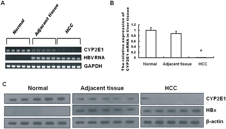
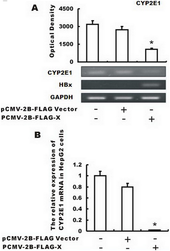
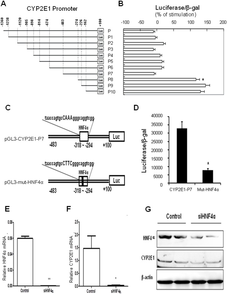
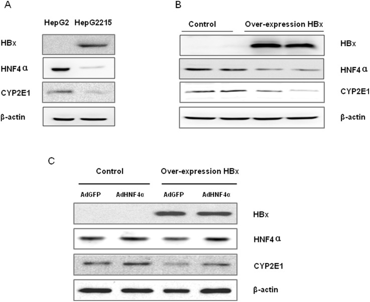
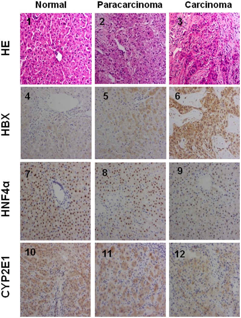
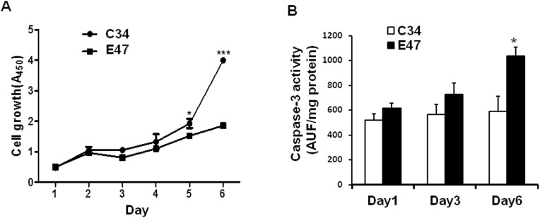
CYP2E1, one of the cytochrome P450 mixed-function oxidases located predominantly in liver, plays a key role in metabolism of xenobiotics including ethanol and procarcinogens. Recently, down-expression of CYP2E1 was found in hepatocellular carcinoma (HCC) with the majority to be chronic hepatitis B virus (HBV) carriers. In this study, we tested a hypothesis that HBx may inhibit CYP2E1 gene expression via hepatocyte nuclear factor 4α (HNF4α). By enforced HBx gene expression in cultured HepG2 cells, we determined the effect of HBx on CYP2E1 mRNA and protein expression. With a bioinformatics analysis, we found a consensus HNF-4α binding sequence located on -318 to -294 bp upstream of human CYP2E1 promoter. Using reporter gene assay and site-directed mutagenesis, we have shown that mutation of this site dramatically decreased CYP2E1 promoter activity. By silencing endogenous HNF-4α, we have further validated knockdown of HNF-4α significantly decreased CYP2E1 expression. Ectopic overexpression of HBx in HepG2 cells inhibits HNF-4α expression, and HNF-4α levels were inversely correlated with viral proteins both in HBV-infected HepG2215 cells and as well as HBV positive HCC liver tissues. Moreover, the HBx-induced CYP2E1 reduction could be rescued by ectopic supplement of HNF4α protein expression. Furthermore, human hepatoma cells C34, which do not express CYP2E1, shows enhanced cell growth rate compared to E47, which constitutively expresses CYP2E1. In addition, the significantly altered liver proteins in CYP2E1 knockout mice were detected with proteomics analysis. Together, HBx inhibits human CYP2E1 gene expression via downregulating HNF4α which contributes to promotion of human hepatoma cell growth. The elucidation of a HBx-HNF4α-CYP2E1 pathway provides novel insight into the molecular mechanism underlining chronic HBV infection associated hepatocarcinogenesis.


Conflict of interest statement

Competing Interests: The authors have declared that no competing interests exist.

Figures

Figure 1
Figure 1. Enhanced HBx expression correlates with lowered CYP2E1 level in livers of HCC patients.
(A) CYP2E1 and HBV RNA were determined by RT-PCR using GAPDH as an internal control in surgically resected human normal hepatic tissue, HCC tissue and its adjacent tissues. (B) CYP2E1 mRNA level was detected by real time quantitative PCR. (C) CYP2E1 and HBx protein were measured by Western blotting using β-actin as an internal control. The values were represented as the mean±standard deviation; *, P<0.001 vs. normal tissue or the adjacent tissue.
Figure 2
Figure 2. Enforced expression of HBx inhibits CYP2E1 gene expression in HepG2 cells.
Plasmid expressing HBx (pCMV-2B–FLAG-X) or a control plasmid (pGL3 empty vector) was transfected into HepG2 cells. (A) Overexpression of HBx significantly inhibited CYP2E1 mRNA compared to the non-transfected cells or pGL3 empty vector transfected cells by RT-PCR with GAPDH as an internal control. (B) Effect of ectopic overexpression of HBx on CYP2E1 mRNA level was determined by real time quantitative PCR in HepG2 cells. The values were represented as the mean standard deviation; *, P<0.001 vs. the control group or the empty vector group.
Figure 3
Figure 3. HNF-4α plays a critical role in CYP2E1 gene expression.
(A) A schematic depiction of different human CYP2E1 promoter regions cloned into the pGL3-basic plasmid. The constructs were designated as CPY2E1-P-luc and its various deletion constructs CPY2E1-P1-luc to -P10. (B) Effects of HBx overexpression on promoter activities of different CYP2E1 promoter constructs and identification of a specific promoter region localized at −483∼−274 bp on the CYP2E1 gene 5′-flanking region upstream of the transcription start site which mediates the inhibition effect of HBx to repress CYP2E1 expression. HepG2 cells were cotransfected with one of the CYP2E1 promoter constructs and HBx expression plasmid or control plasmid. At 24 h post-transfection, cells were harvested for determination of relative luciferase activities. Results are expressed as percent of the corresponding control in the absence of HBx expression and represent mean±standard deviation. Results are expressed as percent of the corresponding untreated control cells and represent as mean±standard deviation; *, P<0.001 vs. construct CPY2E1-P7-luc. (C) Schematic representation of the consensus HNF4α binding element and its mutant forms on the human CYP2E1 gene promoter. Site-directed mutagenesis on HNF4α consensus binding was performed, and the mutant constructs were named pGL3-mut-HNF4α; (D) Effects of HNF4α binding site mutation on CYP2E1 promoter activity. (E–F) Effects of silencing HNF4α on endogenous HNF4α and CYP2E1 mRNA expression by real-time PCR, and (G) on their protein expressions by Western Blotting with β-action as internal control.
Figure 4
Figure 4. HBx inhibits CYP2E1 expression via downregulation of HNF4α.
(A) HBV virus downregulated HNF4α and CYP2E1. HepG2215 cells, a cell line with the HBV genome integrated into the chromosome of HepG2 cells and capable of producing all HBV proteins, was used. Expression of HBx, CYP2E1 and HNF4α levels were measured with Western blotting. (B) Enforced expression of HBx protein downregulates CYP2E1 and HNF4α. HepG2 cells were transiently transfected with HBx-expression plasmid, and 24 hours post transfection, cells were harvested and cell lysate were used to determine HNF4α expression with Western Blotting. (C) HBx-induced CYP2E1 reduction could be rescued by HNF4α. HepG2 cells were transiently transfected with HBx-expression plasmid for 24 hours, then were infected with 1×10⁹ plaque-forming units of AdHNF4α, or the same amount of AdGFP as control for one more day, respectively, in HepG2 cells. Cells were harvested and protein expression of HBx, CYP2E1 and HNF4α levels were measured with Western blotting.
Figure 5
Figure 5. Immunohistochemical staining of HNF4α, CYP2E1 and HBx in human liver tissues.
Row 1, morphology observation with HE staining. Row 2, HBx immunochemical staining. Row 3, HNF4α immunochemical staining. Row 4, CYP2E1 immunochemical staining. All HE staining or immunochemical staining were shown as typical fields from five cases of human normal, and five cases of paracarcinoma or HCC liver tissues.
Figure 6
Figure 6. Cell proliferation and caspase-3 activity assay.
(A) Stable overexpression of CYP2E1 inhibits the cell growth of HepG2 cells. E47 and C34 cells, two stably transfected HepG2 cell lines harboring CYP2E1 recombinant gene or its empty vector respectively, were seeded with the same number in triplicate on 96-well plates and cultured for up to 6 days. Each day, the triplicate of cultured cells was harvested and subjected to CCK-8 assay at 450 nm. The amount of the formazan dye, generated by the activities of dehydrogenases in cells, is directly proportional to the number of living cells. (B) Activities of caspase-3 increased in HepG2 cells which stably expresses ectopic CYP2E1. E47 and C34 cells were seeded in triplicate on 96-well plates. Cells were harvested on day 1, 3 and 6, and cell lysates were used to test the activities of caspase-3 by measuring proteolytic cleavage of the proluminescent substrates AC-DEVD-AMC. The fluorescence was detected to reflect the amount of released AMC (caspase-3, λex = 380, λem = 460). The results were expressed as arbitrary units of fluorescence (AUF) per milligram of lysate protein.

Similar articles

Cited by

References

    1. Schutte K, Bornschein J, Malfertheiner P (2009) Hepatocellular carcinoma epidemiological trends and risk factors. Dig Dis 27: 80–92. - PubMed
    1. El-Serag HB, Rudolph KL (2007) Hepatocellular carcinoma: epidemiology and molecular carcinogenesis. Gastroenterology 132: 2557–2576. - PubMed
    1. Chen M, Therneau T, Orsini LS, Qiao YL (2011) Design and rationale of the HCC BRIDGE study in China: a longitudinal, multicenter cohort trial in hepatocellular carcinoma. BMC Gastroenterol 11: 53–60. - PMC - PubMed
    1. Kew MC (2010) Epidemiology of chronic hepatitis B virus infection, hepatocellular carcinoma, and hepatitis B virus-induced hepatocellular carcinoma. Pathol Biol (Paris) 58: 273–277. - PubMed
    1. Brechot C, Kremsdorf D, Soussan P, Pineau P, Dejean A, et al. (2010) Hepatitis B virus (HBV)-related hepatocellular carcinoma (HCC): molecular mechanisms and novel paradigms. Pathol Biol (Paris) 58: 278–287. - PubMed

Publication types

Substances