. 2014 Sep 19;9(9):e106349.
doi: 10.1371/journal.pone.0106349. eCollection 2014.

Modulation of ErbB2 blockade in ErbB2-positive cancers: the role of ErbB2 Mutations and PHLDA1

Affiliations

Modulation of ErbB2 blockade in ErbB2-positive cancers: the role of ErbB2 Mutations and PHLDA1

Guangyuan Li et al. PLoS One. .

Abstract

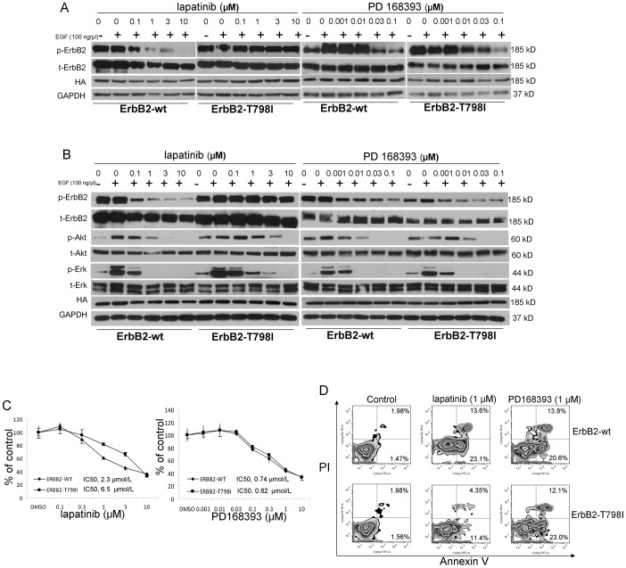
We set out to study the key effectors of resistance and sensitivity to ErbB2 tyrosine kinase inhibitors, such as lapatinib in ErbB2-positive breast and lung cancers. A cell-based in vitro site-directed mutagenesis lapatinib resistance model identified several mutations, including the gatekeeper ErbB2 mutation ErbB2-T798I, as mediating resistance. ErbB2-T798I engineered cell models indeed show resistance to lapatinib but remain sensitive to the irreversible EGFR/ErbB2 inhibitor, PD168393, suggestive of potential alternative treatment strategies to overcome resistance. Gene expression profiling studies identified a select group of downstream targets regulated by ErbB2 signaling and define PHLDA1 as an immediately downregulated gene upon oncogenic ErbB2 signaling inhibition. We find significant down-regulation of PHLDA1 in primary breast cancer and PHLDA1 is statistically significantly less expressed in ErbB2 negative compared with ErbB2 positive tumors consistent with its regulation by ErbB2. Lastly, PHLDA1 overexpression blocks AKT signaling, inhibits cell growth and enhances lapatinib sensitivity further supporting an important negative growth regulator function. Our findings suggest that PHLDA1 might have key inhibitory functions in ErbB2 driven lung and breast cancer cells and a better understanding of its functions might point at novel therapeutic options. In summary, our studies define novel ways of modulating sensitivity and resistance to ErbB2 inhibition in ErbB2-dependent cancers.


Conflict of interest statement

Competing Interests: The authors have declared that no competing interests exist.

Figures

Figure 1
Figure 1. Antitumor effects of lapatinib and PD168393 in the ErbB2 positive lung cancer cell line, Calu3 and the ErbB2 positive breast cancer cell line, SkBr3 after 72 hour treatment.
A. Cellular proliferation of Calu3 and SkBr3 cells treated with different concentrations of lapatinib and PD168393 as determined by the MTS assay. B. Lapatinib and PD168393 induce cellular apoptosis in Calu3 and SkBr3 cells. Representative Annexin V/propidium iodide flow cytometry; the numbers represent percentage of cells in the appropriate quadrant. C. Quantification of apoptosis. D. Lapatinib induces cell cycle arrest in Calu3 cells as detected by BrdU and 7-ADD assay. R1-G1 phase; R2-G2 phase; R3-S phase; R4-G0 phase. E. Dose response of lapatinib and PD168393 on phosphorylation of ErbB2, AKT and ERK in Calu3 and SkBr3 cells in response to EGF treatment. p-ErbB2: phosphorylated ErbB2; t-ErbB2: total-ErbB2; p-AKT: phosphorylated AKT; t-AKT: total AKT; p-ERK: phosphorylated ERK; t-ERK: total ERK.
Figure 2
Figure 2. The irreversible inhibitor PD168393 overcomes lapatinib resistance caused by the ErbB2 T798I mutation.
A. Dose response of lapatinib and PD168393 on phophorylation of ErbB2 in Cos7 cells after 24 hours of transient transfection with ErbB2-wt and ErbB2-T798I vectors. Total ErbB2, HA and GAPDH expression level as control. B. Dose response of lapatinib and PD168393 on phosphorylation of ErbB2, AKT and ERK in ErbB2-wt and ErbB2-T798I stable Calu3 cell clones. Total ErbB2, total AKT, total ERK, HA and GAPDH expression level as control. C. Dose-dependent growth inhibition induced by the treatment of lapatinib and PD168393 for 72 hours in ErbB2-wt and ErbB2-T798I stable Calu3 cell clones as detected by the MTS assay. D. Lapatinib and PD168393 induce cellular apoptosis in ErbB2-wt and ErbB2-T798I stable Calu3 cell clones after 72 hour treatment. E. Quantification of apoptosis.
Figure 3
Figure 3. PHLDA1 is regulated by ErbB2 inhibition at the mRNA level at 6 hours.
Quantitative PCR analysis of gene regulation by lapatinib treatment for 6 hours in SkBr3 and Calu3 cells. Fold induction relative to 0 hour control was plotted after normalization by GAPDH. Δ: p<0.05;×: p<0.01; †: p<0.005; *: p<0.001.
Figure 4
Figure 4. Downregulation of PHLDA1 expression upon ErbB2/EGFR inhibition.
Western blot analysis following treatment with lapatinib, erlotinib and CL387,785 at 1 µm for 6, 24 and 48 hours in ErbB2-positive SkBr3 and EGFR-mutated H1975, HCC827. DMSO treatment was 48 hours. Different cell lines were used with the relevant inhibitor capable of blockade of oncogenic signaling.
Figure 5
Figure 5. PHLDA1 is down-regulated in human breast cancers.
A. Immunohistochemical staining of PHLDA1 in 24 primary breast cancers. The intensity of the staining of PHLDA1 was scored as 0 (no expression), 1+ (weak expression), 2+ (moderate expression) and 3+ (strong expression). SISH was used to determine ErbB2 status in human breast cancer samples. B. Statistical analysis of IHC study. C, PHLDA1 is strongly expressed in normal breast epithelium.
Figure 6
Figure 6. Tumor suppressor activity of PHLDA1 in vitro.
A. Western blotting of whole cell lysates with anti-PHLDA1 shows strong expression of PHLDA1 in pcDNA3.1(-)-PHLDA1 transfected cells. B. Western blotting of cell lysates with pcDNA3.1(-)-PHLDA1 transient transfection shows potent inhibition of AKT phosphorylation. C. MTS cell growth assay of pcDNA3.1(-)-EV and pcDNA3.1(-)-PHLDA1 stable clones. D. Soft Agar assay for anchorage-independent cell growth is done with pcDNA3.1(-)-EV and pcDNA3.1(-)-PHLDA1 stable cell clones. E. Wound-healing assay shows that PHLDA1 overexpression significantly decreased cell mobility. *: p<0.05.
Figure 7
Figure 7. Clonogenic survival assay shows that PHLDA1 overexpression could significantly enhance lapatinib sensitivity in breast cancer cells.
*: p<0.001; Δ: p<0.0001. Western blotting of cell lysates with anti-HA shows equal exogenous expression of PHLDA1 in pcDNA3.1(-)-PHLDA1 transfected cells after 10 days of treatment with DMSO and 0.1 um lapatinib.

References

    1. Pauletti G, Godolphin W, Press MF, Slamon DJ (1996) Detection and quantitation of HER-2/neu gene amplification in human breast cancer archival material using fluorescence in situ hybridization. Oncogene 13: 63–72. - PubMed
    1. Callahan R, Hurvitz S (2011) Human epidermal growth factor receptor-2-positive breast cancer: Current management of early, advanced, and recurrent disease. Curr Opin Obstet Gynecol 23: 37–43. - PMC - PubMed
    1. Slamon DJ, Clark GM, Wong SG, Levin WJ, Ullrich A, et al. (1987) Human breast cancer: correlation of relapse and survival with amplification of the HER-2/neu oncogene. Science 235: 177–182. - PubMed
    1. Slamon DJ, Godolphin W, Jones LA, Holt JA, Wong SG, et al. (1989) Studies of the HER-2/neu proto-oncogene in human breast and ovarian cancer. Science 244: 707–712. - PubMed
    1. Santin AD, Bellone S, Roman JJ, McKenney JK, Pecorelli S (2008) Trastuzumab treatment in patients with advanced or recurrent endometrial carcinoma overexpressing HER2/neu. Int J Gynaecol Obstet 102: 128–131. - PubMed

Publication types

MeSH terms