

Selective targeting of liver cancer with the endothelial marker CD146
- PMID: 25238265
- PMCID: PMC4226708
- DOI: 10.18632/oncotarget.2345
Selective targeting of liver cancer with the endothelial marker CD146
Abstract
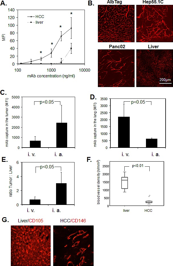
Hepatocellular carcinomas are well-vascularized tumors; the endothelial cells in these tumors have a specific phenotype. Our aim was to develop a new approach for tumor-specific drug delivery with monoclonal antibody targeting of endothelial ligands. CD146, a molecule expressed on the endothelial surface of hepatocellular carcinoma, was identified as a promising candidate for targeting. In the present study, endothelial cells immediately captured circulating anti-CD146 (ME-9F1) antibody, while antibody binding in tumors was significantly higher than in hepatic endothelium. Macroscopically, after intravenous injection, there were no differences in the mean accumulation of anti-CD146 antibody in tumor compared to liver tissue , due to a compensating higher blood vessel density in the liver tissue. Additional blockade of nontumoral epitopes and intra-arterial administration, improved selective antibody capture in the tumor microvasculature and largely prevented antibody distribution in the lung and liver. The potential practical use of this approach was demonstrated by imaging of radionuclide-labeled ME-9F1 antibody, which showed excellent tumor-selective uptake. Our results provide a promising principle for the use of endothelial markers for intratumoral drug delivery. Tumor endothelium-based access might offer new opportunities for the imaging and therapy of hepatocellular carcinoma and other liver malignancies.
Figures


Similar articles
-
Murine CD146 is widely expressed on endothelial cells and is recognized by the monoclonal antibody ME-9F1.Histochem Cell Biol. 2008 Apr;129(4):441-51. doi: 10.1007/s00418-008-0379-x. Epub 2008 Jan 24. Histochem Cell Biol. 2008. PMID: 18214516 Free PMC article.
-
Phenomenon of Endothelial Antibody Capture: Principles and Potential for Locoregional Targeting of Hepatic Tumors.Hepatology. 2018 Nov;68(5):1804-1816. doi: 10.1002/hep.30072. Epub 2018 Sep 21. Hepatology. 2018. PMID: 29734469
-
Endothelial precursor cells promote angiogenesis in hepatocellular carcinoma.World J Gastroenterol. 2012 Sep 21;18(35):4925-33. doi: 10.3748/wjg.v18.i35.4925. World J Gastroenterol. 2012. PMID: 23002366 Free PMC article.
-
Vascular changes in hepatocellular carcinoma.Anat Rec (Hoboken). 2008 Jun;291(6):721-34. doi: 10.1002/ar.20668. Anat Rec (Hoboken). 2008. PMID: 18484619 Review.
-
[Molecular structure and abnormal expression of angiopoietin-2 and antiangiogenic targeting therapy of hepatocelluar carcinoma].Zhonghua Gan Zang Bing Za Zhi. 2009 May;17(5):398-400. Zhonghua Gan Zang Bing Za Zhi. 2009. PMID: 19497214 Review. Chinese. No abstract available.
Cited by
-
Reduced CD146 expression promotes tumorigenesis and cancer stemness in colorectal cancer through activating Wnt/β-catenin signaling.Oncotarget. 2016 Jun 28;7(26):40704-40718. doi: 10.18632/oncotarget.9930. Oncotarget. 2016. PMID: 27302922 Free PMC article.
-
Treatment of Hepatocellular Carcinoma by Intratumoral Injection of 125I-AA98 mAb and Its Efficacy Assessments by Molecular Imaging.Front Bioeng Biotechnol. 2019 Nov 14;7:319. doi: 10.3389/fbioe.2019.00319. eCollection 2019. Front Bioeng Biotechnol. 2019. PMID: 31799244 Free PMC article.
-
Evaluation of CD146 as Target for Radioimmunotherapy against Osteosarcoma.PLoS One. 2016 Oct 24;11(10):e0165382. doi: 10.1371/journal.pone.0165382. eCollection 2016. PLoS One. 2016. PMID: 27776176 Free PMC article.
-
Impact of wall shear stress and ligand avidity on binding of anti-CD146-coated nanoparticles to murine tumor endothelium under flow.Oncotarget. 2015 Nov 24;6(37):39960-8. doi: 10.18632/oncotarget.5662. Oncotarget. 2015. PMID: 26503468 Free PMC article.
-
High-throughput flow cytometry screening of human hepatocellular carcinoma reveals CD146 to be a novel marker of tumor-initiating cells.Biochem Biophys Rep. 2016 Aug 12;8:107-113. doi: 10.1016/j.bbrep.2016.08.012. eCollection 2016 Dec. Biochem Biophys Rep. 2016. PMID: 28955945 Free PMC article.
References
-
- Folkman J. Angiogenesis in cancer, vascular, rheumatoid and other disease. Nat Med. 1995;1:27–31. - PubMed
-
- Jain RK. Normalization of tumor vasculature: an emerging concept in antiangiogenic therapy. Science. 2005;307:58–62. - PubMed
-
- Siemann DW, Bibby MC, Dark GG, Dicker AP, Eskens FA, Horsman MR, Marme D, Lorusso PM. Differentiation and definition of vascular-targeted therapies. Clin Cancer Res. 2005;11:416–20. - PubMed
-
- Giantonio BJ, Catalano PJ, Meropol NJ, O'Dwyer PJ, Mitchell EP, Alberts SR, Schwartz MA, Benson AB, III. Bevacizumab in combination with oxaliplatin, fluorouracil, and leucovorin (FOLFOX4) for previously treated metastatic colorectal cancer: results from the Eastern Cooperative Oncology Group Study E3200. J Clin Oncol. 2007;25:1539–44. - PubMed
-
- Salnikova O, Breuhahn K, Hartmann N, Schmidt J, Ryschich E. Endothelial plasticity governs the site-specific leukocyte recruitment in hepatocellular cancer. Int J Cancer. 2013;133:2372–82. - PubMed
Publication types
MeSH terms
Substances
LinkOut - more resources
Full Text Sources
Other Literature Sources
Medical
