. 2014 Oct;184(10):2840-9.
doi: 10.1016/j.ajpath.2014.06.025.

Novel fusion transcripts associate with progressive prostate cancer

Novel fusion transcripts associate with progressive prostate cancer

Yan P Yu et al. Am J Pathol. 2014 Oct.

Abstract

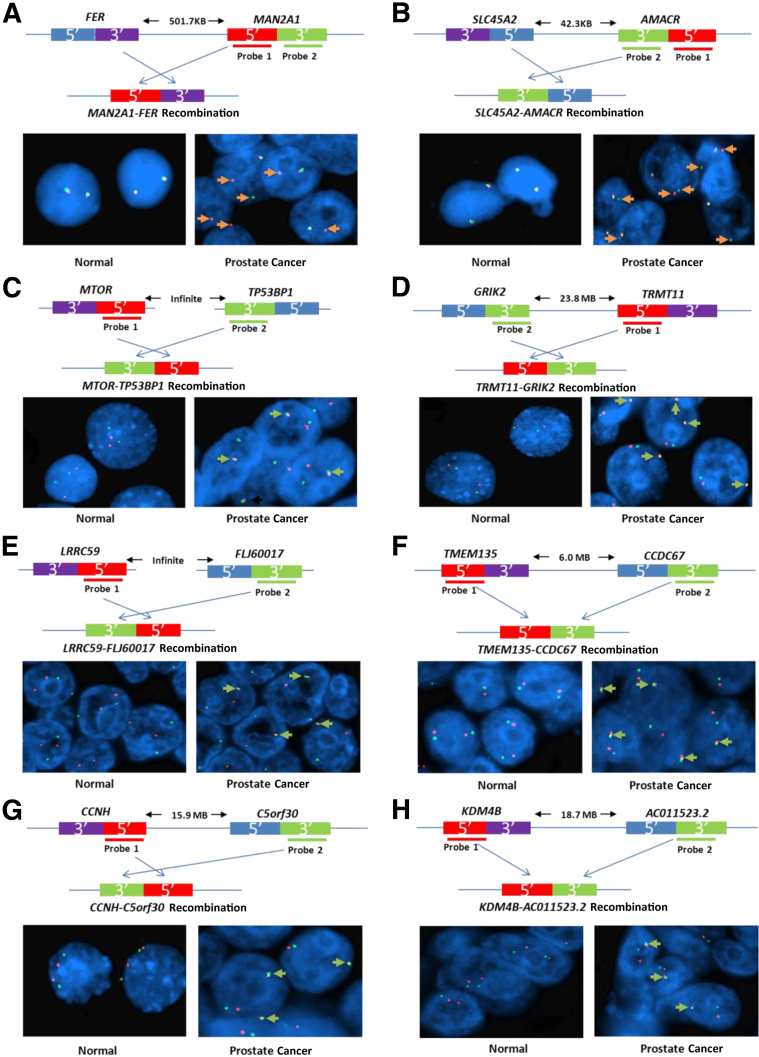
The mechanisms underlying the potential for aggressive behavior of prostate cancer (PCa) remain elusive. In this study, whole genome and/or transcriptome sequencing was performed on 19 specimens of PCa, matched adjacent benign prostate tissues, matched blood specimens, and organ donor prostates. A set of novel fusion transcripts was discovered in PCa. Eight of these fusion transcripts were validated through multiple approaches. The occurrence of these fusion transcripts was then analyzed in 289 prostate samples from three institutes, with clinical follow-up ranging from 1 to 15 years. The analyses indicated that most patients [69 (91%) of 76] positive for any of these fusion transcripts (TRMT11-GRIK2, SLC45A2-AMACR, MTOR-TP53BP1, LRRC59-FLJ60017, TMEM135-CCDC67, KDM4-AC011523.2, MAN2A1-FER, and CCNH-C5orf30) experienced PCa recurrence, metastases, and/or PCa-specific death after radical prostatectomy. These outcomes occurred in only 37% (58/157) of patients without carrying those fusion transcripts. Three fusion transcripts occurred exclusively in PCa samples from patients who experienced recurrence or PCaerelated death. The formation of these fusion transcripts may be the result of genome recombination. A combination of these fusion transcripts in PCa with Gleason's grading or with nomogram significantly improves the prediction rate of PCa recurrence. Our analyses suggest that formation of these fusion transcripts may underlie the aggressive behavior of PCa.


Figures

Figure 1
Figure 1
Unique fusion gene events. Miniature diagrams of genome of the fusion genes, the transcription directions, the distances between the joining genes, and the directions of the fusions. Representative sequencing chromograms of fusion transcripts. The joining gene sequences are indicated. Diagrams of translation products of fusion transcripts are given. Blue head, gene translation product; red tail, gene translation product; orange, novel translation products due to frameshift or translation products from a nongene region. UTR, untranslated region.
Figure 2
Figure 2
FISH suggests genome recombination in prostate cancer cells. Schematic diagram of MAN2A1 and FER (A), SLC45A2 and AMACR (B), MTOR and TP53BP1 (C), GRIK2 and TRMT11 (D), LRRC59 and FLJ60017 (E), TMEM135 and CCDC67 (F), CCNH and C5orf30 (G), and KDM4B and AC011523.2 (H) genome recombination and FISH probe positions. Representative FISH images are shown for normal prostate epithelial cells and cancer cells positive for MAN2A1-FER (A), SLC45A2-AMACR (B), MTOR-TP53BP1 (C), TRMT11-GRIK2 (D), LRRC59-FLJ60017 (E), TMEM135-CCDC67 (F), CCNH-C5orf30 (G), and KDM4B-AC011523.2 (H) fusions. Orange dots denote probe 1; green dots, probe 2. Break-apart signals are indicated by orange arrows; fusion joining signals, green arrows.
Figure 3
Figure 3
Fusion transcripts expressed in prostate cancer. A: Distribution of eight indicated fusion transcripts in 213 prostate cancer samples from UPMC, 30 samples from Stanford University Medical Center, and 36 samples from University of Wisconsin Madison Medical Center. Samples from patients who experienced recurrence were indicated with light gray (PSADT ≥15 months), dark gray (PSADT <4 months), or intermediate gray (PSADT = 5 to 14 months); samples from patients who have no recurrence for at least 5 years, green; and samples from patients whose clinical follow-up is ongoing but <5 years, white (undetermined). B: Correlation of fusion transcript events with prostate cancer recurrence. The percentage of prostate cancer experiencing recurrence from samples positive for fusion transcripts is plotted for each fusion transcript from specified cohorts.
Figure 4
Figure 4
Fusion genes predict recurrence of prostate cancer. A: Scheme of training and validation steps in building fusion gene prediction models for prostate cancer recurrence and short PSADT. (I) The algorithm of fusion gene prediction of prostate cancer recurrence and PSADT <4 months was obtained from 90 randomly assigned prostate cancer samples from UPMC. (II) The algorithm was then applied to 89 samples from UPMC. (III) The algorithm was applied to 21 samples from Stanford University Medical center. (IV) The algorithm was applied to 33 samples from University of Wisconsin Madison Medical Center. B: Prediction rate of prostate cancer recurrence and PSADT ≤4 months using prostate cancer sample cohorts from UPMC, Stanford Medical Center, and University of Wisconsin Madison Medical Center, on the basis of the algorithm obtained from the 90–training sample cohort. C: Kaplan-Meier analysis of patients who were positive for any of TRMT11-GRIK2, SLC45A2-AMACR, MTOR-TP53BP1, LRRC59-FLJ60017, TMEM135-CCDC67, and CCNH-C5orf30 versus those who were negative for these fusion events. Kaplan-Meier analysis of prostate cancer sample cohort from UPMC and Stanford University Medical Center; P value is indicated for the significant difference in survival between the group that is positive for at least one fusion transcript and the group that is negative for at least one fusion transcript.
Figure 5
Figure 5
Combining status of fusion transcript and clinical/pathological parameter to improve prediction of prostate cancer recurrence. A: Combining Gleason's grading and the status of eight fusion transcripts in prostate cancer samples using LDA technique to predict the recurrence of prostate cancer. ROC curve analysis of Gleason alone or Gleason plus the presence of fusion transcripts using LDA technique in the prediction of prostate cancer recurrence; P value (permutation test) is indicated for the significant difference between the ROC curve generated by Gleason alone and the curve generated by Gleason plus the presence of fusion transcripts using LDA technique. Kaplan-Meier analysis of PSA-free survival of prostate cancer patients with Gleason score ≥8 versus <8 from combined UPMC testing, Wisconsin, and Stanford data sets; P value (log-rank test) is indicated for the significant difference in survival between the group that has Gleason score ≥8 and the group that has score <8. Kaplan-Meier analysis of PSA-free survival of prostate cancer patients with Gleason score ≥8 or positive for any of the eight fusion transcripts in the prostate cancer samples versus those with Gleason score <8 and negative for fusion transcripts using LDA from combined UPMC testing, Wisconsin, and Stanford data sets. P value (log-rank test) is indicated for the significant difference in survival between the group that is positive for at least one fusion transcript or has Gleason score ≥8 and the group that is negative for fusion transcript and has Gleason score <8. B: Combining nomogram and the status of eight fusion transcripts in prostate cancer samples using LDA technique to predict the recurrence of prostate cancer. ROC analysis of nomogram alone or nomogram plus the presence of fusion transcripts using LDA technique in the prediction of prostate cancer recurrence. P value (permutation test) is indicated for the significant difference between the ROC curve generated by nomogram alone and the curve generated by nomogram plus the presence of fusion transcripts using LDA technique. Kaplan-Meier analysis of PSA-free survival of prostate cancer patients with probability >88 versus ≤88 from combined UPMC testing, Wisconsin, and Stanford data sets; P value (log-rank test) is indicated for the significant difference in survival between the group that has probability >88 PSA-free survival and the group that has ≤88 probability. Kaplan-Meier analysis of PSA-free survival of prostate cancer patients with nomogram ≤88 or positive for any of the eight fusion transcripts in the prostate cancer samples versus those >88 and negative for fusion transcripts using LDA from combined UPMC testing, Wisconsin, and Stanford data sets. P value (log-rank test) is indicated for the significant difference in survival between the group that is negative for fusion transcript and has probability >88 PSA-free survival and the group that is positive for fusion transcript or has ≤88 probability.

Comment in

Similar articles

Cited by

References

    1. Jemal A., Bray F., Center M.M., Ferlay J., Ward E., Forman D. Global cancer statistics. CA Cancer J Clin. 2011;61:69–90. - PubMed
    1. Siegel R., Naishadham D., Jemal A. Cancer statistics, 2012. CA Cancer J Clin. 2012;62:10–29. - PubMed
    1. Li H., Durbin R. Fast and accurate short read alignment with Burrows-Wheeler transform. Bioinformatics. 2009;25:1754–1760. - PMC - PubMed
    1. Yu Y.P., Ding Y., Chen R., Liao S.G., Ren B.G., Michalopoulos A., Michalopoulos G., Nelson J., Tseng G.C., Luo J.H. Whole-genome methylation sequencing reveals distinct impact of differential methylations on gene transcription in prostate cancer. Am J Pathol. 2013;183:1960–1970. - PMC - PubMed
    1. Luo J.H., Ding Y., Chen R., Michalopoulos G., Nelson J., Tseng G., Yu Y.P. Genome-wide methylation analysis of prostate tissues reveals global methylation patterns of prostate cancer. Am J Pathol. 2013;182:2028–2036. - PMC - PubMed

Publication types

MeSH terms