

CD8+ lymphocyte intratumoral infiltration as a stage-independent predictor of Merkel cell carcinoma survival: a population-based study
- PMID: 25239411
- PMCID: PMC4174450
- DOI: 10.1309/AJCPIKDZM39CRPNC
CD8+ lymphocyte intratumoral infiltration as a stage-independent predictor of Merkel cell carcinoma survival: a population-based study
Abstract
Objectives: Intratumoral CD8+ lymphocytes (IT-CD8s) have shown promise as a prognostic indicator for Merkel cell carcinoma (MCC). We tested whether IT-CD8s predict survival among a population-based MCC cohort.
Methods: One hundred thirty-seven MCC cases that had not previously been analyzed for IT-CD8s were studied.
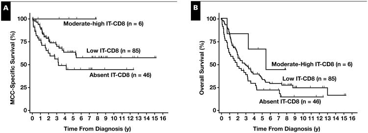
Results: Three-year MCC-specific survival rates were 56%, 72%, and 100% for patients with absent (n = 46), low (n = 85), and moderate or strong (n = 6) IT-CD8s, respectively. Increased IT-CD8s were associated with improved MCC-specific survival in a multivariate competing risk-regression analysis including stage, age, and sex (hazard ratio [HR] = 0.5; 95% confidence interval [CI] = 0.3-0.9). Although a similar trend was observed for overall survival, statistical significance was not reached (HR = 0.8; 95% CI = 0.6-1.0), likely because of the high rate of non-MCC deaths among older patients.
Conclusions: This study of prospectively captured MCC cases supports the concept that cellular immunity is important in MCC outcome and that CD8+ lymphocyte infiltration adds prognostic information to conventional staging.
Keywords: Merkel cell carcinoma; Survival; T cells.
Copyright© by the American Society for Clinical Pathology.
Figures
Similar articles
-
Transcriptome-wide studies of merkel cell carcinoma and validation of intratumoral CD8+ lymphocyte invasion as an independent predictor of survival.J Clin Oncol. 2011 Apr 20;29(12):1539-46. doi: 10.1200/JCO.2010.30.6308. Epub 2011 Mar 21. J Clin Oncol. 2011. PMID: 21422430 Free PMC article.
-
Prognostic Role of Tumoral PD-L1 and IDO1 Expression, and Intratumoral CD8+ and FoxP3+ Lymphocyte Infiltrates in 132 Primary Cutaneous Merkel Cell Carcinomas.Int J Mol Sci. 2021 May 23;22(11):5489. doi: 10.3390/ijms22115489. Int J Mol Sci. 2021. PMID: 34071045 Free PMC article.
-
Merkel cell carcinomas infiltrated with CD33+ myeloid cells and CD8+ T cells are associated with improved outcome.J Am Acad Dermatol. 2018 May;78(5):973-982.e8. doi: 10.1016/j.jaad.2017.12.029. Epub 2017 Dec 19. J Am Acad Dermatol. 2018. PMID: 29273486
-
Complete spontaneous regression of primary Merkel cell carcinoma with tumoural infiltration: a systematic review.Eur J Dermatol. 2021 Jun 1;31(3):381-391. doi: 10.1684/ejd.2021.4065. Eur J Dermatol. 2021. PMID: 34080974
-
Predictors of survival in neurometastatic Merkel cell carcinoma.Eur J Cancer. 2018 Sep;101:152-159. doi: 10.1016/j.ejca.2018.07.002. Epub 2018 Jul 30. Eur J Cancer. 2018. PMID: 30071443
Cited by
-
Response rates and durability of chemotherapy among 62 patients with metastatic Merkel cell carcinoma.Cancer Med. 2016 Sep;5(9):2294-301. doi: 10.1002/cam4.815. Epub 2016 Jul 19. Cancer Med. 2016. PMID: 27431483 Free PMC article.
-
Merkel Cell Polyoma Viral Load and Intratumoral CD8+ Lymphocyte Infiltration Predict Overall Survival in Patients With Merkel Cell Carcinoma.Front Oncol. 2019 Jan 24;9:20. doi: 10.3389/fonc.2019.00020. eCollection 2019. Front Oncol. 2019. PMID: 30733932 Free PMC article.
-
Virus-positive Merkel Cell Carcinoma Is an Independent Prognostic Group with Distinct Predictive Biomarkers.Clin Cancer Res. 2021 May 1;27(9):2494-2504. doi: 10.1158/1078-0432.CCR-20-0864. Epub 2021 Feb 5. Clin Cancer Res. 2021. PMID: 33547200 Free PMC article.
-
Density, Distribution, and Composition of Immune Infiltrates Correlate with Survival in Merkel Cell Carcinoma.Clin Cancer Res. 2016 Nov 15;22(22):5553-5563. doi: 10.1158/1078-0432.CCR-16-0392. Epub 2016 May 10. Clin Cancer Res. 2016. PMID: 27166398 Free PMC article.
-
Merkel Cell Carcinoma Therapeutic Update.Curr Treat Options Oncol. 2016 Jul;17(7):36. doi: 10.1007/s11864-016-0409-1. Curr Treat Options Oncol. 2016. PMID: 27262710 Free PMC article. Review.
References
-
- Albores-Saavedra J, Batich K, Chable-Montero F, et al. Merkel cell carcinoma demographics, morphology, and survival based on 3870 cases: a population based study. J Cutan Pathol. 2009;37:20–27. - PubMed
-
- Engels EA, Frisch M, Goedert JJ, et al. Merkel cell carcinoma and HIV infection. Lancet. 2002;359:497–498. - PubMed
-
- Penn I, First MR. Merkel's cell carcinoma in organ recipients: report of 41 cases. Transplantation. 1999;68:1717–1721. - PubMed
Publication types
MeSH terms
Grants and funding
LinkOut - more resources
Full Text Sources
Other Literature Sources
Medical
Research Materials
