. 2015 Apr;23(4):403-415.
doi: 10.1016/j.jagp.2014.08.005. Epub 2014 Aug 15.

Worsening cognitive impairment and neurodegenerative pathology progressively increase risk for delirium

Affiliations

Worsening cognitive impairment and neurodegenerative pathology progressively increase risk for delirium

Daniel H J Davis et al. Am J Geriatr Psychiatry. 2015 Apr.

Abstract

Background: Delirium is a profound neuropsychiatric disturbance precipitated by acute illness. Although dementia is the major risk factor this has typically been considered a binary quantity (i.e., cognitively impaired versus cognitively normal) with respect to delirium risk. We used humans and mice to address the hypothesis that the severity of underlying neurodegenerative changes and/or cognitive impairment progressively alters delirium risk.

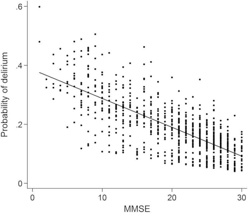
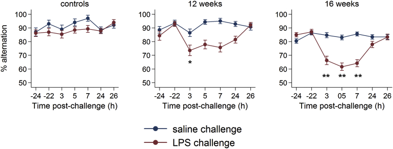
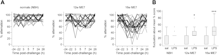
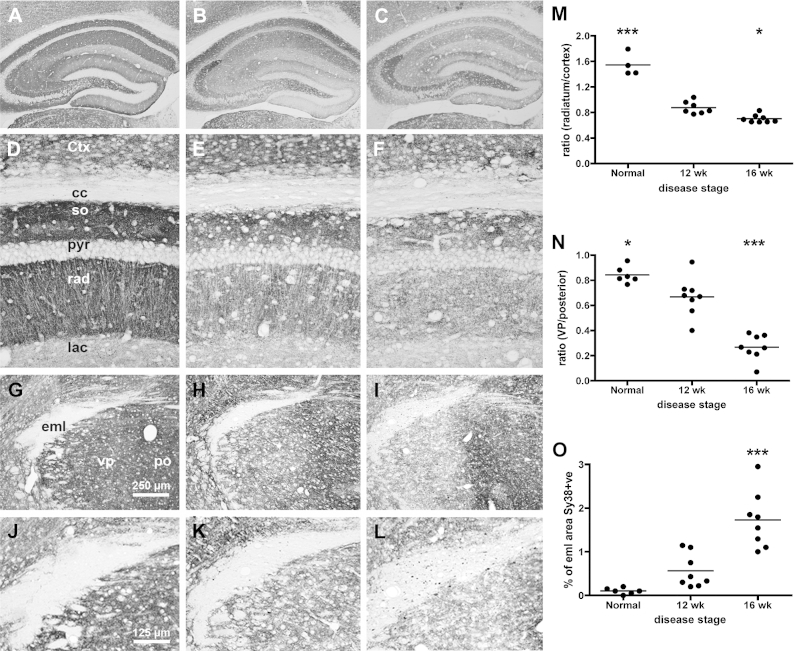
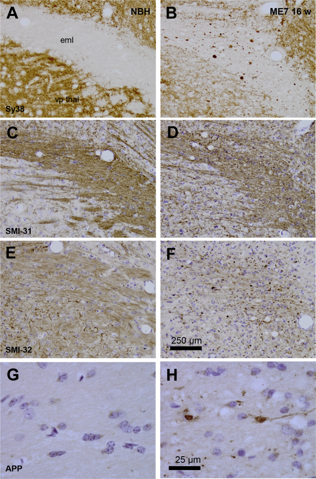
Methods: Humans in a population-based longitudinal study, Vantaa 85+, were followed for incident delirium. Odds for reporting delirium at follow-up (outcome) were modeled using random-effects logistic regression, where prior cognitive impairment measured by Mini-Mental State Exam (MMSE) (exposure) was considered. To address whether underlying neurodegenerative pathology increased susceptibility to acute cognitive change, mice at three stages of neurodegenerative disease progression (ME7 model of neurodegeneration: controls, 12 weeks, and 16 weeks) were assessed for acute cognitive dysfunction upon systemic inflammation induced by bacterial lipopolysaccharide (LPS; 100 μg/kg). Synaptic and axonal correlates of susceptibility to acute dysfunction were assessed using immunohistochemistry.

Results: In the Vantaa cohort, 465 persons (88.4 ± 2.8 years) completed MMSE at baseline. For every MMSE point lost, risk of incident delirium increased by 5% (p = 0.02). LPS precipitated severe and fluctuating cognitive deficits in 16-week ME7 mice but lower incidence or no deficits in 12-week ME7 and controls, respectively. This was associated with progressive thalamic synaptic loss and axonal pathology.

Conclusion: A human population-based cohort with graded severity of existing cognitive impairment and a mouse model with progressing neurodegeneration both indicate that the risk of delirium increases with greater severity of pre-existing cognitive impairment and neuropathology.

Keywords: Delirium; ageing; axonal; basal forebrain; cognitive decline; dementia; hippocampus; inflammation; neurodegeneration; neuroinflammation; neuropathology; susceptibility; synaptic; systemic; thalamus.


Figures

Figure 1
Figure 1
Delirium risk in relation to prior MMSE. The probability of delirium is plotted against previous MMSE score, where each point is an observation of delirium risk at follow-up. The model-predicted coefficient for delirium risk is described as a straight line with a negative slope, showing an inverse relationship.
Figure 2
Figure 2
Acute cognitive impairments induced by LPS are more severe in animals with more advanced neurodegenerative disease. Performance of NBH (normal) mice, ME7 animals 12 weeks post-inoculation, or ME7 animals 16 weeks post-inoculation upon challenge with saline or LPS (100 μg/kg), plotted with intervals showing the standard error of the mean. Criterion baseline performance (≥70%) was ascertained for all challenged animals, and these were then tested 15 times (every 20 minutes for >5 hours: 3h–9h post LPS) post-challenge as well as for 10 further trials 24 hours post-challenge. Statistically significantly different RR for errors are denoted by *p <0.05 and **p <0.01.
Figure 3
Figure 3
Systemic inflammation induces fluctuating cognitive deficits only in animals with existing neurodegenerative disease. [A] Individual trajectories of performance in mice given LPS challenge, given in each experimental group. Plots are jittered on the y-axis to distinguish overlapping plots. [B] Fluctuation about the mean correct performance (% trials) according to experimental group. NBH normal brain homogenate (controls), 12w ME7 mice, and 16w ME7 mice. Mann-Whitney: NBH-LPS versus NBH-saline, p = 0.16; ME7 12w-saline versus ME7 12w-LPS, p = 0.03 (denoted *); ME7 16w saline versus ME7 16w-LPS, p <0.001 (denoted ***).
Figure 4
Figure 4
Synaptic integrity deteriorates as a function of disease progression. Formalin fixed, paraffin-embedded brains from NBH and ME7 animals, at 12 and 16 weeks post-inoculation, were sectioned (10 μm) and labelled with antibodies against synaptophysin (Sy38). [A–C] Gross view of hippocampus of normal, 12w, and 16w, showing severe synaptic loss in the strata of the CA1 and dentate gyrus, but preservation of the DG -> CA3 projection. [D–F] Higher power pictures of these strata, used for quantification in the hippocampus (M) according to the equation Tcc − Trad/Tcc − TCtx (G–I; see Supplementary Methods; available online) 10x pictures of the thalamus at approximately bregma −2.5 (AP), showing the external medullary lamina (eml), ventroposterior nucleus (VP) and the posterior nucleus (Po) and clear synaptic loss in the VP at 16 weeks. [J–L] Higher power pictures of these areas, used for quantification of thalamic synaptic density according to the equation Teml − TVP/Teml − TPo (N; see Supplementary Methods; available online) and % sy38-positive area within the eml (O). Pairwise comparisons to ME7 12 week animals by Bonferroni post-hoc tests after significant main effect of disease stage by one-way ANOVA. *p <0.05, ***p <0.001.
Figure 5
Figure 5
Axonal pathology in animals at 16 weeks of disease progression. Formalin fixed, paraffin-embedded brains from NBH and ME7 animals, 16 weeks post-inoculation, were sectioned (10 μm) and labelled with antibodies against synaptophysin (Sy38 [A,B]), phosphorylated neurofilament heavy chain (SMI-31 [C,D]); unphosphorylated neurofilament heavy chain (SMI-32 [E,F]) and amyloid precursor protein (APP695 [G,H]). Photograph with 10x objectives, except [G,H]: photograph under 100x oil immersion objective. Scale bar for [A–F] shown in [F] = 250 μm, and that in [H] serves for panels [G] and [H] (25 μm). vp thal: ventroposterior thalamic nucleus; eml: external medullary lamina.

References

    1. Inouye S.K., Westendorp R.G., Saczynski J.S. Delirium in elderly people. Lancet. 2014;383:911–922. - PMC - PubMed
    1. Maclullich A.M., Anand A., Davis D.H., et al. New horizons in the pathogenesis, assessment and management of delirium. Age Ageing. 2013;42:667–674. - PMC - PubMed
    1. Siddiqi N., House A.O., Holmes J.D. Occurrence and outcome of delirium in medical in-patients: a systematic literature review. Age Ageing. 2006;35:350–364. - PubMed
    1. Vasilevskis E.E., Han J.H., Hughes C.G., et al. Epidemiology and risk factors for delirium across hospital settings. Best Pract Res Clin Anaesthesiol. 2012;26:277–287. - PMC - PubMed
    1. Partridge J.S., Martin F.C., Harari D., et al. The delirium experience: what is the effect on patients, relatives and staff and what can be done to modify this? Int J Geriatr Psychiatry. 2013;28:804–812. - PubMed

Publication types

MeSH terms

Substances