. 2014 Dec 15;307(12):H1691-704.
doi: 10.1152/ajpheart.00281.2014. Epub 2014 Sep 19.

Sirtuin 1 ablation in endothelial cells is associated with impaired angiogenesis and diastolic dysfunction

Affiliations

Sirtuin 1 ablation in endothelial cells is associated with impaired angiogenesis and diastolic dysfunction

Julien Maizel et al. Am J Physiol Heart Circ Physiol. .

Abstract

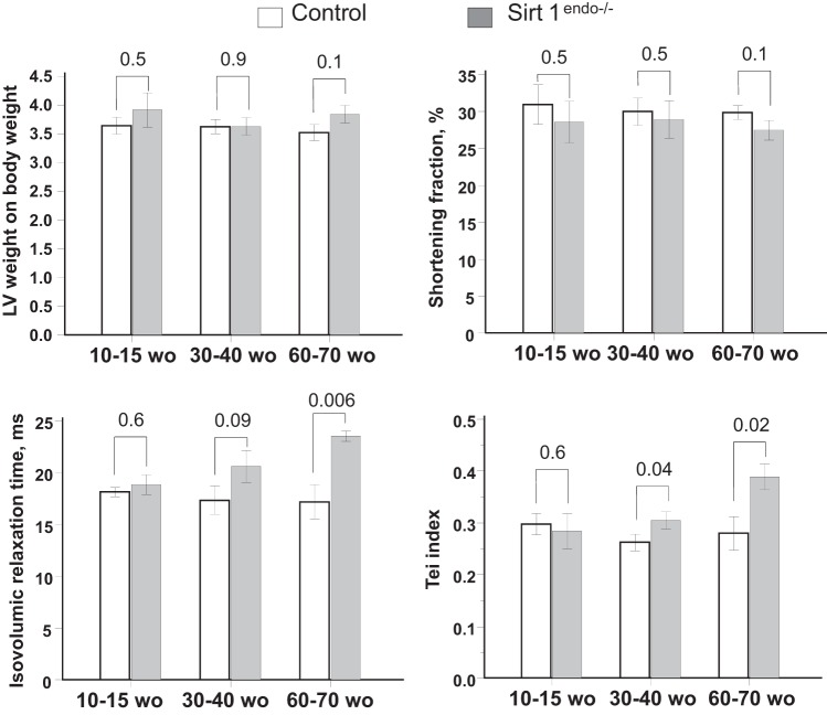
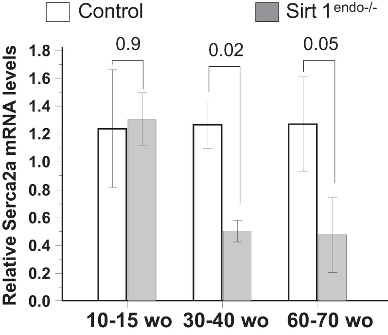
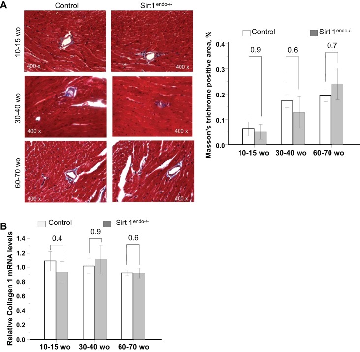
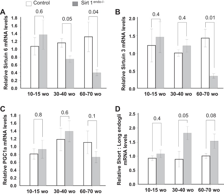
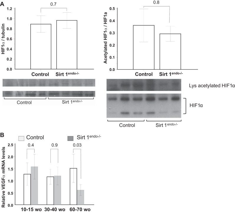
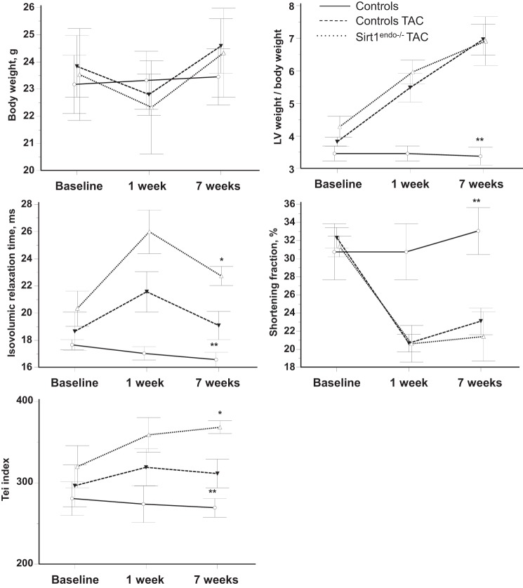
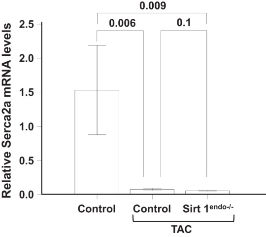
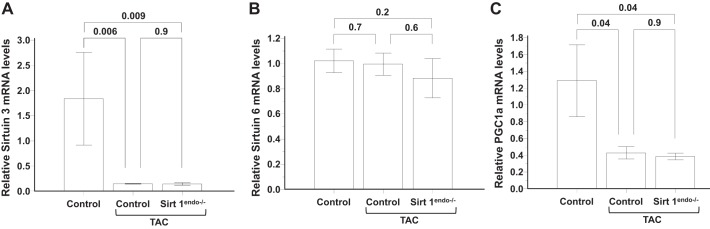
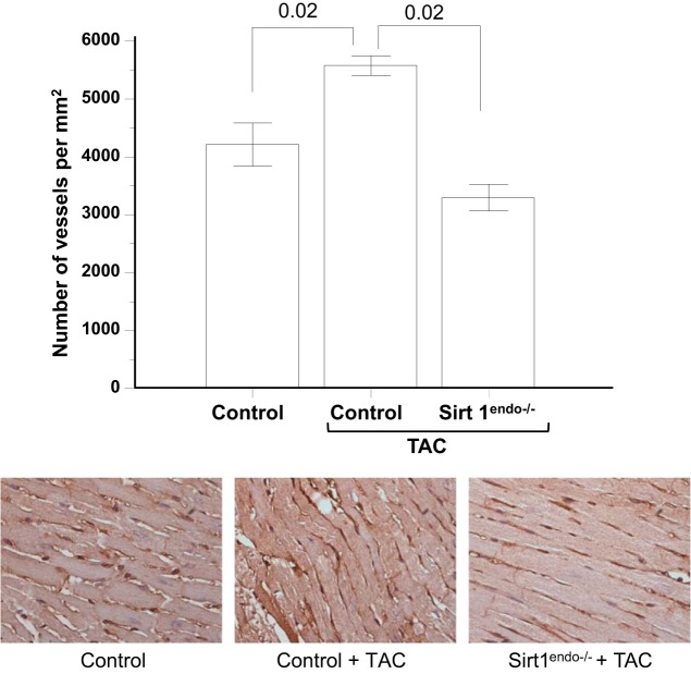
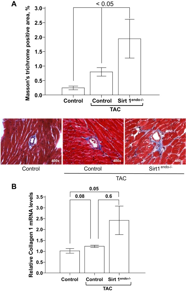
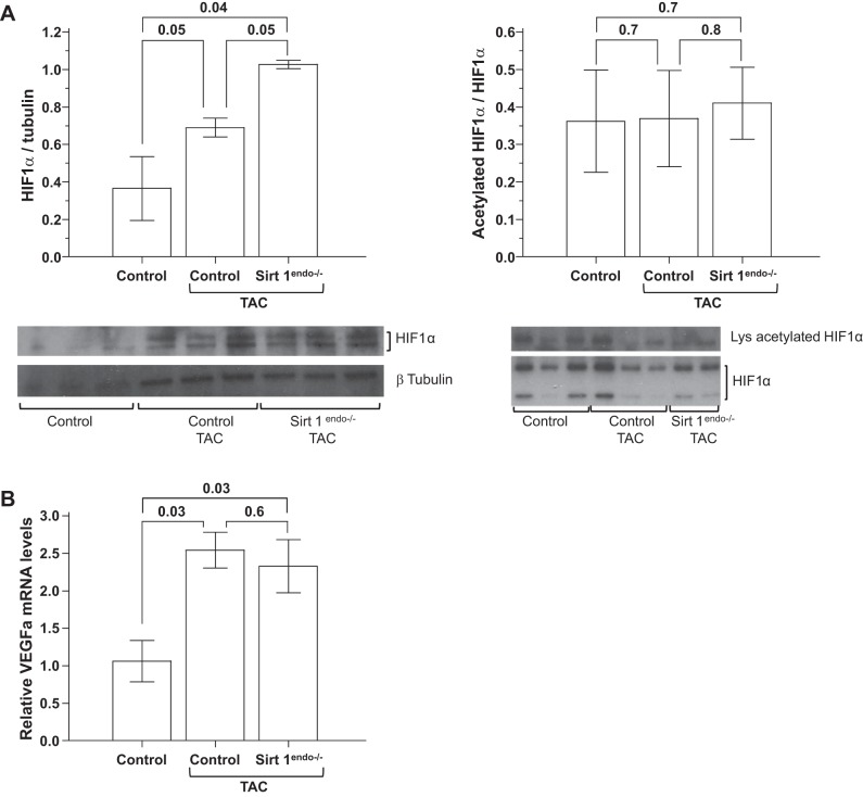
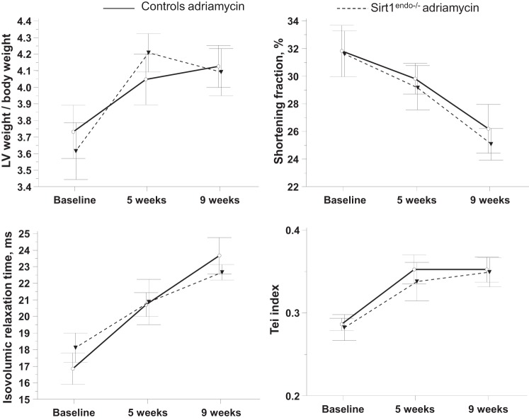
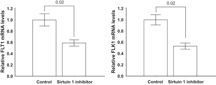
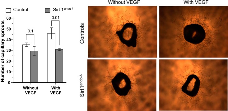
Discordant myocardial growth and angiogenesis can explain left ventricular (LV) hypertrophy progressing toward heart failure with aging. Sirtuin 1 expression declines with age; therefore we explored the role played by angiogenesis and Sirtuin 1 in the development of cardiomyopathy. We compared the cardiac function of 10- to 15-wk-old (wo), 30-40 wo, and 61-70 wo endothelial Sirtuin 1-deleted (Sirt1(endo-/-)) mice and their corresponding knockout controls (Sirt1(Flox/Flox)). After 30-40 wk, Sirt1(endo-/-) animals exhibited diastolic dysfunction (DD), decreased mRNA expression of Serca2a in the LV, and decreased capillary density compared with control animals despite a similar VEGFa mRNA expression. However, LV fibrosis and hypoxia-inducible factor (HIF)1α expression were not different. The creation of a transverse aortic constriction (TAC) provoked more severe DD and LV fibrosis in Sirt1(endo-/-) compared with control TAC animals. Although the VEGFa mRNA expression was not different and the protein expression of HIF1α was higher in the Sirt1(endo-/-) TAC animals, capillary density remained reduced. In cultured endothelial cells administration of Sirtuin 1 inhibitor decreased mRNA expression of VEGF receptors FLT 1 and FLK 1. Ex vivo capillary sprouting from aortic explants showed impaired angiogenic response to VEGF in the Sirt1(endo-/-) mice. In conclusion, the data demonstrate 1) a defect in angiogenesis preceding development of DD; 2) dispensability of endothelial Sirtuin 1 under unstressed conditions and during normal aging; and 3) impaired angiogenic adaptation and aggravated DD in Sirt1(endo-/-) mice challenged with LV overload.

Keywords: Adriamycin cardiomyopathy; VEGF; angiogenesis; echocardiography; hypoxia; transverse aortic constriction.


Figures

Fig. 1.
Fig. 1.
Cardiac consequences of endothelial Sirtuin 1 inactivation at different ages: comparisons of left ventricular (LV) weight/body weight, shortening fraction, isovolumic relaxation time (IVRT), and Tei index at different ages between control and Sirt1endo−/− mice measured by echocardiography. Echocardiography shows a diastolic dysfunction after 30–40 wk of age (wo) with increased IVRT and Tei index. Shortening fraction and LV weight/body weight were similar between the groups of experimental animals.
Fig. 2.
Fig. 2.
Cardiac expression of Serca2a measured by quantitative PCR in the LV of control and Sirt1endo−/− animals at different ages. Expression of Serca2a mRNA in the LV of Sirt1endo−/− animals decreased after 30–40 wk of age compared with control animals.
Fig. 3.
Fig. 3.
CD-31 staining (brown stain) in the LV of control and Sirt1endo−/− animals at different ages. Quantification of the CD31 staining is presented in the bar graph as the number of vessels per square millimeter. Note that the capillary density in the LV myocardium of the Sirt1endo−/− mice was decreased after 30–40 wk of age compared with control mice.
Fig. 4.
Fig. 4.
A: Masson's trichrome staining (blue stain) in the LV of control and Sirt1endo−/− animals at different ages. Quantification of the fibrosis is presented in the bar graph as % of Masson's trichrome-positive area. No evidence of exaggerated fibrosis in Sirt1endo−/− animals is seen. Masson's trichrome staining did not show any significant differences of LV fibrosis between Sirt1endo−/− and control animals at different ages. B: cardiac expression of Collagen 1 measured by quantitative PCR in the LV of control and Sirt1endo−/− animals at different ages. mRNA expression of collagen 1 in the LV of Sirt1endo−/− and control animals was not different.
Fig. 5.
Fig. 5.
Cardiac expression of metabolic (Sirtuin 3, Sirtuin 6, and PGC1a) and senescence (short:long endoglin) markers measured by quantitative PCR in the LV of control and Sirt1endo−/− animals at different ages. mRNA expression of Sirtuin 6 (A) and Sirtuin 3 (B) decreased with age in the myocardium of Sirt1endo−/− animals. PGC1a mRNA abundance (C) was not different from control animals. Compared with control mice, the mRNA expression of short:long endoglin (D) increased in the Sirt1endo−/− mice with aging.
Fig. 6.
Fig. 6.
A: expression of total protein HIF1α and immunoprecipitated acetylated HIF1α measured by immunoblotting in the LV of control and Sirt1endo−/− animals. B: cardiac expression of VEGFa measured by quantitative PCR in the LV of control and Sirt1endo−/− animals at different ages.
Fig. 7.
Fig. 7.
Comparisons of body weight, LV weight/body weight, shortening fraction, IVRT, and Tei index between control animals without surgery, control animals with transverse aortic constriction (TAC), and Sirt1endo−/− TAC animals measured by echocardiography. Measurements were performed before the surgery (baseline) and 1 and 7 wk after the surgery. Inactivation of Sirtuin 1 in the coronary endothelial cells worsens the cardiac consequences due to pressure overload provoked by TAC. The diastolic dysfunction (IVRT and Tei index) of the Sirt1endo−/− animals subjected to TAC was more profound on echocardiography than in control TAC animals. LV weight/body weight increased and shortening fraction decreased similarly after surgery between the 2 groups. *P < 0.05, Sirt1endo−/− TAC vs. controls TAC; **P < 0.05, controls vs. Sirt1endo−/− TAC and controls TAC.
Fig. 8.
Fig. 8.
Cardiac expression of Serca2a measured by quantitative PCR in the LV of control animals without surgery, control animals with TAC, and Sirt1endo−/− TAC animals 7 wk after surgery. Expression of Serca2a mRNA in the LV decreased in both constricted TAC groups but was not different between Sirt1endo−/− and control animals.
Fig. 9.
Fig. 9.
Cardiac expression of metabolic (Sirtuin 3, Sirtuin 6, and PGC1a) markers measured by quantitative PCR in the LV of control animals without surgery, control animals with TAC, and Sirt1endo−/− TAC animals 7 wk after surgery. A: mRNA expression of Sirtuin 3 in the LV decreased in both TAC groups. B: Sirtuin 6 mRNA expression levels in the LV remained similar between the different groups. C: mRNA expression of PGC1a in the LV decreased in both constricted groups.
Fig. 10.
Fig. 10.
CD31 staining (brown stain) in the LV of control animals without surgery, control animals with TAC, and Sirt1endo−/− TAC animals 7 wk after surgery. Quantification of the CD31 staining is presented in the bar graph as the number of vessels per square millimeter. Note that the capillary density in the control TAC animals increased, but the opposite occurred in the Sirt1endo−/− TAC group of mice.
Fig. 11.
Fig. 11.
A: Masson's trichrome staining (blue stain) in the LV of control animals without surgery, control animals with TAC, and Sirt1endo−/− TAC animals 7 wk after surgery. Quantification of the fibrosis is presented in the bar graph as % of Masson's trichrome-positive area. LV fibrosis increased in both groups of TAC animals, but the extent of fibrosis was significantly higher in the Sirt1endo−/− animals. B: cardiac expression of Collagen 1 measured by quantitative PCR in the LV of control animals without surgery, control animals with TAC, and Sirt1endo−/− TAC animals 7 wk after surgery. Collagen 1 mRNA expression in the myocardium of the Sirt1endo−/− constricted animals significantly increased, but there was no significant difference with the control group.
Fig. 12.
Fig. 12.
A: expression of total protein HIF1α and immunoprecipitated acetylated HIF1α measured by immunoblotting in the LV of control animals without surgery, control animals with TAC, and Sirt1endo−/− TAC animals 7 wk after surgery. Immunoblotting of HIF1α in the LV shows the increased expression of total HIF1α in the LV of Sirt1endo−/− of TAC animals. Expression of lysine-acetylated HIF1α was similar between the TAC groups; therefore the proportion of deacetylated HIF1α was higher in the Sirt1endo−/− animals. B: cardiac expression of VEGFa measured by quantitative PCR in the LV of control animals without surgery, control animals with TAC, and Sirt1endo−/− TAC animals 7 wk after surgery. mRNA expression of VEGFa increased in the LV of both constricted groups.
Fig. 13.
Fig. 13.
Comparisons of LV weight/body weight, shortening fraction, IVRT, and Tei index measured by echocardiography between Sirt1endo−/− and control animals subjected to Adriamycin injections. Echocardiography was performed at baseline and 5 wk and 9 wk after the beginning of treatment.
Fig. 14.
Fig. 14.
A: Masson's trichrome staining (blue stain) in the LV of control animals without Adriamycin, control animals with Adriamycin, and Sirt1endo−/− Adriamycin animals 9 wk after the beginning of treatment. Quantification of fibrosis is presented in the bar graph as % of Masson's trichrome-positive area. B: CD31 staining (brown stain) in the LV of control animals without Adriamycin, control animals with Adriamycin, and Sirt1endo−/− Adriamycin animals 9 wk after the beginning of treatment. Quantification of CD31 staining is presented in the bar graph as the number of vessels per square millimeter.
Fig. 15.
Fig. 15.
Expression of the VEGF receptors FLT1 and FLK1 measured by quantitative PCR in cultured immortalized human umbilical vein endothelial cells (HUVECs) exposed to 5 days of Sirtuin 1 inhibitor or placebo (Control).
Fig. 16.
Fig. 16.
Ex vivo capillary sprouting from aortic explants of control and Sirt1endo−/− mice after 6 days of culture in medium with or without VEGF. The numbers of capillary sprouts per aortic explant are presented in the bar graph.

Similar articles

Cited by

References

    1. Arany Z, Foo SY, Ma Y, Ruas JL, Bommi-Reddy A, Girnun G, Cooper M, Laznik D, Chinsomboon J, Rangwala SM, Baek KH, Rosenzweig A, Spiegelman BM. HIF-independent regulation of VEGF and angiogenesis by the transcriptional coactivator PGC-1alpha. Nature 451: 1008–1012, 2008. - PubMed
    1. Bertazzoli C, Bellini O, Magrini U, Tosana MG. Quantitative experimental evaluation of adriamycin cardiotoxicity in the mouse. Cancer Treat Rep 63: 1877–1883, 1979. - PubMed
    1. Brodsky S, Chen J, Lee A, Akassoglou K, Norman J, Goligorsky MS. Plasmin-dependent and -independent effects of plasminogen activators and inhibitor-1 on ex vivo angiogenesis. Am J Physiol Heart Circ Physiol 281: H1784–H1792, 2001. - PubMed
    1. Chen J, Xavier S, Moskowitz-Kassai E, Chen R, Lu CY, Sanduski K, Špes A, Turk B, Goligorsky MS. Cathepsin cleavage of sirtuin 1 in endothelial progenitor cells mediates stress-induced premature senescence. Am J Pathol 180: 973–983, 2012. - PMC - PubMed
    1. Cheng HL, Mostoslavsky R, Saito S, Manis JP, Gu Y, Patel P, Bronson R, Appella E, Alt FW, Chua KF. Developmental defects and p53 hyperacetylation in Sir2 homolog (SIRT1)-deficient mice. Proc Natl Acad Sci USA 100: 10794–10799, 2003. - PMC - PubMed

Publication types

MeSH terms

Substances

LinkOut - more resources