Review
. 2015 Jan:88:10-23.
doi: 10.1016/j.neuropharm.2014.09.011. Epub 2014 Sep 17.

Tasks for inhibitory interneurons in intact brain circuits

Affiliations
Review

Tasks for inhibitory interneurons in intact brain circuits

Lisa Roux et al. Neuropharmacology. 2015 Jan.

Abstract

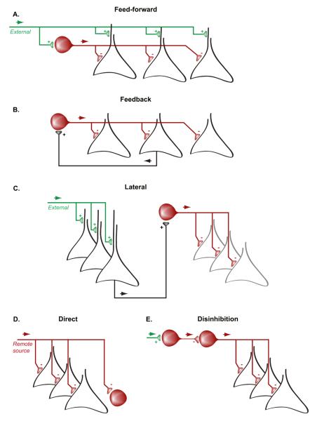
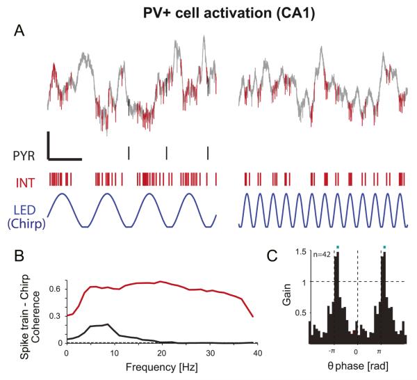
Synaptic inhibition, brought about by a rich variety of interneuron types, counters excitation, modulates the gain, timing, tuning, bursting properties of principal cell firing, and exerts selective filtering of synaptic excitation. At the network level, it allows for coordinating transient interactions among the principal cells to form cooperative assemblies for efficient transmission of information and routing of excitatory activity across networks, typically in the form of brain oscillations. Recent techniques based on targeted expression of neuronal activity modulators, such as optogenetics, allow physiological identification and perturbation of specific interneuron subtypes in the intact brain. Combined with large-scale recordings or imaging techniques, these approaches facilitate our understanding of the multiple roles of inhibitory interneurons in shaping circuit functions.

Keywords: Circuits; Inhibition; Interneurons; Optogenetics; Oscillations; Pharmacogenetics.


Figures

Figure 1
Figure 1
Main forms of inhibitory microcircuits. A. In a feed-forward inhibitory circuit, interneurons (red) receive excitatory inputs from an external source (green) and in turn inhibit the local principal neurons (black). These latter neurons are often also targeted by the external excitatory input and the relative strength of excitation on the principal cells and interneurons as well as the interneuron-induced inhibition determine the firing discharge of principal cells. B. In a feed-back inhibitory circuit, interneurons receive excitation from principal cells and, in turn they inhibit the principal cells. Thus, local excitation is a condition for inhibition. C. Lateral inhibition allows a first assembly of principal cells (black) to suppress the activity of another assembly of principal cells (grey) through the excitation of inhibitory interneurons. In real networks, lateral inhibition is usually reciprocal and such connectivity allows for assembly competition (an exclusive OR operation in Boolean terms). D. Direct inhibition involves the suppression of local principal cell or interneuron activity by long-range interneurons from remote brain regions. E. Disinhibition of the principal cells occurs when their direct inhibitory inputs are suppressed by another population of inhibitory interneurons.
Figure 2
Figure 2
Dendrite-targeting interneurons control principal cell bursting. A-B. Dendritic recording from a CA1 pyramidal cell performed in vivo with a sharp electrode showed that inhibition modulates dendritic spike invasion. The recording configuration is shown in B. A. (a) Intradendritic depolarization (0.4 nA) evoked fast sodium spikes and a slow spike, likely a calcium spike. (b-d) Dendritic depolarization was paired with commissural stimulation (short arrows) in order to activate inhibitory interneurons. Bottom traces: extracellular recordings from the pyramidal layer. Weak commissural stimulation delayed (b, 30 μA), abolished (c, 50 μA) or aborted the slow spike (d, 50μA). (c) [triangleup], inhibitory postsynaptic potential evoked by the commissural stimulus. B. After the recordings, the cell was injected with biocytin for morphological reconstruction with a drawing tube. Adapted from Buzsaki et al., 1996. Copyright (1996) National Academy of Sciences, U.S.A. C-D. Current-clamp recordings from the distal apical dendrites of CA1 pyramidal cells performed in vitro, combined with pharmacogenetic manipulations (via the expression of the ligand-gated Cl channel, PSAML141F-GlyR in the SOM+ cells, and application of its ligand PSEM308), showed that silencing the dendrite-targeting SOM+ cells increases dendritic spike generation during Schaffer Collateral photostimulation, whereas silencing of the soma-targeting PV+ cells does not. Adapted by permission from Macmillan Publishers Ltd: Nature Neuroscience (Lovett-Barron et al. 2012), copyright (2012). E. Left: Example whole-cell recording from an excitatory neuron during optogenetic inhibition of nearby dendrite-targeting SOM+ neurons in the somatosensory cortex of anesthetized mice. Right: Optogenetic silencing (Light ON) of the SOM+ cells increased burst firing in the nearby principal cells. Each thin line represents an individual neuron and filled circles with error bars connected by thick lines represent mean ± s.e.m. Adapted by permission from Macmillan Publishers Ltd: Nature Neuroscience (Gentet et al. 2012), copyright (2012). F. In vivo extracellular recordings combined with optogenetics in freely moving mice showed that SOM+ silencing (but not PV+ cell silencing) increases burst firing in the putative pyramidal cells, as shown by the relative increase (mean ± s.e.m.) of occurrence for different burst lengths (comparison between PV+ and SOM+ cell silencing, *P < 0.05, #P < 0.0005). Adapted from Royer et al. 2012.
Figure 3
Figure 3
Theta-resonance of pyramidal cell spiking induced by PV+ cell activation in the hippocampal CA1 region. A. Example traces of local field potentials (LFP) and spikes (1-5000 Hz; calibration: 100 ms, 200 μV) in the CA1 pyramidal layer during two distinct periods (~8Hz - left- and ~25Hz -right-) of a chirp pattern photostimulation of PV+ interneurons (chirp between 0 and 40 Hz;, blue trace). Red: PV+ interneuron spikes (INT). Black: pyramidal cell (PYR) spikes. Note that during PV+ cell activation, the pyramidal cell tended to spike specifically at theta frequency. B. Coherence between chirp pattern and spiking; dashed line shows chance coherence. Note the narrow-band coherence of the PYR at theta frequency (black) and the wide-band coherence of the interneuron (red). During theta-band chirp pattern PV+ cell activation, the PYR spikes specifically at chirp troughs (not shown). C. Mean theta spiking gain for the PYR during PV+ cell activation. For each PYR, firing rates resolved by chirp phase (top) were computed and divided by the baseline rate (in the lack of any light stimulation). Gain=1 thus indicates no change relative to spontaneous activity. Blue bars indicate phase bins for which the number of units with increased spiking (gain>1) exceeds chance level (exact Binomial test, p<0.001). The mean gain is >1 at the chirp theta trough indicating that PYR exhibit excess (“rebound”) spiking. Figure adapted from Stark et al., 2013.

References

    1. Adesnik H, Bruns W, Taniguchi H, Huang ZJ, Scanziani M. A neural circuit for spatial summation in visual cortex. Nature. 2012;490:226–231. - PMC - PubMed
    1. Alonso A, Kohler C. Evidence for separate projections of hippocampal pyramidal and nonpyramidal neurons to different parts of the septum in the rat brain. Neuroscience letters. 1982;31:209–214. - PubMed
    1. Alonso A, Kohler C. A study of the reciprocal connections between the septum and the entorhinal area using anterograde and retrograde axonal transport methods in the rat brain. The Journal of comparative neurology. 1984;225:327–343. - PubMed
    1. Alonso M, Lepousez G, Sebastien W, Bardy C, Gabellec MM, Torquet N, Lledo PM. Activation of adult-born neurons facilitates learning and memory. Nat Neurosci. 2012;15:897–904. - PubMed
    1. Anderson JS, Carandini M, Ferster D. Orientation tuning of input conductance, excitation, and inhibition in cat primary visual cortex. J Neurophysiol. 2000;84:909–926. - PubMed

Publication types