. 2014 Nov;13(11):2595-606.
doi: 10.1158/1535-7163.MCT-14-0422. Epub 2014 Sep 19.

Development of targeted near-infrared imaging agents for prostate cancer

Affiliations

Development of targeted near-infrared imaging agents for prostate cancer

Xinning Wang et al. Mol Cancer Ther. 2014 Nov.

Abstract

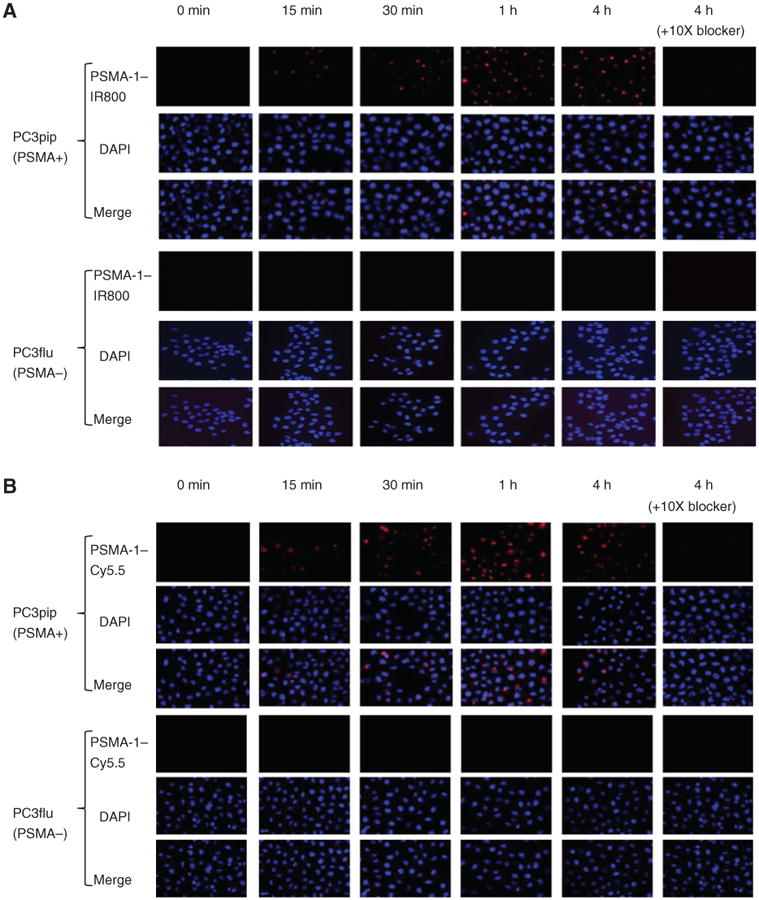
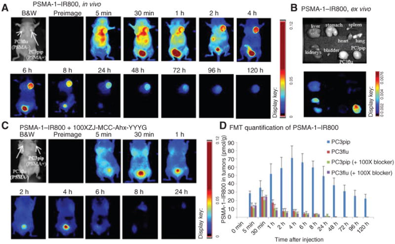
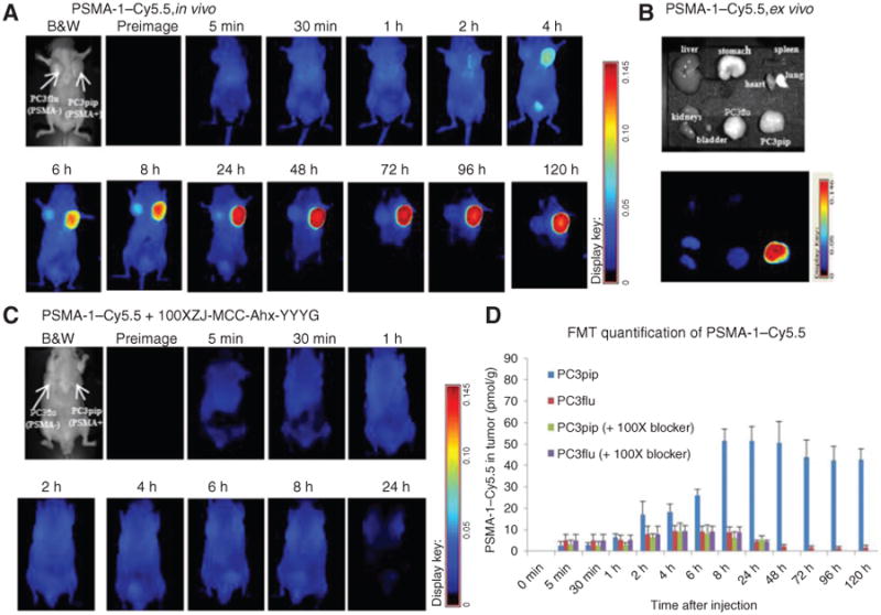
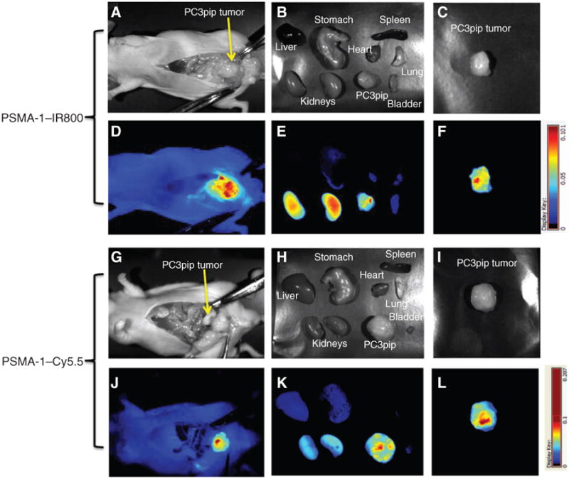
Prostate cancer is the most common noncutaneous malignancy affecting men in North America. Radical prostatectomy remains a definitive treatment for prostate cancer. However, prostate surgeries are still performed "blindly" with the extent of tumor infiltration past the margins of the surgery only being determined postoperatively. An imaging modality that can be used during surgery is needed to help define the tumor margins. With its abundant expression in prostate cancer, prostate-specific membrane antigen (PSMA) is an ideal target for detection of prostate cancer. The purpose of this study was to develop PSMA-targeted near-infrared (NIR) optical imaging probes for intraoperative visualization of prostate cancer. We synthesized a high-affinity PSMA ligand (PSMA-1) with low molecular weight and further labeled it with commercially available NIR dyes IRDy800 and Cy5.5. PSMA-1 and PSMA-1-NIR conjugates had binding affinities better than the parent ligand Cys-CO-Glu. Selective binding was measured for each of the probes in both in vitro and in vivo studies using competitive binding and uptake studies. Interestingly, the results indicated that the pharmacokinetics of the probes was dependent of the fluorophore conjugated to the PSMA-1 ligand and varied widely. These data suggest that PSMA-targeted probes have the potential to be further developed as contrast agents for clinical intraoperative fluorescence-guided surgery.


Conflict of interest statement

Disclosure of Potential Conflicts of Interest: No potential conflicts of interest were disclosed.

Figures

Figure 1
Figure 1
Structures of PSMA ligands and PSMA–NIR conjugates. d-isomers of glutamic acid were used in PSMA-1 to provide better in vivo stability. IRDye800 and Cy5.5 are conjugated to PSMA-1 through the γ-NH2 group of C-terminal lysine.
Figure 2
Figure 2
In vitro cellular uptake results of PSMA-1–IR800 and PSMA-1–Cy5.5. PSMA-positive PC3pip cells and PSMA-negative cells PC3flu cells on coverslips were incubated with no probe (0 minutes, A and B) or 1 μmol/L of PSMA-1–IR800 (A) or 1 μmol/L of PSMA-1–Cy5.5 (B) for 15 minutes, 30 minutes, 1 hour, and 4 hours. The nucleus was stained by DAPI (false color blue), and uptake of PSMA-1–IR800 and PSMA-1–Cy5.5 was assessed by fluorescence microscopy (false color red). Specificity of PSMA-1–NIR conjugates to PSMA was evaluated by incubation of PC3pip and PC3flu cells with 1 μmol/L of PSMA-1–NIR conjugates and 10 μmol/L of Cys-CO-Glu, last column in each panel. Signal in PC3pip cells was significantly competed by Cys-CO-Glu, suggesting that the binding of PSMA-1–IR800 and PSMA-1–Cy5.5 to PSMA is specific. Images are taken at 40×. Representative images are shown from 3 independent experiments.
Figure 3
Figure 3
Imaging of PSMA-1–IR800 in mice bearing flank PC3pip and PC3flu tumors. A, in vivo Maestro imaging of a typical mouse treated with PSMA-1–IR800. Mice received 1 nmol of PSMA-1–IR800 via tail vein injection and then were imaged at the designated times. Representative images are shown of n = 5. B, ex vivo imaging of mice organs at 120 hours postinjection of PSMA-1–IR800. The fluorescent signal in PC3pip tumor was significantly higher than in other organs. C, in vivo Maestro imaging of mice injected with 1 nmol of PSMA-1–IR800 and 100 nmol of a selective PSMA receptor–binding molecule, ZJ-MCC-Ahx-YYYG (34). Images are on the same scale as in A. Blockade of fluorescent uptake in PC3pip tumors was observed. D, FMT 3D quantification of PSMA-1–IR800 in PC3pip and PC3flu tumors from the mice used in A and C. Values represent mean ± SD of 5 animals.
Figure 4
Figure 4
Imaging of PSMA-1–Cy5.5 in mice bearing flank PC3pip and PC3flu tumors. A, in vivo Maestro imaging of mice treated with PSMA-1–Cy5.5. Mice received 1 nmol of PSMA-1–Cy5.5 through tail vein injection and then were imaged at the indicated times. Representative images of n = 5 mice are shown. Selective uptake in PC3pip tumors was observed. Highest PC3pip tumor uptake was observed 24 hours postinjection. B, ex vivo imaging of mice organs at 120 hours postinjection of PSMA-1–Cy5.5. The fluorescent signal in PC3pip tumor was significantly higher than in other organs. C, in vivo Maestro imaging of mice injected with 1 nmol of PSMA-1–Cy5.5 and 100 nmol of a selective PSMA receptor–binding molecule, ZJ-MCC-Ahx-YYYG (34). Images are on the same scale as in A. Blockade of fluorescent uptake in PC3pip tumors was observed. D, FMT 3D quantification of PSMA-1–Cy5.5 in PC3pip and PC3flu tumors from mice used in A and C. Values represent mean ± SD of 5 animals.
Figure 5
Figure 5
PSMA-1–NIR probes can selectively target orthotopic PSMA-positive PC3pip tumors as shown by Maestro images. Mice received 1 nmol of PSMA–IR800 (A–F) or 1 nmol of PSMA-1–Cy5.5 (G–L) via a tail vein injection. Mice were sacrificed at 4 hours postinjection of PSMA-1–IR800, the abdomen opened to expose the tumor, and both black and white images (A) and fluorescent images (D) were taken. Organs were then harvested for ex vivo images (B and E) and finally tumors were imaged separately ex vivo (C and F). Mice that were administered PSMA-1–Cy5.5 were sacrificed at 24 hours postinjection; the abdomen was opened to expose tumor and then imaged. Both black and white images (G) and fluorescent images (J) were taken, organs were harvested for imaging (H and K), and finally tumors were imaged separately ex vivo (I and L). Pictures are representative images of 4 mice for each probe. Bright fluorescent signal was observed in PC3pip tumor.

References

    1. Siegel R, Ma J, Zou Z, Jemal A. Cancer statistics, 2014. CA Cancer J Clin. 2014;64:9–29. - PubMed
    1. Theiss M, Wirth MP, Manseck A, Frohmuller HG. Prognostic significance of capsular invasion and capsular penetration in patients with clinically localized prostate cancer undergoing radical prostatectomy. Prostate. 1995;27:13–7. - PubMed
    1. Swanson GP, Lerner SP. Positive margins after radical prostatectomy: implications for failure and role of adjuvant treatment. Urol Oncol. 2013;31:531–41. - PubMed
    1. Wright JL, Dalkin BL, True LD, Ellis WJ, Stanford JL, Lange PH, et al. Positive surgical margins at radical prostatectomy predict prostate cancer specific mortality. J Urol. 2010;183:2213–8. - PMC - PubMed
    1. Horoszewicz JS, Kawinski E, Murphy GP. Monoclonal antibodies to a new antigenic marker in epithelial prostatic cells and serum of prostatic cancer patients. Anticancer Res. 1987;7:927–35. - PubMed

Publication types