. 2014 Nov;13(11):1450-64.
doi: 10.1128/EC.00183-14. Epub 2014 Sep 19.

Ultrastructure and composition of the Nannochloropsis gaditana cell wall

Affiliations

Ultrastructure and composition of the Nannochloropsis gaditana cell wall

Matthew J Scholz et al. Eukaryot Cell. 2014 Nov.

Abstract

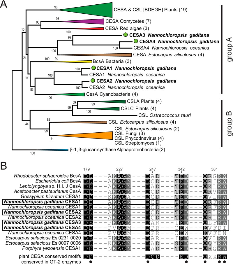
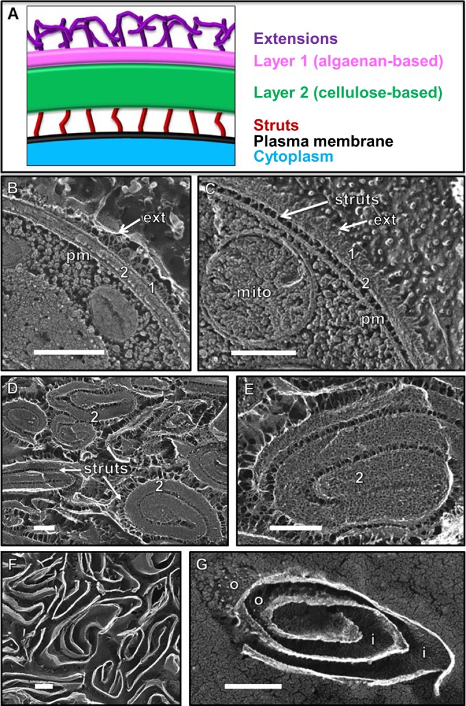
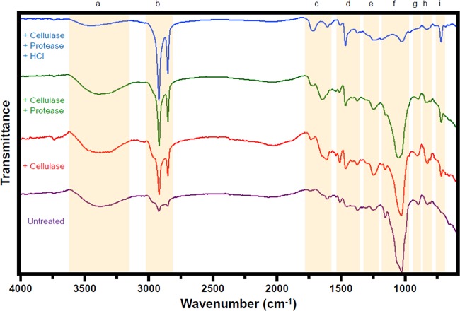
Marine algae of the genus Nannochloropsis are promising producers of biofuel precursors and nutraceuticals and are also harvested commercially for aquaculture feed. We have used quick-freeze, deep-etch electron microscopy, Fourier transform infrared spectroscopy, and carbohydrate analyses to characterize the architecture of the Nannochloropsis gaditana (strain CCMP 526) cell wall, whose recalcitrance presents a significant barrier to biocommodity extraction. The data indicate a bilayer structure consisting of a cellulosic inner wall (~75% of the mass balance) protected by an outer hydrophobic algaenan layer. Cellulase treatment of walls purified after cell lysis generates highly enriched algaenan preparations without using the harsh chemical treatments typically used in algaenan isolation and characterization. Nannochloropsis algaenan was determined to comprise long, straight-chain, saturated aliphatics with ether cross-links, which closely resembles the cutan of vascular plants. Chemical identification of >85% of the isolated cell wall mass is detailed, and genome analysis is used to identify candidate biosynthetic enzymes.


Figures

FIG 1
FIG 1
(A) Phylogenetic tree of cellulose synthases (CESA) and cellulose synthase-like proteins (CSL). The tree was constructed using the maximum-likelihood (ML) approach. N. gaditana sequences are marked with green circles. Numbers indicate bootstrap values in the ML analysis. Numbers in parentheses indicate the numbers of sequences compressed in subtrees. Groups A and B are described in the text. The full list of accession numbers from the proteins used can be found in Table S1 in the supplemental material. (B) Multiple sequence alignments of the motifs common to cellulose synthases. Numbers indicate the amino acid positions in the Rhodobacter sphaeroides BcsA protein (58). Dashes do not represent gaps in the alignment, and not all aligned amino acids are shown. Dots indicate the conserved residues (D, D, D, and QXXRW) found in characterized cellulose synthases within the glycosyl transferase 2 (GT-2) family, as cataloged by the Carbohydrate-Active enZymes (CAZy) database.
FIG 2
FIG 2
(A) Model of the Nannochloropsis wall based on quick-freeze deep-etch EM (QFDE-EM) images. The native layer 1 adopts a TLS configuration in thin section (2) but forms a single layer in replicas and is overlain with fibrous extensions. Layer 2 is thicker and often associates with the plasma membrane via narrow struts. (B) Cross fracture through the native wall showing its two layers (1, 2), distal extensions (ext), and close association with the plasma membrane (pm). (C) Tangential fracture through the two native wall layers showing distal extensions (ext) and proximal struts attaching to the plasma membrane (pm). mito, mitochondrion. (D and E) Pressed cell walls, with the prominent fibrillar layer 2 masking the presence of layer 1. (F and G) Same pressed wall preparation as shown in panels D and E after 24 h of incubation in cellulase. Fibrillar layer 2 and struts were digested, exposing layer 1, whose outer (o) and inner (i) faces are granular. All scale bars, 250 nm.
FIG 3
FIG 3
FTIR spectra of CCMP 526 pressed walls. Spectra are provided for pressed walls and following stepwise cellulase, protease, and HCl digestion. Nine notable wavenumber regions (a to i) have been highlighted.

References

    1. Andersen RA, Brett RW, Potter D, Sexton JP. 1998. Phylogeny of the Eustigmatophyceae based upon 18S rDNA, with emphasis on Nannochloropsis. Protist 149:61–74. 10.1016/S1434-4610(98)70010-0. - DOI - PubMed
    1. Murakami R, Hashimoto H. 2009. Unusual nuclear division in Nannochloropsis oculata (Eustigmatophyceae, Heterokonta) which may ensure faithful transmission of secondary plastids. Protist 160:41–49. 10.1016/j.protis.2008.09.002. - DOI - PubMed
    1. Rodolfi L, Zittelli GC, Barsanti L, Rosati G, Tredici MR. 2003. Growth medium recycling in Nannochloropsis sp mass cultivation. Biomol. Eng. 20:243–248. 10.1016/S1389-0344(03)00063-7. - DOI - PubMed
    1. Bondioli P, Della Bella L, Rivolta G, Chini Zittelli G, Bassi N, Rodolfi L, Casini D, Prussi M, Chiaramonti D, Tredici MR. 2012. Oil production by the marine microalgae Nannochloropsis sp. F&M-M24 and Tetraselmis suecica F&M-M33. Bioresour. Technol. 114:567–572. 10.1016/j.biortech.2012.02.123. - DOI - PubMed
    1. Jinkerson RE, Radakovits R, Posewitz MC. 2013. Genomic insights from the oleaginous model alga Nannochloropsis gaditana. Bioengineered 4:37–43. 10.4161/bioe.21880. - DOI - PMC - PubMed

Publication types