A mistaken identity: rhabdomyosarcoma of the middle ear cleft misdiagnosed as chronic suppurative otitis media with temporal lobe abscess
- PMID: 25240007
- PMCID: PMC4170300
- DOI: 10.1136/bcr-2014-206615
A mistaken identity: rhabdomyosarcoma of the middle ear cleft misdiagnosed as chronic suppurative otitis media with temporal lobe abscess
Abstract
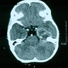
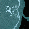
A 5-year-old girl presented with a 3-month history of left side facial palsy, followed sequentially by purulent ear discharge, complete external ophthalmoplaegia and blurred vision. On clinical examination she was febrile with left-sided conductive hearing loss. She was clinically diagnosed to have chronic suppurative otitis media of the unsafe type with petrous apicitis, middle cranial fossa abscess and cavernous sinus involvement. Preliminary CT scan findings were reported as a large left temporal lobe abscess and left otitis media with cholesteatoma. MRI of the brain obtained later corroborated the abnormalities detected on the CT scan. Ten days after admission, a mass was seen protruding from the external auditory canal. A biopsy of the mass was obtained and sent for histopathological examination. Meanwhile, review of the MRI suggested an aggressive neoplasm such as sarcoma/rhabdomyosarcoma. Histopathology clinched the final diagnosis of an anaplastic type of embryonal rhabdomyosarcoma of the middle ear cleft.
2014 BMJ Publishing Group Ltd.
Figures


Similar articles
-
[Cholesteatoma of the external and middle ear in the childhood].Vestn Otorinolaringol. 2015;(1):25-27. doi: 10.17116/otorino201580125-27. Vestn Otorinolaringol. 2015. PMID: 25909669 Russian.
-
Tuberculous otitis media: a case presentation and review of the literature.BMJ Case Rep. 2010 Dec 1;2010:bcr0220102721. doi: 10.1136/bcr.02.2010.2721. BMJ Case Rep. 2010. PMID: 22798297 Free PMC article. Review.
-
[Clinical analysis of complications of suppurative otitis media in children].Lin Chuang Er Bi Yan Hou Tou Jing Wai Ke Za Zhi. 2020 Jul;34(7):587-591. doi: 10.13201/j.issn.2096-7993.2020.07.003. Lin Chuang Er Bi Yan Hou Tou Jing Wai Ke Za Zhi. 2020. PMID: 32791630 Free PMC article. Chinese.
-
[Choristoma of the middle ear].Vestn Otorinolaringol. 2023;88(3):73-77. doi: 10.17116/otorino20228803173. Vestn Otorinolaringol. 2023. PMID: 37450395 Russian.
-
[Case of diagnosis and surgical treatment of intracranial cholesteatoma].Vestn Otorinolaringol. 2022;87(4):89-94. doi: 10.17116/otorino20228704189. Vestn Otorinolaringol. 2022. PMID: 36107187 Review. Russian.
Cited by
-
Embryonal Rhabdomyosarcoma - A Mimicker of Squamosal Otitis Media.Iran J Otorhinolaryngol. 2020 Jan;32(108):57-61. doi: 10.22038/ijorl.2019.38807.2280. Iran J Otorhinolaryngol. 2020. PMID: 32083033 Free PMC article.
-
Diagnosis and Treatment of Nodular Fasciitis of Ear Region in Children: A Case Report and Review of Literature.Healthcare (Basel). 2022 Oct 7;10(10):1962. doi: 10.3390/healthcare10101962. Healthcare (Basel). 2022. PMID: 36292409 Free PMC article.
References
-
- McCarville MB, Spunt SL, Pappo AS. Rhabdomyosarcoma in pediatric patients: the good, the bad, and the unusual. AJR Am J Roentgenol 2001;176:1563–9 - PubMed
-
- Durve DV, Kanegaonkar RG, Albert D, et al. Paediatric rhabdomyosarocma of the ear and temporal bone. Clin Otolaryngol Allied Sci 2004;29:32–7 - PubMed
-
- Jan MM. Facial paralysis: a presenting feature of rhabdomyosarcoma. Int J Pediatr Otorhinolaryngol 1998;46:221–4 - PubMed
-
- Ayachea D, Darrouzetb V, Dubrullec F, et al. Imaging of non-operated cholesteatoma: clinical practice guidelines. Eur Ann Otorhinolaryngol Head Neck Dis 2012;129:148–52 - PubMed
Publication types
MeSH terms
LinkOut - more resources
Full Text Sources
Other Literature Sources
Medical
