. 2014 Nov;55(11):2296-308.
doi: 10.1194/jlr.M050633. Epub 2014 Sep 19.

Macrophage deficiency of Akt2 reduces atherosclerosis in Ldlr null mice

Affiliations

Macrophage deficiency of Akt2 reduces atherosclerosis in Ldlr null mice

Vladimir R Babaev et al. J Lipid Res. 2014 Nov.

Abstract

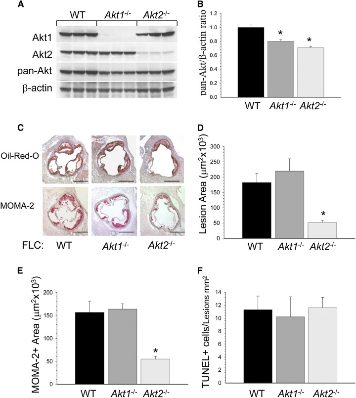
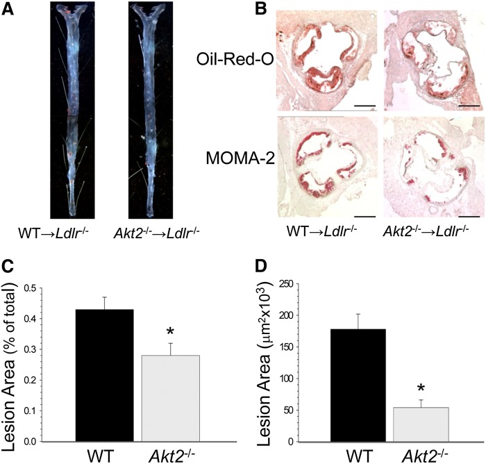
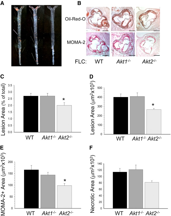
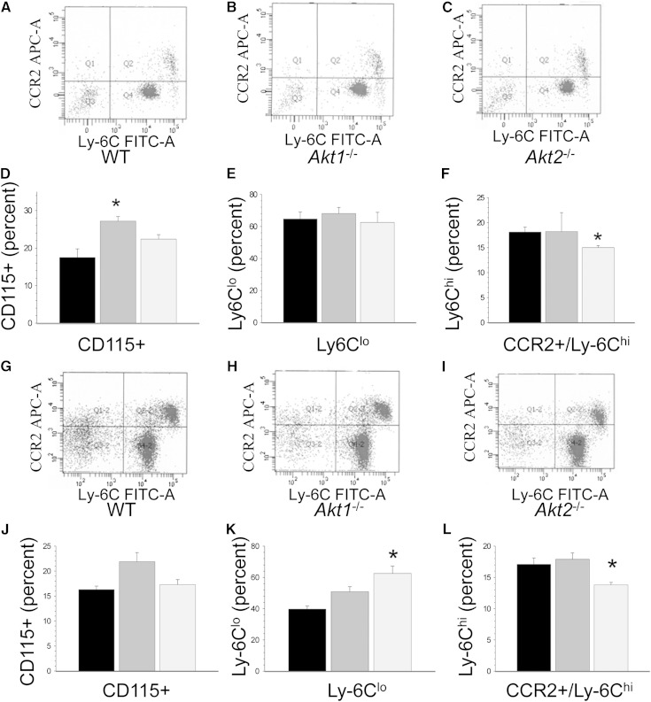
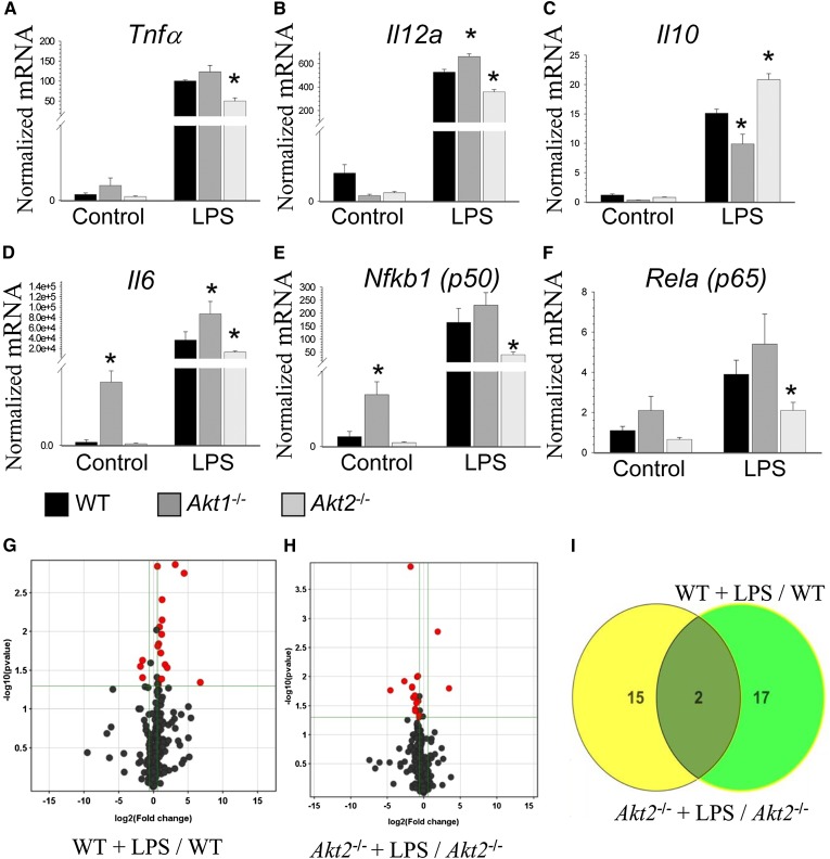
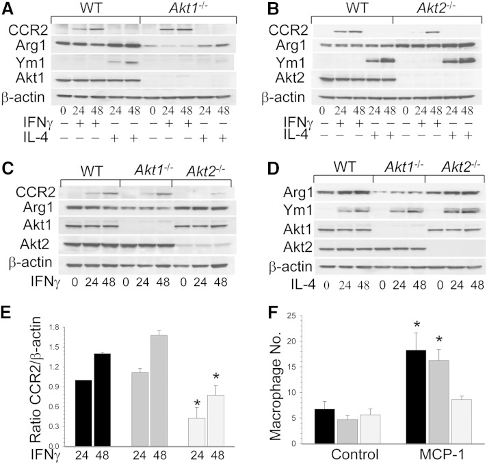
Macrophages play crucial roles in the formation of atherosclerotic lesions. Akt, a serine/threonine protein kinase B, is vital for cell proliferation, migration, and survival. Macrophages express three Akt isoforms, Akt1, Akt2, and Akt3, but the roles of Akt1 and Akt2 in atherosclerosis in vivo remain unclear. To dissect the impact of macrophage Akt1 and Akt2 on early atherosclerosis, we generated mice with hematopoietic deficiency of Akt1 or Akt2. After 8 weeks on Western diet, Ldlr(-/-) mice reconstituted with Akt1(-/-) fetal liver cells (Akt1(-/-)→Ldlr(-/-)) had similar atherosclerotic lesion areas compared with control mice transplanted with WT cells (WT→Ldlr(-/-)). In contrast, Akt2(-/-)→Ldlr(-/-) mice had dramatically reduced atherosclerotic lesions compared with WT→Ldlr(-/-) mice of both genders. Similarly, in the setting of advanced atherosclerotic lesions, Akt2(-/-)→Ldlr(-/-) mice had smaller aortic lesions compared with WT→Ldlr(-/-) and Akt1(-/-)→Ldlr(-/-) mice. Importantly, Akt2(-/-)→Ldlr(-/-) mice had reduced numbers of proinflammatory blood monocytes expressing Ly-6C(hi) and chemokine C-C motif receptor 2. Peritoneal macrophages isolated from Akt2(-/-) mice were skewed toward an M2 phenotype and showed decreased expression of proinflammatory genes and reduced cell migration. Our data demonstrate that loss of Akt2 suppresses the ability of macrophages to undergo M1 polarization reducing both early and advanced atherosclerosis.

Keywords: apoptosis; cell signaling; foam cells; macrophages/monocytes.


Figures

Fig. 1.
Fig. 1.
Loss of Akt1 and Akt2 decreases Akt levels in macrophages, but only Akt2 deficiency in hematopoietic cells reduces early atherosclerosis in female Ldlr−/− mice. A, B: Macrophage Akt1 and Akt2 deficiency significantly reduces total Akt protein levels in the proportion of WT > Akt1−/− > Akt2−/− (* P < 0.05 by one-way ANOVA, multiple comparison procedures, Holm-Sidak method). Peritoneal macrophages were isolated from Ldlr−/− mice reconstituted with WT (black), Akt1−/− (dark gray), and Akt2−/− (light gray) FLCs (n = 3/group), then proteins were extracted, resolved (50 μg/well) and analyzed by Western blot using an Akt isoform sampler kit. The graph (B) presents an average (mean ± SEM) of the ratio of pan-Akt/β-actin in macrophages. C: Akt2−/−Ldlr−/− mice had significantly smaller atherosclerotic lesions in the aortic sinus than WT→Ldlr−/− or Akt1−/−Ldlr−/− mice after 8 weeks on the Western diet. Serial cryostat sections were stained with Oil Red O to detect neutral lipids or with MOMA-2 antibody to reveal macrophages. Scale bars, 200 μm. D–F: The extent of atherosclerotic lesions in the aortic sinus (D), MOMA-2-positive area (E), and apoptotic cell numbers (F) in the aortic lesions of Ldlr−/− mice reconstituted with WT (black), Akt1−/−(dark gray), or Akt2−/− (light gray) FLCs (* P < 0.05 vs. control WT→Ldlr−/− mice by a one-way ANOVA on ranks, Tukey test).
Fig. 2.
Fig. 2.
Hematopoietic Akt2 deficiency suppresses early atherosclerosis in male Ldlr−/−mice. A, B: Atherosclerotic lesions in the distal (A) and proximal (B) aortas of mice reconstituted with WT and Akt2−/− FLCs. Aortas were pinned out en face and stained with Sudan IV (A); serial sections of the aortic sinus were stained with Oil Red O or macrophage stain, MOMA-2 (B). Scale bars, 200 μm; a pin size, 10 μm. C, D: The extent of atherosclerotic lesions in the distal (C) and proximal (D) aortas of Ldlr−/− mice reconstituted with WT (black) or Akt2−/− (light gray) FLCs (* P < 0.05 by Mann-Whitney rank sum test).
Fig. 3.
Fig. 3.
Loss of Akt2 in hematopoietic cells suppresses formation of advanced atherosclerotic lesions in Ldlr−/− mice. A, B: Atherosclerotic lesions in the distal and proximal aortas of mice reconstituted with WT, Akt1−/−, or Akt2−/− FLCs after 16 weeks on the Western diet. Mouse aortas pinned out en face (A) and aortic sinus sections stained with Oil Red O or MOMA-2 (B). Scale bars, 200 μm; a pin size, 10 μm. C–F: The extent of atherosclerotic lesions in the distal (C) and proximal (D) aortas, MOMA-2-positive area (E), and necrotic area (F) in atherosclerotic lesions of Ldlr−/− mice reconstituted with WT (black), Akt1−/− (dark gray), or Akt2−/− (light gray) FLCs (* P < 0.05 vs. control group by one-way ANOVA, Holm-Sidak method).
Fig. 4.
Fig. 4.
Akt2−/−Ldlr−/− mice have decreased levels of CCR2+/Ly6Chi monocytes on a chow diet (A–F) and the Western diet (G–L). A–C, G–I: Flow cytometry analysis of CCR2 and Ly-6C expression in CD115-gated blood monocytes of Ldlr−/− mice reconstituted with WT, Akt1−/−, and Akt2−/− FLCs. D–F, J–L: Percent of CD115, Ly-6Clo, and CCR2+/Ly-6Chi monocytes in mice transplanted with WT (black), Akt1−/− (dark gray), and Akt2−/− (light gray) FLCs (* P < 0.05 compared with WT cells by one-way ANOVA, Dunnett’s method).
Fig. 5.
Fig. 5.
Distinct responses of Akt1−/− and Akt2−/− macrophages to LPS treatment compared with WT cells. A–F: Real-time PCR analysis of Tnf, Il12a, Il10, Il6, Nfkb1 (p50), and Rela(p60) gene expression in WT (black), Akt1−/− (dark gray), or Akt2−/− (light gray) macrophages in response to LPS. Cells were incubated with media alone (control) or together with LPS (50 ng/ml) for 6 h. The graphs represent data (mean ± SEM) obtained from the same numbers (n = 3/group) of mice (* P < 0.05 compared with WT cells untreated or treated with LPS by one-way ANOVA). G, H: Volcano plots demonstrating (red dots) significantly (P < 0.05) altered (>1.5-fold absolute fold change) miRNA changes, reported as log2 (fold change) and –log10 (P value by unpaired t-test; n = 3). OpenArrays WT + LPS/ WT (G) and Akt2−/− + LPS/ Akt2−/− (H). I: Venn diagram highlighting 17 miRNAs that are altered by LPS and dependent on Akt2 in macrophages (green).
Fig. 6.
Fig. 6.
Akt2−/− macrophages are resistant to M1 priming, and Akt2−/− cells express less CCR2 and were slower in the migration assay than Akt1−/− and WT cells. A–D: Expression of M1 and M2 markers in WT, Akt1−/−, and Akt2−/− macrophages in response to IFNγ or IL-4. Peritoneal macrophages were untreated (0) or treated with the recombinant mouse IFNγ (50 ng/ml) or the recombinant mouse IL-4 (20 ng/ml) for 24 or 48 h. Macrophage proteins were extracted, resolved (40 μg/well), and analyzed by Western blot. E: The graphs exhibit the averages (mean ± SEM) of CCR2 expression in WT (black), Akt1−/− (dark gray), or Akt2−/− (light gray) macrophages after treatment with IFNγ (* P < 0.05 compared with control WT cells at the same time point by one-way ANOVA). F: Peritoneal macrophages were cultured in transwell inserts in triplicate for 2 h and, after addition of MCP-1 (100 ng/ml) into the lower chamber, incubated for 2 h at 37°C. The cells were stained with DAPI and analyzed under a fluorescent microscope (* P < 0.05 compared with control WT cells by one-way ANOVA, Tukey test).

References

    1. Moore K. J., Sheedy F. J., Fisher E. A. 2013. Macrophages in atherosclerosis: a dynamic balance. Nat. Rev. Immunol. 13: 709–721. - PMC - PubMed
    1. Manning B. D., Cantley L. C. 2007. AKT/PKB signaling: navigating downstream. Cell. 129: 1261–1274. - PMC - PubMed
    1. Liu H., Perlman H., Pagliari L. J., Pope R. M. 2001. Constitutively activated Akt-1 is vital for the survival of human monocyte-differentiated macrophages. Role of Mcl-1, independent of nuclear factor (NF)-kappaB, Bad, or caspase activation. J. Exp. Med. 194: 113–126. - PMC - PubMed
    1. Chen W. S., Xu P-Z., Gottlob K., Chen M-L., Sokol K., Shiyanova T., Roninson I., Weng W., Suzuki R., Tobe K., et al. 2001. Growth retardation and increased apoptosis in mice with homozygous disruption of the Akt1 gene. Genes Dev. 15: 2203–2208. - PMC - PubMed
    1. Cho H., Thorvaldsen J. L., Chu Q., Feng F., Birnbaum M. J. 2001. Akt1/PKBalpha is required for normal growth but dispensable for maintenance of glucose homeostasis in mice. J. Biol. Chem. 276: 38349–38352. - PubMed

Publication types

MeSH terms

LinkOut - more resources