High-risk plaque in the superficial femoral artery of people with peripheral artery disease: prevalence and associated clinical characteristics
- PMID: 25240112
- PMCID: PMC5117640
- DOI: 10.1016/j.atherosclerosis.2014.08.034
High-risk plaque in the superficial femoral artery of people with peripheral artery disease: prevalence and associated clinical characteristics
Abstract
Objective: We used magnetic resonance imaging (MRI) to study the prevalence and associated clinical characteristics of high-risk plaque (defined as presence of lipid-rich necrotic core [LRNC] and intraplaque hemorrhage) in the superficial femoral arteries (SFA) among people with peripheral artery disease (PAD).
Background: The prevalence and clinical characteristics associated with high-risk plaque in the SFA are unknown.
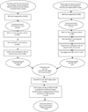
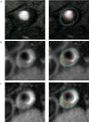
Methods: Three-hundred-three participants with PAD underwent MRI of the proximal SFA using a 1.5 T S platform. Twelve contiguous 2.5 mm cross-sectional images were obtained.
Results: LRNC was present in 68 (22.4%) participants. Only one had intra-plaque hemorrhage. After adjusting for age and sex, smoking prevalence was higher among adults with LRNC than among those without LRNC (35.9% vs. 21.4%, p = 0.02). Among participants with vs. without LRNC there were no differences in mean percent lumen area (31% vs. 33%, p = 0.42), normalized mean wall area (0.71 vs. 0.70, p = 0.67) or maximum wall area (0.96 vs. 0.92, p = 0.54) in the SFA. Among participants with LRNC, cross-sectional images containing LRNC had a smaller percent lumen area (33% ± 1% vs. 39% ± 1%, p < 0.001), greater normalized mean wall thickness (0.25 ± 0.01 vs. 0.22 ± 0.01, p < 0.001), and greater normalized maximum wall thickness (0.41 ± 0.01 vs. 0.31 ± 0.01, p < 0.001), compared to cross-sectional images without LRNC.
Conclusions: Fewer than 25% of adults with PAD had high-risk plaque in the proximal SFA using MRI. Smoking was the only clinical characteristic associated with presence of LRNC. Further study is needed to determine the prognostic significance of LRNC in the SFA.
Clinical trial registration-url: http://www.clinicaltrials.gov. Unique identifier: NCT00520312.
Keywords: Atherosclerosis; Magnetic resonance imaging; Peripheral vascular disease; Plaque.
Copyright © 2014 Elsevier Ireland Ltd. All rights reserved.
Figures


References
-
- Virmani R, Burke AP, Farb A, Kolodgie FD. Pathology of the vulnerable plaque. J Am Coll Cardiol. 2006;47(8 Suppl):C13–C18. - PubMed
-
- Naghavi M, Libby P, Falk E, Casscells SW, Litovsky S, Rumberger J, et al. From vulnerable plaque to vulnerable patient: a call for new definitions and risk assessment strategies: Part I. Circulation. 2003;108(14):1664–1672. - PubMed
-
- Takaya N, Yuan C, Chu B, Saam T, Underhill H, Cai J, et al. Association between carotid plaque characteristics and subsequent ischemic cerebrovascular events: a prospective assessment with MRI--initial results. Stroke. 2006;37(3):818–823. - PubMed
-
- Virmani R, Kolodgie FD, Burke AP, Finn AV, Gold HK, Tulenko TN, et al. Atherosclerotic plaque progression and vulnerability to rupture: angiogenesis as a source of intraplaque hemorrhage. Arterioscler Thromb Vasc Biol. 2005;25(10):2054–2061. - PubMed
Publication types
MeSH terms
Substances
Associated data
Grants and funding
LinkOut - more resources
Full Text Sources
Other Literature Sources
Medical

