. 2014 Nov:468-470:421-443.
doi: 10.1016/j.virol.2014.08.024.

Understanding the enormous diversity of bacteriophages: the tailed phages that infect the bacterial family Enterobacteriaceae

Affiliations

Understanding the enormous diversity of bacteriophages: the tailed phages that infect the bacterial family Enterobacteriaceae

Julianne H Grose et al. Virology. 2014 Nov.

Abstract

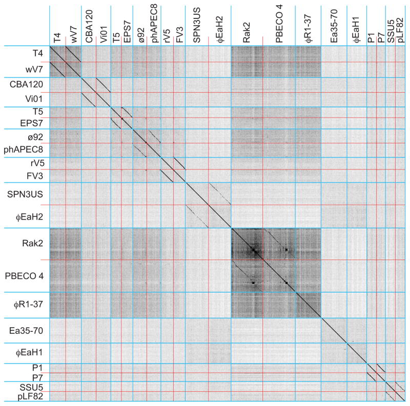
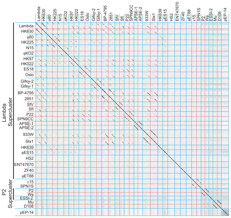
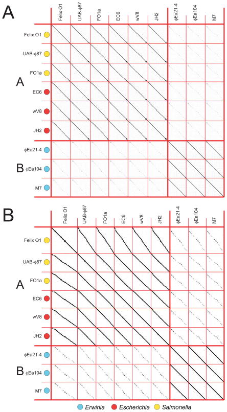
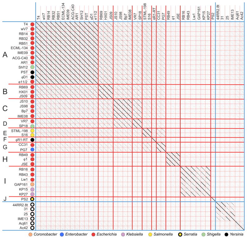
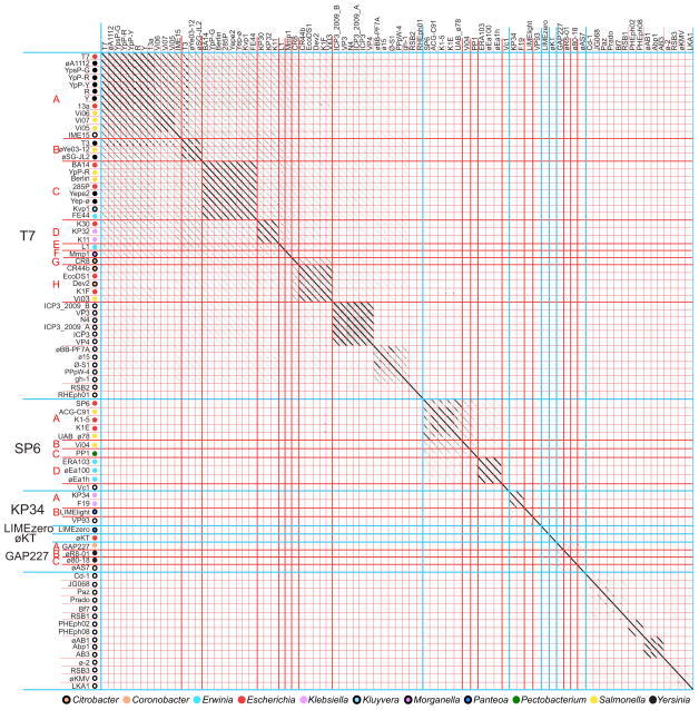
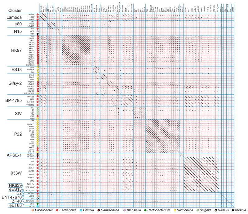
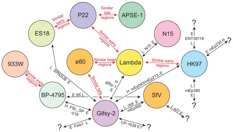
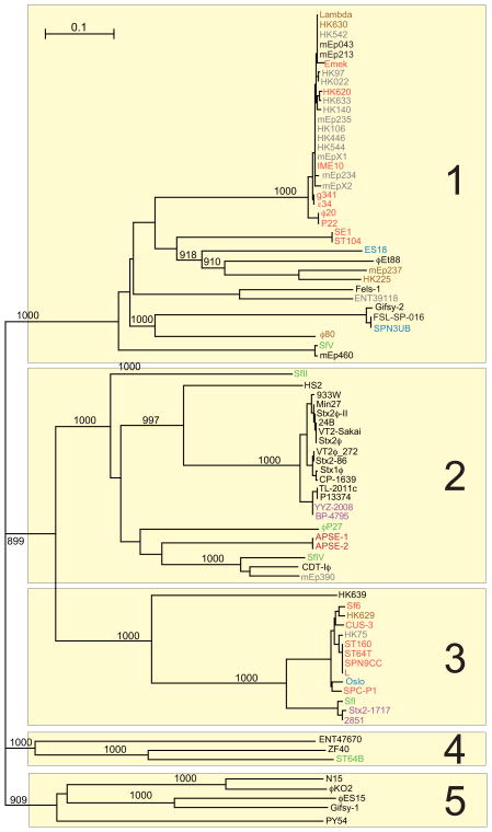
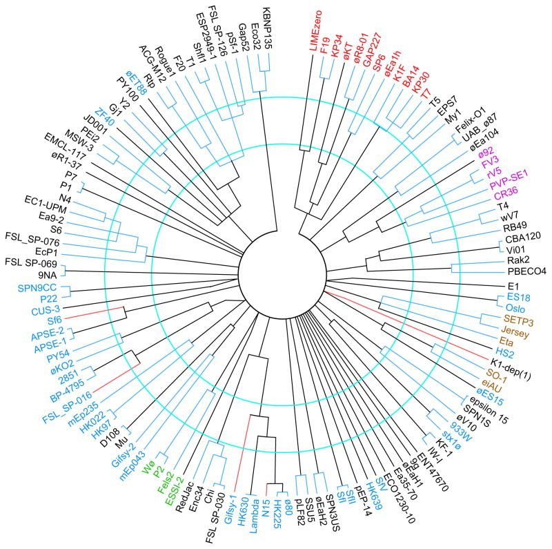
Bacteriophages are the predominant biological entity on the planet. The recent explosion of sequence information has made estimates of their diversity possible. We describe the genomic comparison of 337 fully sequenced tailed phages isolated on 18 genera and 31 species of bacteria in the Enterobacteriaceae. These phages were largely unambiguously grouped into 56 diverse clusters (32 lytic and 24 temperate) that have syntenic similarity over >50% of the genomes within each cluster, but substantially less sequence similarity between clusters. Most clusters naturally break into sets of more closely related subclusters, 78% of which are correlated with their host genera. The largest groups of related phages are superclusters united by genome synteny to lambda (81 phages) and T7 (51 phages). This study forms a robust framework for understanding diversity and evolutionary relationships of existing tailed phages, for relating newly discovered phages and for determining host/phage relationships.


Figures

Figure 1
Figure 1. Dot plot analysis of 21 Enterobacteriaceae tailed phages with genomes larger than 90 kbp
Blue lines separate phage clusters and red lines separate genomes within the clusters. Dot plot was produced using Gepard (Krumsiek et al., 2007) at a word size setting of 10.
Figure 2
Figure 2. Dot plot analysis of 37 lytic Enterobacteriaceae tailed phages with genomes smaller than 90 kbp
Blue lines separate clusters and red lines separate genomes within the clusters. Dot plots were produced using Gepard (Krumsiek et al., 2007) at a word size of 10.
Figure 3
Figure 3. Dot plot analysis of temperate Enterobacteriaceae tailed phages with genomes smaller than 90 kbp
Blue lines separate clusters and red lines separate genomes within the clusters. Dot plots were produced using Gepard (Krumsiek et al., 2007) at a word size of 10.
Figure 4
Figure 4. Whole genome nucleotide (A) and gene product (B) dot plots of known Felix-O1-like phages reveals two subclusters
Phage genomes and subclusters are separated by thin and thick red lines, respectively. Subcluster names are indicated on the left by red letters, and a key for the phage hosts is shown below. Dot plots were produced using Gepard (Krumsiek et al., 2007) at a word size of 11 for the genome dot plot and 6 for the gene product dot plot. Amino acid sequences for the gene product dot plot consisted of tandem sequences of all the annotated predicted encoded proteins aligned in the order their genes occur in the genome. The reported circular genome sequence assembly of Felix-O1 (Accession No. AF320576) was linearized at bp 16830 in order to align it with the known ends of cluster member phage M7’s linear genome (Born et al., 2011). Other cluster member genomes were oriented to align with these genomes.
Figure 5
Figure 5. Whole genome nucleotide dot plot of known T4-like phages reveals ten subclusters
The dot plot is presented as described in the legend to figure 4. Genomes are aligned with the T4 sequence reported in accession No. AF158101 and compared with a Gepard word size of 12. Open circles indicate phages that infect hosts outside the Enterobacteriaceae family (see text).
Figure 6
Figure 6. Whole genome nucleotide dot plot of 84 T7 supercluster phages reveals six clusters
The dot plot is presented as described in the legend to figure 4. Genomes are aligned with the T7 sequence reported in accession No. V01146 and compared with a Gepard word size of 12. Open circles indicate phages that infect hosts outside the Enterobacteriaceae family (see text).
Figure 7
Figure 7. Whole genome nucleotide dot plot of 81 lambda supercluster phages reveals 17 clusters
The dot plot is presented as described in the legend to figure 4 (Gepard word size of 12). Phage genomes are separated by red lines and clusters by blue lines. Hosts from which the phages were isolated are indicated on the vertical axis. The genomes shown are all oriented according to the standard phage lambda virion chromosome map with the head genes on the left and lysis on the right.
Figure 8
Figure 8. Relationships among the non-singleton clusters of the lambda supercluster reveals inter-cluster hybridization events
Circles represent the non-singleton clusters within the lambda supercluster. Red arrows indicated cluster pairs in which most members have significant (usually mosaically related and always divergent) homologies in the indicated regions. Black arrows indicate examples of regions that were rather recently exchanged between these clusters that generated apparently ‘hybrid’ phages (see text). On each black arrow a particular ‘hybrid’ phage(s) is indicated with its genome sections that are closely related to other members of the two clusters connected by the arrow; these sections are labeled L (late region), H (head region) and E (early region), and the source of each region is indicated by the cluster circle nearest to the indicated region (e.g., phage SPN3UB has an early region similar to phages in the gifsy-2-like cluster and a late region similar to phages in the ES18-like cluster. Black question marks (?) indicate that the source of the indicated region is currently unknown.
Figure 9
Figure 9. A neighbor-joining tree of the lambda supercluster Q proteins
A CLUSTAL tree of the different lambda supercluster Q protein types shows bootstrap values (out of 1000 trials; values less than 900 and those on very short branches are not shown). The five major Q protein sequence types are numbered on the right. Four of the five major types have weak but recognizable sequence similarity to phage lambda Q protein, but type 4 does not. None of the four ‘type 4’ phages carry a gene with Q homology, so the type 4 putative late operon activator proteins were chosen because they are each encoded by a gene that lie between recognizable nin region genes and recognizable terminase genes (like the other true Q homologues).
Figure 10
Figure 10. Neighbor-joining tree of representative Enterobacteriaceae tailed phage major capsid proteins
A representative set of MCP types was aligned and a tree constructed by ClustalX; representative MCPs from all clusters and many subclusters are shown. The phages that encode the MCPs are indicated outside of the tree. The members of the following superclusters are indicated by label color as follows: T7, red; lambda, blue; rV5, magenta; SETP3, brown, P2, green. Nodes with bootstrap values less than 985 (out of 1000 trials) were collapsed; all the nodes shown thus have >985 bootstrap support, except slanted lines near the center which denote convincing similarities in the 25–30% identity range in pairwise alignments that fail to show this in CLUSTAL multiple alignments (probably because an MCP in another branch has weak similarity). The red branch lines indicate MCPs that are very different from others in their cluster and were most likely obtained by horizontal transfer (see text), and contiguous blue lines connect members of the same cluster. The inner and outer blue circles indicate the locations of approximately 50% and 75% AA sequence identity. An apparent frameshift in the phage Stx1ø MCP gene was ‘fixed’ for purposes of comparison.

References

    1. Adriaenssens EM, Ceyssens PJ, Dunon V, Ackermann HW, Van Vaerenbergh J, Maes M, De Proft M, Lavigne R. Bacteriophages LIMElight and LIMEzero of Pantoea agglomerans, belonging to the “phiKMV-like viruses”. Appl Environ Microbiol. 2011;77:3443–50. - PMC - PubMed
    1. Altschul SF, Gish W, Miller W, Myers EW, Lipman DJ. Basic local alignment search tool. J Mol Biol. 1990;215:403–10. - PubMed
    1. Anderson TF. The activity of a bacteriostatic substance in the reaction between bacterial virus and host. Science. 1945;101:565–6. - PubMed
    1. Andres D, Roske Y, Doering C, Heinemann U, Seckler R, Barbirz S. Tail morphology controls DNA release in two Salmonella phages with one lipopolysaccharide receptor recognition system. Mol Microbiol. 2012;83:1244–53. - PubMed
    1. Baker J, Limberger R, Schneider SJ, Campbell A. Recombination and modular exchange in the genesis of new lambdoid phages. New Biol. 1991;3:297–308. - PubMed

Publication types

Substances