. 2014 Oct;32(10):1026-35.
doi: 10.1038/nbt.3002. Epub 2014 Sep 21.

Generation of the epicardial lineage from human pluripotent stem cells

Affiliations

Generation of the epicardial lineage from human pluripotent stem cells

Alec D Witty et al. Nat Biotechnol. 2014 Oct.

Abstract

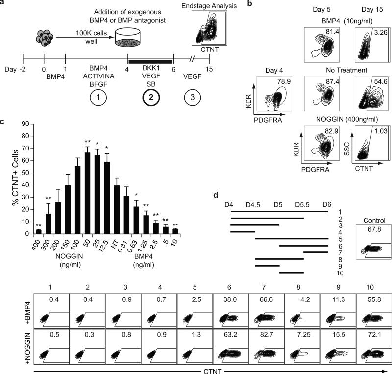
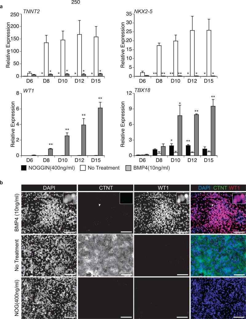
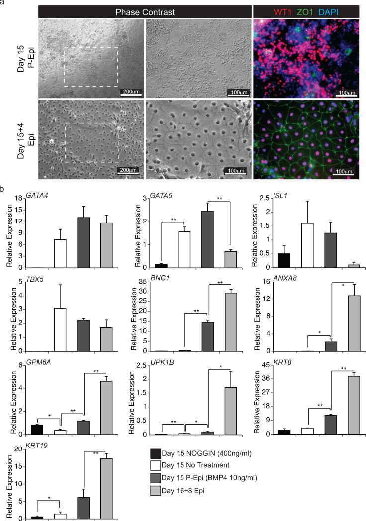
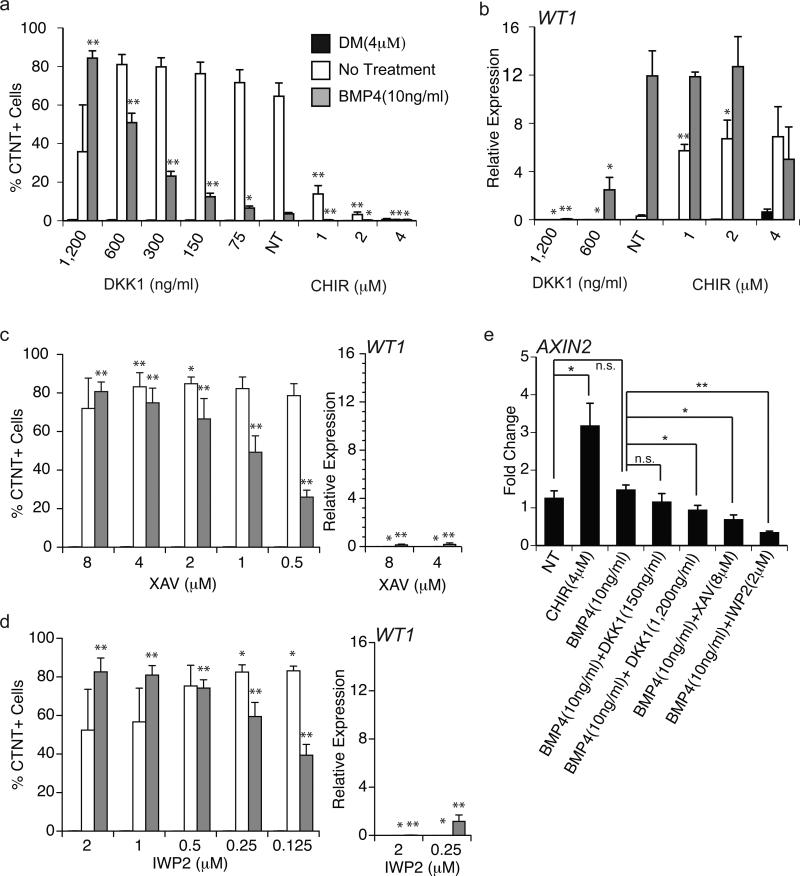
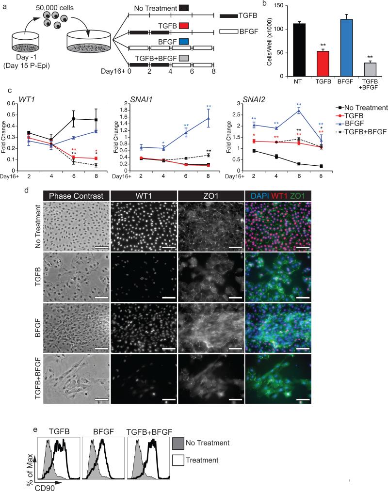
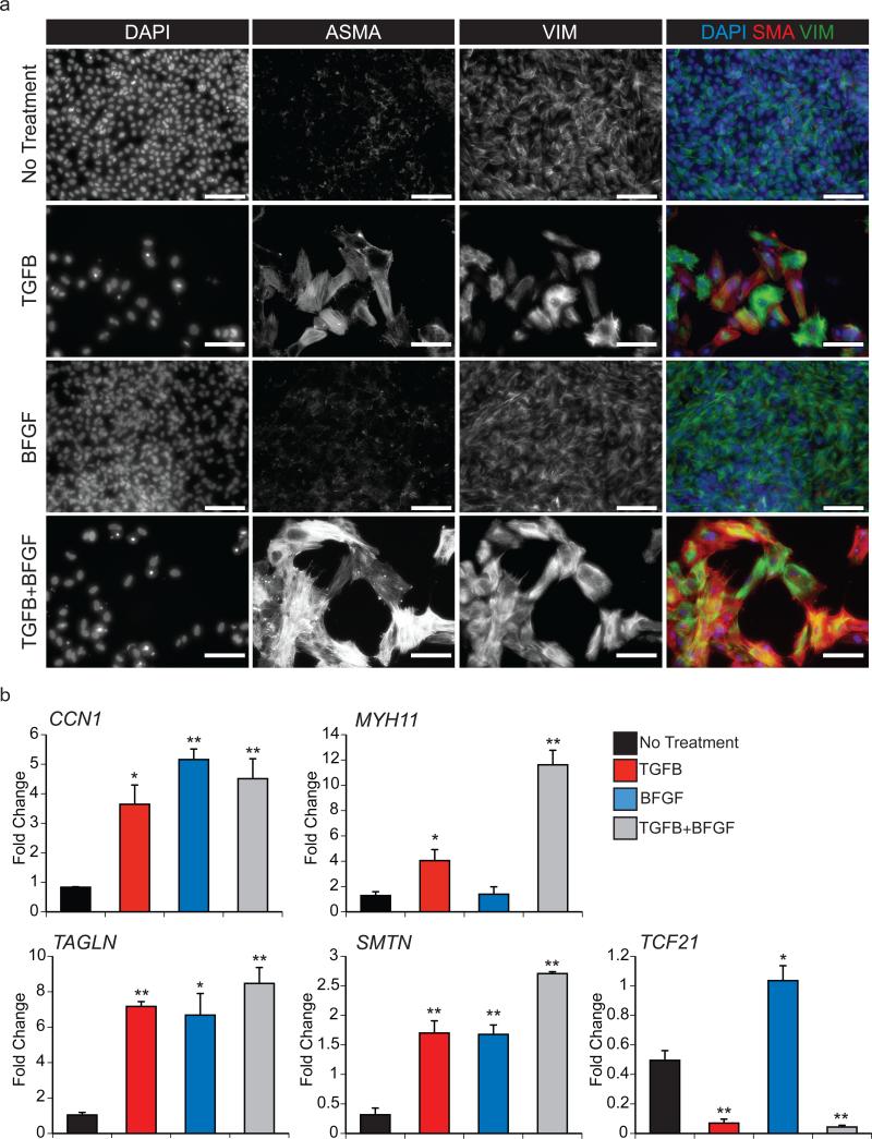
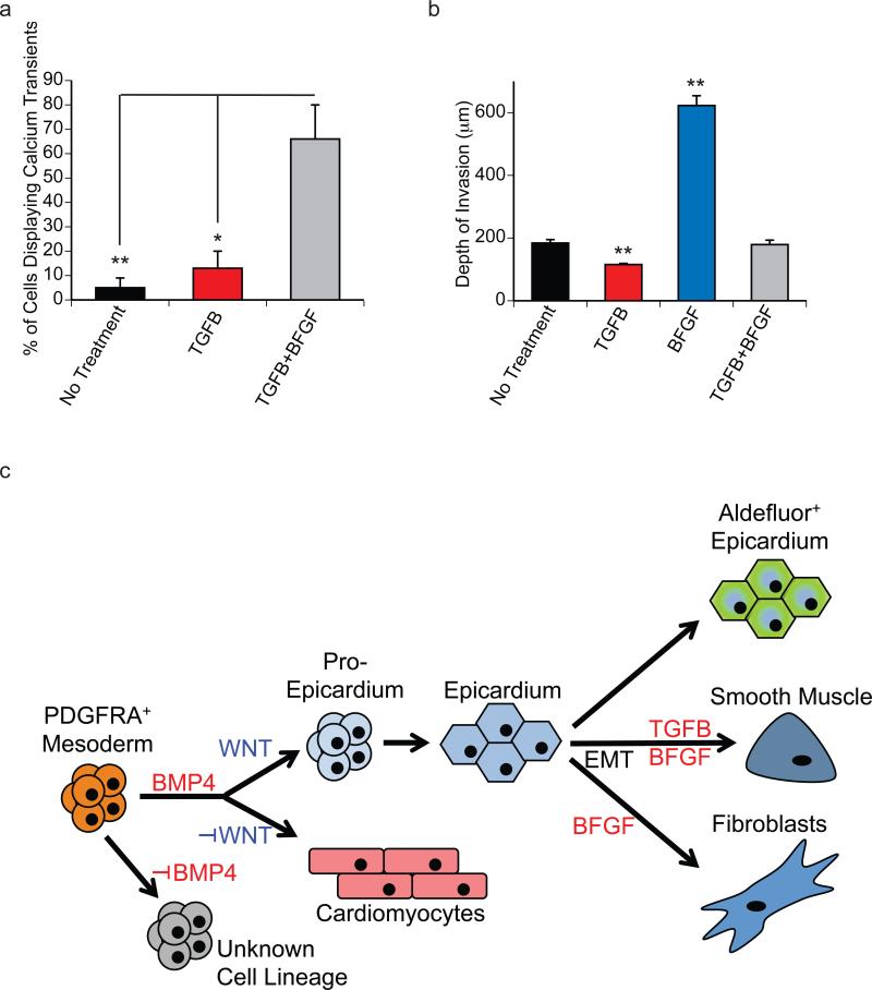
The epicardium supports cardiomyocyte proliferation early in development and provides fibroblasts and vascular smooth muscle cells to the developing heart. The epicardium has been shown to play an important role during tissue remodeling after cardiac injury, making access to this cell lineage necessary for the study of regenerative medicine. Here we describe the generation of epicardial lineage cells from human pluripotent stem cells by stage-specific activation of the BMP and WNT signaling pathways. These cells display morphological characteristics and express markers of the epicardial lineage, including the transcription factors WT1 and TBX18 and the retinoic acid-producing enzyme ALDH1A2. When induced to undergo epithelial-to-mesenchymal transition, the cells give rise to populations that display characteristics of the fibroblast and vascular smooth muscle lineages. These findings identify BMP and WNT as key regulators of the epicardial lineage in vitro and provide a model for investigating epicardial function in human development and disease.


Figures

Figure 1
Figure 1. BMP specifies the differentiation of cardiomyocytes from hESC-derived mesoderm
(a) Scheme of the protocol used to differentiate hESCs towards the CM lineage highlighting the three main stages of development: 1) mesoderm induction, 2) cardiovascular specification and 3) maturation. Cells from ACTIVINA/BMP4-induced D4 EBs are plated as a monolayer on gelatin coated wells. The BMP pathway was manipulated for a 48-hour period (D4-D6) in the presence of VEGF (5 ng/ml), SB (5.4 μM) and DKK1 (150ng/ml). Following specification, the cultures were maintained in VEGF (5ng/ml) for 9 days and then analyzed for the presence of CTNT+ cells by flow cytometry. (b) Flow cytometric analyses of the frequency of KDR+PDGFRA+ cells in D4 and D5 mesoderm populations and of CTNT+ cells in D15 populations generated from D4 mesoderm treated with: no factors (no treatment), BMP4 (10ng/ml) or NOGGIN (400ng/ml). (c) Flow cytometric analyses of the frequency of CTNT+ cells in D15 populations generated from D4 mesoderm treated with the indicated amounts of BMP4 or NOGGIN. NT = no treatment. Bars represent standard error of the mean of the values from three independent experiments (N=3); *P≤0.05, **P≤0.01 compared to NT as analyzed by Student's T-test. (d) Scheme indicating the time intervals of BMP4 or NOGGIN treatment between D4 and D6. Ten different intervals were evaluated. On D15 the resulting populations were analyzed for the presence of CTNT+ cells by flow cytometry. Control cultures were not treated with either the agonist or antagonist during the 48-hour period.
Figure 2
Figure 2. BMP4 treatment specifies WT1+ cells
(a) qRT-PCR-based expression analyses of the cardiac genes TNNT2 and NKX2-5 and the epicardial genes WT1 and TBX18 on the indicated days (D) of culture from cell populations generated by treatment with BMP4 (10ng/ml), NOGGIN (400ng/ml) or no additional factors. Values shown are relative to the housekeeping gene TBP. Error bars represent standard error of the mean from the values of three independent experiments (N=3); *P≤0.05, **P≤0.01 compared to non-treated cells as analyzed by Student's T-test. (b) Fluorescent immunostaining for the presence of CTNT and WT1 protein in D15 populations treated with no additional factors, BMP4 (10ng/ml) or NOGGIN (400ng/ml). DAPI staining shows cell nuclei. Insert is high magnification showing nuclear staining of WT1 where the region of interest is indicated by the white arrowhead. Scale bars represent 100μm.
Figure 3
Figure 3. WT1+ cells generate epithelial sheets following passage
(a) Phase contrast microscopy and fluorescent immunostaining showing morphology of D15 P-Epi and D15+4 Epi populations and the presence of ZO1 (green) and WT1 (red) protein. DAPI (blue) staining shows cell nuclei. Scale bar are as indicated. White dashed box in first panel depicts region of view at higher magnification in subsequent panels. (b) qRT-PCR-based expression analyses of the indicated genes in D15 populations generated from untreated cells (control) or cells treated with NOGGIN (400ng/ml), or BMP4 (P-Epi, 10ng/ml) as well as in D16+8 Epi cells derived from the BMP-4 treated PEpi cells. Values shown are relative to the housekeeping gene TBP. Error bars represent standard error of the mean of the values from three independent experiments (N=3); *P≤0.05, **P≤0.01 compared to non-treated cells or D15 P-Epi cells as analyzed by Student's T-test.
Figure 4
Figure 4. BMP and WNT signaling modulate cardiomyocyte and pre-epicardium specification
(a) Flow cytometric analyses of the frequency of CTNT+ cells in D15 cultures generated from D4 mesoderm treated with BMP4 (10ng/ml), DM (4μM) or no treatment (control) in combination with the indicated amounts of DKK1 or CHIR. Bars represent standard error of the mean of the values from three independent experiments (N=3); *P≤0.05, **P≤0.01 when compared to the ‘no WNT treatment’ (NT) control in the context of the indicated manipulation of the BMP pathway as analyzed by Student's T-test. (b) qRT-PCR-based analyses of WT1 expression in D15 cultures generated from D4 mesoderm treated BMP4 (10ng/ml), DM (4μM) or no additional factors (control) in combination with the indicated amounts of DKK1 or CHIR. Values shown are relative to the housekeeping gene TBP. Bars represent standard error of the mean of the values from three independent experiments (N=3); *P≤0.05, **P≤0.01 when compared to the ‘no WNT treatment’ (NT) control in the context of the indicated manipulation of the BMP pathway as analyzed by Student's T-test. (c) The frequency of CTNT+ cells determined by flow cytometry and the expression levels of WT1 evaluated by qRT-PCR analyses in D15 cultures generated from D4 mesoderm treated BMP4 (10ng/ml), DM (4μM) or no additional factors (control) in combination with the indicated amounts of XAV. Bars represent standard error of the mean of the values from three independent experiments (N=3) *P≤0.05, **P≤0.01 when compared to no WNT treatment (see Figure 4a and 4b) in context of specific BMP treatment as analyzed by Student's T-test. (d) The frequency of CTNT+ cells determined by flow cytometry and the expression levels of WT1 evaluated by qRT-PCR analyses in D15 cultures generated from D4 mesoderm treated with BMP4 (10ng/ml), DM (4μM) or no additional factors (control) in combination with the indicated amounts of IWP2. Bars represent standard error of the mean of the values from three independent experiments (N=3) *P≤0.05, **P≤0.01 when compared to no WNT treatment (see Figure 4a and 4b) in context of specific BMP treatment as analyzed by Student's T-test. (e) qRT-PCR-based expression analyses of AXIN2 in D5 populations, 24 hours following the indicated treatments. Values shown are fold change relative to D4 pre-treated cultures. Bars represent standard error of the mean of the values from three independent experiments (N=3); *P≤0.05, **P≤0.01 as analyzed by Student's T-test.
Figure 5
Figure 5. Epicardial cells undergo EMT in response to TGFB1 and BFGF treatment
(a) Scheme of the protocol used for the EMT analyses. D15 P-Epi cultures were passaged, allowed to settle for 1 day and then subjected to the following treatments: TGFB1 (5ng/ml) for 4 days followed by no treatment (TGFB, red), BFGF (10ng/ml) for 8 days (BFGF, blue), TGFB1 (5ng/ml) for 4 days followed by BFGF (10ng/ml) for 4 day (TGFB+BFGF, green) or no additional factors throughout the 8 day culture (control, black). (b) Total cell numbers per well 8 days following EMT initiation with the indicated treatments. Cultures were initiated with 5x10⁴ cells. Bars represent standard error of the mean of the values from three independent experiments (N=3); **P≤0.01 compared to the non-treated control as analyzed by Student's T-test. (c) qRT-PCR-based expression analyses of the epicardial gene WT1 and the EMT-regulated genes SNAI1 and SNAI2 on days (D) 2, 4, 6 and 8 following EMT initiation with the indicated factors. Values are expressed as fold change relative to experiment-matched pre-passaged D15 P-Epi cultures. Bars represent standard error of the mean of the values from three independent experiments (N=3); *P≤0.05, **P≤0.01 compared to non-treated control as analyzed by Student's T-test. (d) Phase contrast and fluorescent immunostaining showing cell morphology and the presence of ZO1 and WT1 proteins in Epi derived cultures 8 days following EMT initiation with the indicated factors. DAPI staining shows cell nuclei. Scale bar represents 100μm. (e) Flow cytometric analyses for the expression of CD90 in the cultures 8 days following EMT initiation with the indicated factors.
Figure 6
Figure 6. Epicardium-derived cells display characteristics of fibroblasts and vascular smooth muscle cells
(a) Fluorescent immunostaining for ASMA and VIM proteins in the cultures 8 days following EMT initiation with the indicated treatments. DAPI staining shows cell nuclei. Scale bar represents 100μm. (b) qRT-PCR-based expression analyses of the smooth muscle genes CCN1, MYH11, TAGLN and SMTN and the epicardial/cardiac fibroblast gene TCF21 in cultures 8 days following EMT initiation with the indicated factors. Values are expressed as fold change relative to experiment-matched pre-passaged D15 P-Epi cells. Bars represent standard error of the mean of the values from three independent experiments (N=3); *P≤0.05, **P≤0.01 compared to the non-treated cells as analyzed by Student's T-test.
Figure 7
Figure 7. Functional assessment of EPDCs
(a) The total proportion of actively cycling cells in EMT-induced cultures was measured for the indicated treatments. Treatment with TGFB+BFGF generated populations with the largest proportion of actively cycling cells in response to agonists. Bars represent standard error of the mean; N=3/group; *P<0.05, **P<0.01 compared by one-way ANOVA with Tukey post hoc test. (e) Maximum matrigel invasion depth on D8 following EMT initiation. Bars represent standard error of the mean of the values from three independent experiments (N=3); **P≤0.01 compared to non-treated controls as analyzed by Student's T-test. (e) Model highlighting the specification of PDGFRA+ mesoderm to the cardiomyocyte and pro-epicardial lineages by BMP4 and canonical WNT signaling. When passaged in the absence of factors, the pro-epicardial cells form epithelial sheets resembling the epicardium that can be identified through the Aldefluor assay. In the presence of TGFB+BFGF or BFGF, the passaged cells undergo EMT and give rise to smooth muscle-like and fibroblast-like cells.

Comment in

References

    1. DeRuiter MC, Poelmann RE, VanderPlas-de Vries I, Mentink MM, Gittenberger-de Groot AC. The development of the myocardium and endocardium in mouse embryos. Fusion of two heart tubes? Anatomy and embryology. 1992;185:461–473. - PubMed
    1. Limana F, Capogrossi MC, Germani A. The epicardium in cardiac repair: from the stem cell view. Pharmacology & therapeutics. 2011;129:82–96. - PubMed
    1. Komiyama M, Ito K, Shimada Y. Origin and development of the epicardium in the mouse embryo. Anatomy and embryology. 1987;176:183–189. - PubMed
    1. Austin AF, Compton LA, Love JD, Brown CB, Barnett JV. Primary and immortalized mouse epicardial cells undergo differentiation in response to TGFbeta. Dev Dyn. 2008;237:366–376. - PubMed
    1. Bax NA, et al. In vitro epithelial-to-mesenchymal transformation in human adult epicardial cells is regulated by TGFbeta-signaling and WT1. Basic research in cardiology. 2011;106:829–847. - PMC - PubMed

Publication types

MeSH terms

Substances