. 2014 Oct 3;452(4):1040-5.
doi: 10.1016/j.bbrc.2014.09.037. Epub 2014 Sep 18.

CD4 aptamer-RORγt shRNA chimera inhibits IL-17 synthesis by human CD4(+) T cells

Affiliations

CD4 aptamer-RORγt shRNA chimera inhibits IL-17 synthesis by human CD4(+) T cells

Pingfang Song et al. Biochem Biophys Res Commun. .

Abstract

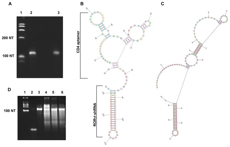
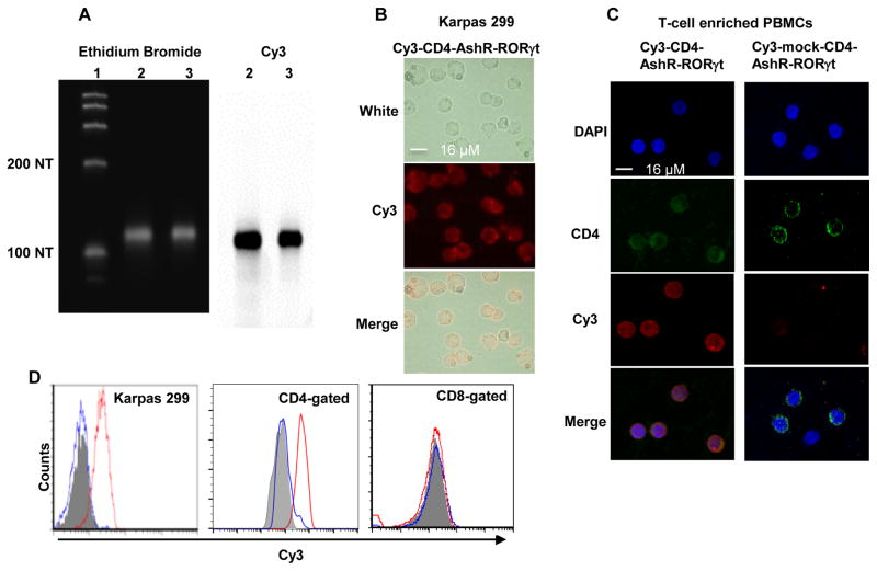
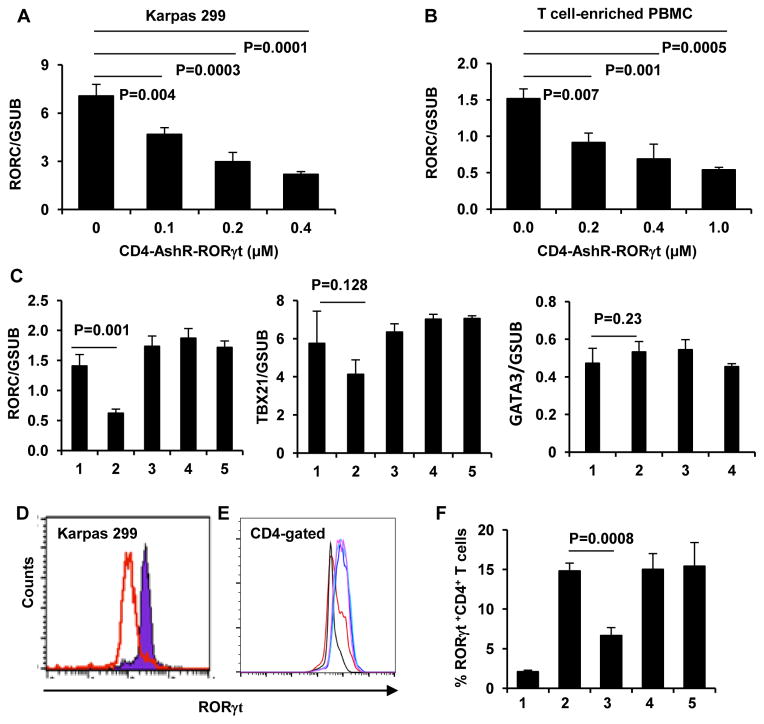
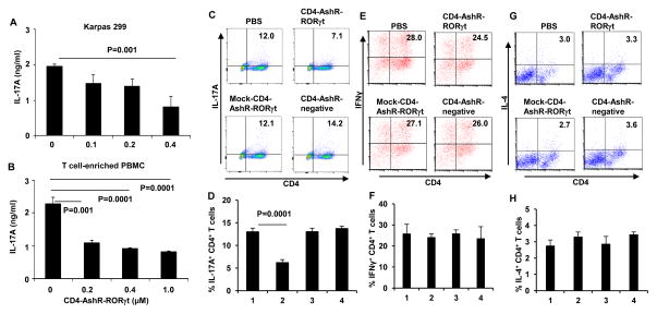
Cell type specific delivery of RNAi to T cells has remained to be a challenge. Here we describe an aptamer mediated delivery of shRNA to CD4(+) T cells targeting RORγt to suppress Th17 cells. A cDNA encoding CD4 aptamer and RORγt shRNA was constructed and the chimeric CD4 aptamer-RORγt shRNA (CD4-AshR-RORγt) was generated using in vitro T7 RNA transcription. 2'-F-dCTP and 2'-F-dUTP were incorporated into CD4-AshR-RORγt for RNase resistance. CD4-AshR-RORγt was specifically uptaken by CD4(+) Karpas 299 cells and primary human CD4(+) T cells. The RORγt shRNA moiety of CD4-AshR-RORγt chimera was cleaved and released by Dicer. Furthermore, CD4-AshR-RORγt suppressed RORγt gene expression in Karpas 299 cells and CD4(+) T cells and consequently inhibited Th17 cell differentiation and IL-17 production. These results demonstrate that aptamer-facilitated cell specific delivery of shRNA represents a novel approach for efficient RNAi delivery and is potentially to be developed for therapeutics targeting specific T cells subtypes.

Keywords: Aptamer–shRNA; RORγt; Th17.


Conflict of interest statement

Disclosures: The authors have no financial conflicts of interest.

Figures

Figure 1
Figure 1. CD4-AshR-RORγt chimera
(A) Chimera in vitro transcribed by T7 RNA polymerase was analyzed by denatured PAGE and ethidium bromide staining. Lane 1, ssRNA ladder; Lane 2, CD4-AshR-RORγt chimera; Lane 3, mock-CD4-AshR-RORγt chimera. (B and C) Predicted secondary structure of CD4-AshR-RORγt chimera (B) and mock-CD4-AshR-RORγt chimera (C). The region of the CD4 aptamer (clone 9 [26]) responsible for binding to CD4 is outlined. The shRNA portion of the chimera consists of targeted RORγt siRNA with 2 overhang nucleotides at its 3′end and a 7 nucleotide loop. (D) Cleavage analysis of synthesized chimeras by Dicer. Lane 1, ssRNA ladder; Lane 2, antisense siRNA to RORγt; Lane 3, intact CD4-AshR-RORγt chimera; Lane 4–6, chimeras were digested with Dicer: Lane 4, Mock CD4-AshR-RORγt chimera; Lane 5, CD4-AshR-RORγt chimera; Lane 6, CD4-AshR-scrambled control chimera (representative of two experiments).
Figure 2
Figure 2. CD4-AshR-RORγt chimera efficiently entered CD4+ human T cells
(A) CD4- and mock-CD4-AshR-RORγt chimeras were labeled by incorporating Cy3-CTP during in vitro transcription. Cy3 scanning (right panel) showed strong Cy3-signaling bands that were at an appropriate size of transcripts shown in ethidium bromide imaging (left panel). Lane 1, ssRNA ladder; Lane 2, CD4-AshR-RORγt chimera; Lane 3, mock-CD4-AshR-RORγt chimera. (B and C) Uptake of Cy3-labeld CD4-AshR-RORγt chimera by CD4+ human T cell line Karpas 299 cells and CD4+ T cells in PBMCs. (D) Flow cytometric analysis showed that Cy3-labeled CD4-AshR-RORγt chimera was significantly internalized in CD4+ Karpas 299 cells and CD4+ T cells but not in CD8+ T cells. There is no uptake of Cy3-labeled mock-CD4-AshR-RORγt chimera by Karpas 299 cells or T-cell enriched PBMCs. Gray: PBS; Red line: Cy3-labeled CD4-AshR-RORγt chimera; Blue line: Cy3-labeled mock-CD4-AshR-RORγt chimera (representative of 2–5 experiments).
Figure 3
Figure 3. Specific silencing of RORγt in human CD4+ T cells by CD4-AshR-RORγt chimera
Karpas 299 cells and PBMCs were treated as described in methods. (A through C) Quantitative real-time PCR assay for RORγt gene expression. RORγt gene expression was significantly reduced by CD4-AshR-RORγt chimera in a concentration-dependent manner in Kapas 299 cells (A) and T-cell enriched PBMCs (B). Mock-CD4-AshR-RORγt chimera, CD4-AshR-scrambled control or CD4-AshR-CCR5 chimera had no effect on RORγt gene expression in T-cell enriched PBMCs (C). Additionally, all the chimeras lacked a significant inhibition on TBX21or GATA3 in T-cell enriched PBMCs (C). (Data are presented as mean ± SD of three experiments). 1, PBS; 2, CD4-AshR-RORγt chimera; 3, mock-CD4-AshR-RORγt chimera; 4, CD4-AshR-scrambled control chimera; 5, CD4-AshR-CCR5 chimera. (D and E) RORγt protein expression was analysed by flow cytometry. (D) Karpas 299 cells were stimulated with PMA 50 ng/ml for 24 h. Red line, CD4-AshR-RORγt chimera; Blue line, Mock-CD4-AshR-RORγt chimera (representative of three experiments). (E) PBMCs were stimulated with anti-CD3/CD28 and LPS for 48 h. RORγt expression was reduced by CD4-AshR-RORγt chimera (red line), but not by mock-CD4-AshR-RORγt chimeras (blue line) or CD4-AshR-scrambled control chimera (purple line). Black line, PBMCs without stimulation; green line, PBMCs with stimulation but without chimeras (representative of three experiments). (F) The percentage of RORγt+ cells in stimulated T-cell enriched PBMCs was reduced by CD4-AshR-RORγt chimeras, but not by mock-CD4-AshR-RORγt chimera or CD4-AshR-scrambled control chimera. 1, PBMCs without stimulation; 2, PBMCs with stimulation but without chimeras; 3, Stimulated PBMCs were treated with CD4-AshR-RORγt chimeras; 4, Stimulated PBMCs were treated with mock-CD4-AshR-RORγt chimera; 5, Stimulated PBMCs were treated with CD4-AshR-scrambled control chimera (Data are presented as mean ± SD of three experiments).
Figure 4
Figure 4. Reduction of IL-17A synthesis in CD4+ T cells by CD4-AshR-RORγt chimera
Karpas 299 cells were stimulated with PMA and T-cell enriched PBMCs were stimulated with anti-CD3/CD28 and cytokines with anti-cytokine antibodies or LPS for Th1, Th2 or Th17 polarization respectively. (A and B) IL-17A in the supernatant was measured by ELISA. IL-17A production was significantly decreased by CD4-AshR-RORγt chimera in a concentration-dependent fashion (Data are presented as mean ± SD of three experiments). (C and D) Reduction of IL-17A-producing CD4+ T cells by CD4-AshR-RORγt chimera, but not by mock- CD4-AshR-RORγt or CD4-AshR-scrambled control chimeras. (E, F, G and H) CD4-AshR-RORγt chimera had no significant impacts on IFN-γ- and IL-4-producing CD4+ T cells. 1, PBS; 2; CD4-AshR-RORγt chimera; 3, mock-CD4-AshR-RORγt chimera; 4, CD4-AshR-scrambled control chimera (Data are presented as mean ± SD of three experiments).

References

    1. Freeley M, Long A. Advances in siRNA delivery to T-cells: potential clinical applications for inflammatory disease, cancer and infection. Biochem J. 2013;455:133–147. - PubMed
    1. Gehl J. Electroporation: theory and methods, perspectives for drug delivery, gene therapy and research. Acta Physiologica Scandinavica. 2003;177:437–447. - PubMed
    1. Lai W, Chang CH, Farber DL. Gene transfection and expression in resting and activated murine CD4 T cell subsets. J Immunol Methods. 2003;282:93–102. - PubMed
    1. Gomez-Valades AG, Llamas M, Blanch S, Perales JC, Roman J, Gomez-Casajus L, Mascaro C. Specific Jak3 Downregulation in Lymphocytes Impairs gammac Cytokine Signal Transduction and Alleviates Antigen-driven Inflammation In Vivo. Mol Ther Nucleic Acids. 2012;1:e42. - PMC - PubMed
    1. Rangachari M, Zhu C, Sakuishi K, Xiao S, Karman J, Chen A, Angin M, Wakeham A, Greenfield EA, Sobel RA, Okada H, McKinnon PJ, Mak TW, Addo MM, Anderson AC, Kuchroo VK. Bat3 promotes T cell responses and autoimmunity by repressing Tim-3-mediated cell death and exhaustion. Nat Med. 2012;18:1394–1400. - PMC - PubMed

Publication types

MeSH terms