Review
. 2015 Feb;165(2):255-69.
doi: 10.1016/j.trsl.2014.08.005. Epub 2014 Sep 1.

The meninges: new therapeutic targets for multiple sclerosis

Affiliations
Review

The meninges: new therapeutic targets for multiple sclerosis

Abigail E Russi et al. Transl Res. 2015 Feb.

Abstract

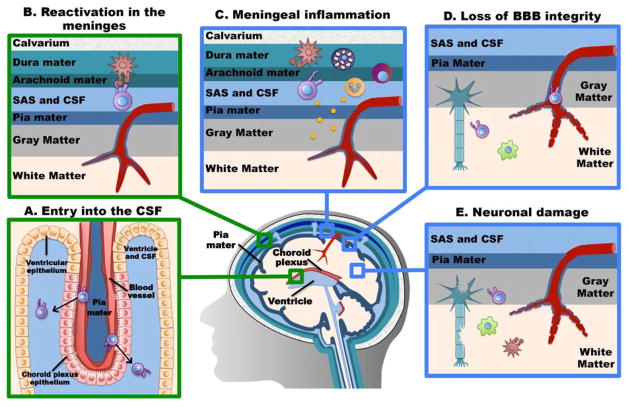
The central nervous system (CNS) largely comprises nonregenerating cells, including neurons and myelin-producing oligodendrocytes, which are particularly vulnerable to immune cell-mediated damage. To protect the CNS, mechanisms exist that normally restrict the transit of peripheral immune cells into the brain and spinal cord, conferring an "immune-specialized" status. Thus, there has been a long-standing debate as to how these restrictions are overcome in several inflammatory diseases of the CNS, including multiple sclerosis (MS). In this review, we highlight the role of the meninges, tissues that surround and protect the CNS and enclose the cerebral spinal fluid, in promoting chronic inflammation that leads to neuronal damage. Although the meninges have traditionally been considered structures that provide physical protection for the brain and spinal cord, new data have established these tissues as sites of active immunity. It has been hypothesized that the meninges are important players in normal immunosurveillance of the CNS but also serve as initial sites of anti-myelin immune responses. The resulting robust meningeal inflammation elicits loss of localized blood-brain barrier (BBB) integrity and facilitates a large-scale influx of immune cells into the CNS parenchyma. We propose that targeting the cells and molecules mediating these inflammatory responses within the meninges offers promising therapies for MS that are free from the constraints imposed by the BBB. Importantly, such therapies may avoid the systemic immunosuppression often associated with the existing treatments.


Figures

Figure 1
Figure 1. The anatomical relationship between the meninges, cerebral blood vessels, and CNS parenchyma
The meninges are composed of three layers: the dura mater, arachnoid mater, and pia mater. These tissues surround the brain and spinal cord and house the cerebrospinal fluid (CSF) located within the subarachnoid space. CSF provides buoyancy to the brain, regulates cerebral blood flow but also serves as a passageway from the peripheral blood circulation into the meninges. CSF is produced by the choroid plexus, which lines the ventricles of the brain. In the brain, the gray matter, containing the less myelinated nerve cell bodies, is located directly adjacent to the meninges with in direct contact with the pia mater. The white matter contains the highly myelinated nerve axons of and is more centrally located. In the spinal cord, the gray matter is located at the center and the white matter comprises the lateral aspects of the cord (not depicted). Pia-lined blood vessels form the blood-brain barrier as they transition into the CNS parenchyma.
Figure 2
Figure 2. CSF flows from the ventricles to the subarachnoid space of the meninges
The choroid plexus is a specialized epithelium lining all four ventricles of the brain (third and fourth ventricles depicted above) that produces cerebrospinal fluid (CSF). The CSF freely flows from the ventricles to the subarachnoid space of the meninges before resorption via the arachnoid villi into the dural sinuses.
Figure 3
Figure 3. Damage by the “second-wave” cells is hypothesized to be executed by pro-inflammatory mediator diffusion from the meninges into the CNS parenchyma
Infiltrating and resident meningeal cells are activated to produce harmful mediators (i.e. IFNγ, TNF, and likely GM-CSF, IL-17, etc.). These mediators diffuse into the Virchow-Robin spaces and across the pia mater to damage neurons and oligodendrocytes directly or by activating resident microglia and infiltrating CNS cells.
Figure 4
Figure 4. The “two-step” hypothesis
Proposed sequence of events that mediate T cell access to the uninflamed CNS. The first wave depicts the presumed physiological immune surveillance of the CNS by T cells, while the second wave occurs only if autoreactive T cells interact with their cognate antigen.

Similar articles

Cited by

References

    1. Carson MJ, et al. CNS immune privilege: hiding in plain sight. Immunol Rev. 2006;213:48–65. - PMC - PubMed
    1. Sedgwick JD, et al. Isolation and direct characterization of resident microglial cells from the normal and inflamed central nervous system. Proc Natl Acad Sci U S A. 1991;88(16):7438–42. - PMC - PubMed
    1. Malipiero U, et al. TGFbeta receptor II gene deletion in leucocytes prevents cerebral vasculitis in bacterial meningitis. Brain. 2006;129(Pt 9):2404–15. - PubMed
    1. Strle K, et al. Interleukin-10 in the brain. Crit Rev Immunol. 2001;21(5):427–49. - PubMed
    1. Noseworthy JH, et al. Multiple sclerosis. N Engl J Med. 2000;343(13):938–52. - PubMed

Publication types

Substances