. 2014 Oct 13;26(4):509-20.
doi: 10.1016/j.ccr.2014.08.001. Epub 2014 Sep 18.

Role of casein kinase 1A1 in the biology and targeted therapy of del(5q) MDS

Affiliations

Role of casein kinase 1A1 in the biology and targeted therapy of del(5q) MDS

Rebekka K Schneider et al. Cancer Cell. .

Abstract

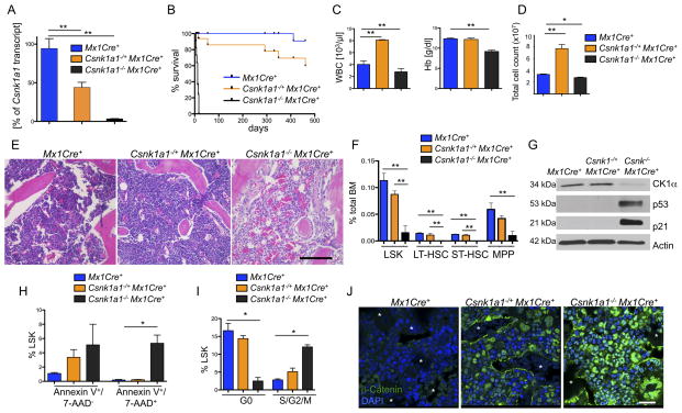
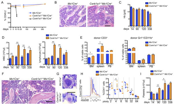
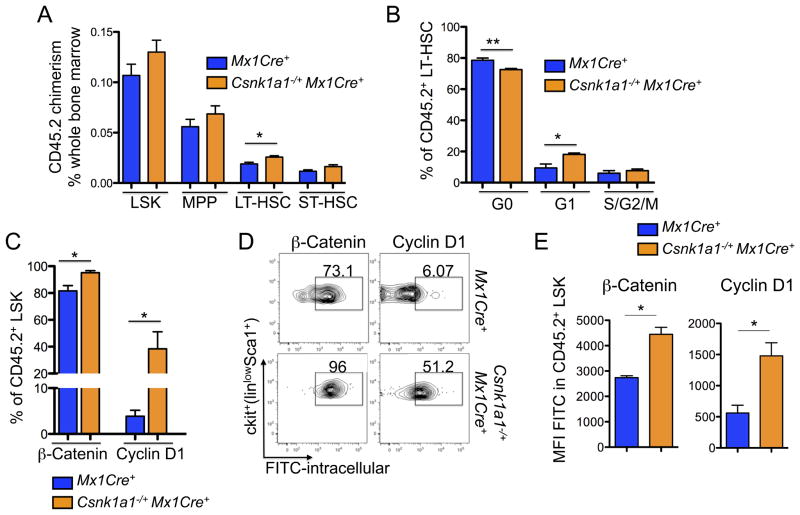
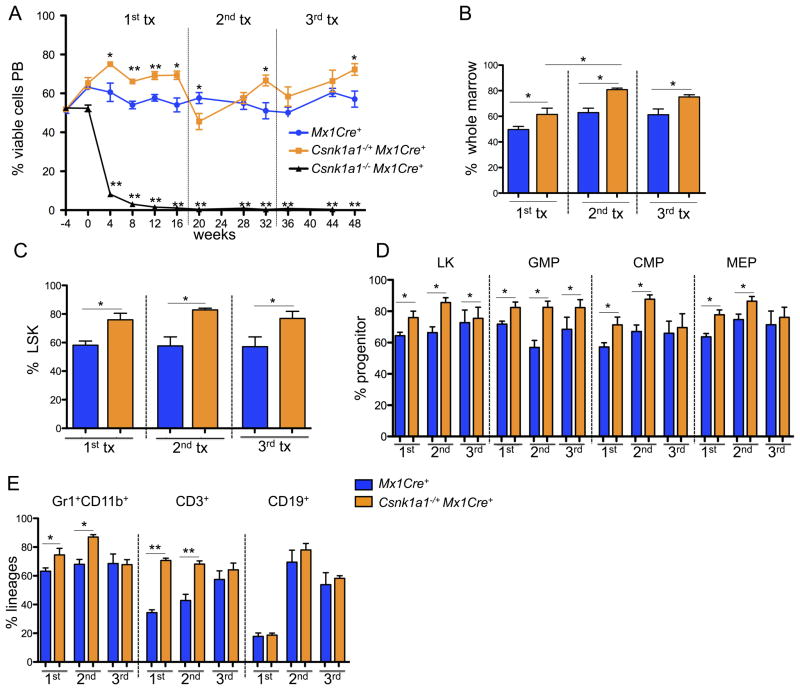
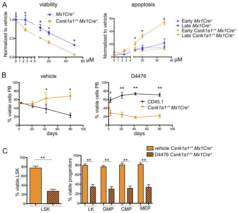
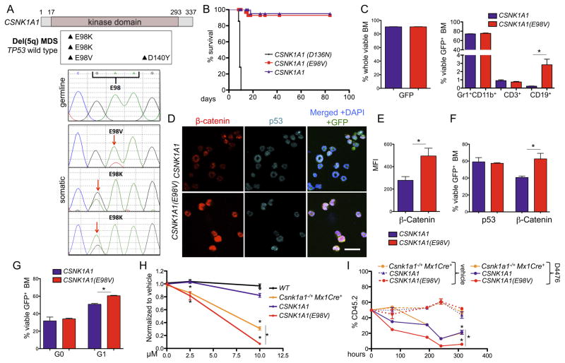
The casein kinase 1A1 gene (CSNK1A1) is a putative tumor suppressor gene located in the common deleted region for del(5q) myelodysplastic syndrome (MDS). We generated a murine model with conditional inactivation of Csnk1a1 and found that Csnk1a1 haploinsufficiency induces hematopoietic stem cell expansion and a competitive repopulation advantage, whereas homozygous deletion induces hematopoietic stem cell failure. Based on this finding, we found that heterozygous inactivation of Csnk1a1 sensitizes cells to a CSNK1 inhibitor relative to cells with two intact alleles. In addition, we identified recurrent somatic mutations in CSNK1A1 on the nondeleted allele of patients with del(5q) MDS. These studies demonstrate that CSNK1A1 plays a central role in the biology of del(5q) MDS and is a promising therapeutic target.


Figures

Figure 1
Figure 1. Conditional homozygous inactivation of Csnk1a1 results in hematopoietic stem and progenitor cell ablation
(A) Deletion of Csnk1a1 in whole bone marrow cells was determined 7 days after poly(I:C) induction by quantification of Csnk1a1 transcript levels by qt-RT-PCR. Data is presented as remaining Csnk1a1 transcript expression in percent relative to Mx1Cre+-control mice (mean±SD, n=3). (B) Kaplan-Meier survival curve of Csnk1a1−/+Mx1Cre+ (n=10), Csnk1a1−/ −Mx1Cre+ (n=10) and Mx1Cre+ (n=10) control mice. Time point 0 is the day of the first of 3 poly(I:C) inductions. (C) Absolute numbers of white blood cells (WBC) and hemoglobin (Hb) levels in peripheral blood from Csnk1a1−/+Mx1Cre+, Csnk1a1−/−Mx1Cre+ and Mx1Cre+ controls 10 days after poly(I:C) induction (mean±SD, n=3, *p<0.05, **p<0.001). (D) Numbers of whole bone marrow cells collected from tibias, femurs and pelvis of Csnk1a1−/+MxCre+, Csnk1a1/ MxCre+ 10 days after poly(I:C) induction (mean ±SD; n=3, *p<0.001). (E) Histological analysis of HE-stained spine from Csnk1a1−/+Mx1Cre+, Csnk1a1−/ −Mx1Cre+ and Mx1Cre+ controls 10 days after poly(I:C). Scale bar: 200 μm. (F) Analysis of the HSC compartment, defined as LinlowSca1+ckit+ (LSK), long-term (LT; linlowSca1+ckit+CD48CD150+), short-term (ST; linlowSca1+ckit+CD48CD150) HSC and multipotent progenitor cells (MPP, linlowSca1+ckit+CD48+CD150) in the bone marrow from Csnk1a1−/+Mx1Cre+, Csnk1a1−/ −Mx1Cre+ and Mx1Cre+ controls 10 days after poly(I:C), (mean±SD, n=5, *p<0.05, **p<0.001). (G) Western blot of whole bone marrow lysate 8 days after induction of poly(I:C). (H) Apoptosis was assessed in the LSK fraction from BM by Annexin V and 7-AAD staining (early apoptosis: Annexin V+/7-AAD; late apoptosis: Annexin V+/7-AAD+, (mean±SD, n=3, *p<0.05, **p<0.001). (I) Cell cycle was analyzed by combined proliferation (Ki67) and cell cycle (Hoechst 33342) staining in permeabilized LSK from bone marrow (G0: Ki67/Hoechst; S/G2/M: Ki67+/Hoechst+), (mean±SD, n=5, *p<0.05). (J) Immunofluorescence staining of paraffin-embedded bone marrow with an antibody against β-catenin (DAPI counterstaining). Asterisks highlight erythrocyte filled sinusoids. Scale bar: 20 μm. See also Figure S1.
Figure 2
Figure 2. Homozygous Csnk1a1 inactivation causes cell intrinsic hematopoietic stem cell ablation, while Csnk1a1 heterozygous inactivation causes cell-intrinsic lineage expansion
(A) Donor chimerism (CD45.2) of Csnk1a1−/+Mx1Cre+, Csnk1a1−/−Mx1Cre+ and Mx1Cre+ derived hematopoietic cells was monitored over time (mean±SD, n=7, **p<0.001). (B) Histomorphological analysis of transplanted Csnk1a1−/+Mx1Cre+ and Mx1Cre+ cells 8 weeks after poly(I:C) induction. Scale bar: 200 μm. (C) Hemoglobin (Hb) levels were followed over time (mean±SD, n=7, non-significant). (D) White blood cell and lymphocyte count were monitored over time (mean±SD, n=7, *p<0.05, **p<0.001). (E) Distribution of donor derived (CD45.2+) myeloid (Gr1+/CD11b+) and T-cells (CD3+) was analyzed by flow cytometry in bone marrow, spleen and peripheral blood (mean±SD, n=5, *p<0.05). (F) Histomorphological analysis of megakaryocyte dysplasia in transplanted Mx1Cre+ and Csnk1a1−/+Mx1Cre+ 8 weeks after poly(I:C) induction. Scale bar: 100 μm. (G) Detailed megakaryocyte morphology on cytospin preparations (May-Gruenwald-Giemsa staining, Oil immersion, Scale bar: 20 μm). (H) Representative ploidy analysis and quantification on CD45.2+, CD41+ megakaryocytes using Hoechst33342 staining on fixed and permeabilized cells. (mean±SD, n=4, *p<0.05). (I) Platelet counts were taken over time (mean±SD, n=7, *p<0.05). See also Figure S2.
Figure 3
Figure 3. Haploinsufficiency of Csnk1a1 leads to cell-intrinsic expansion of transplanted hematopoietic stem cells
(A) HSC chimerism (CD45.2) was analyzed in CD45.1 mice repopulated with Csnk1a1−/+Mx1Cre+ and Mx1Cre+ cells 8 weeks after induction with poly(I:C) in the LSK, MPP, LT-HSC, and ST-HSC (mean±SD, n=5, *p<0.05). (B) Cell cycle was analyzed by combined proliferation (Ki67) and cell cycle (Hoechst 33342) staining in permeabilized LSK and LT-HSC from bone marrow (G0: Ki67/Hoechst; S/G2/M: Ki67+/Hoechst+), (mean±SD, n=5, *p<0.05). (C) Intracellular flow cytometry for β-catenin and cyclin D1 (FITC-labeled secondary antibody each) on the CD45.2+ viable LSK population (mean±SD, n=3, *p<0.05). (D) Corresponding representative flow blots to the quantitative analysis of intracellular β-catenin and cyclin D1, accumulation. (E) Mean fluorescence intensity (MFI) of intracellular β-catenin and cyclin D1 in LSK (mean±SD, n=3, *p<0.05). See also Figure S3.
Figure 4
Figure 4. Csnk1a1 haploinsufficient hematopoietic stem cells show increased repopulating ability consistent with increased self-renewal
(A) Competitive repopulation assays were performed by mixing CD45.2-expressing cells (Csnk1a1−/+Mx1Cre+, Csnk1a1−/ −Mx1Cre+, or Mx1Cre+) with CD45.1 competitor cells at an approximately 50:50 ratio, and transplanting the cells into lethally irradiated CD45.1 recipients. The percentage of CD45.2 donor cell chimerism in the whole peripheral blood from peripheral blood of lethally irradiated recipient animals is shown. Time (weeks) denotes the time relative to termination of the poly(I:C) injections (poly(I:C)=timepoint 0). After 16 weeks, bone marrow was harvested and transplanted for secondary transplants, and 16 weeks later for tertiary transplants in lethally irradiated mice (mean±SD, n=5, *p<0.05; **p<0.001). (B) Donor chimerism of total bone marrow cells performed at 16 (first competitive transplant), 32 (secondary competitive transplant, 16 weeks after transplantation) or 48 (tertiary competitive transplant) weeks after poly(I:C) induction (mean±SD, n=5, *p<0.05). (C, D) Donor chimerism of the hematopoietic stem (LSK) (C) and progenitor cell compartments: LK, linlowSca1ckit+; common-myeloid progenitors (CMP), LK CD34+CD16/32; granulocyte-macrophage progenitors (GMP), LK CD34+CD16/32+; myeloerythroid progenitors (MEP), LK CD34CD16/32 (D) performed at 16 (first competitive transplant), 32 (secondary competitive transplant), or 48 (tertiary competitive transplant) weeks after poly(I:C) induction (mean±SD, n=5, *p<0.05). (E) Chimerism of hematopoietic lineages in the bone marrow each 16 weeks after the first, second and third competitive transplant. Composite data of donor (CD45.2+) granulocytes (Gr1+CD11b+), B-cells (CD19+) or T-cells (CD3+) are shown (mean±SD, n=5, *p<0.05).
Figure 5
Figure 5. Csnk1a1 haploinsufficiency provides a therapeutical window for the specific treatment of disease-propagating hematopoietic stem cells
(A) Sorted hematopoietic progenitor cells (LK) were pre-stimulated for 24 hours after the sort and treated for 72 hours with varying concentrations of D4476. Viability of cells was analyzed after 72 hours with the CellTitre glo assay, apoptosis by combined Annexin V and 7AAD staining discriminating early (Annexin V+7AAD) and late apoptosis (AnnexinV+7AAD+) using flow cytometry. (mean±SD, n=5, *p<0.05). (B) 21 days after poly(I:C) treatment, Csnk1a1−/+Mx1Cre+ or CD45.1 bone marrow was harvested and LSK cells were sort-purified. Equal ratios of Csnk1a1−/+Mx1Cre+ and CD45.1+ LSK were mixed and treated for 72 hours ex vivo with either D4476 or DMSO, followed by transplantation into lethally irradiated CD45.1 mice. The chimerism was followed over time in the peripheral blood (mean±SD, n=6, *p<0.05; **p<0.001). (C) The chimerism of Csnk1a1−/+Mx1Cre+ in the bone marrow was analyzed in the LSK and progenitor fractions after 12 weeks under DMSO or D4476 treatment conditions (mean±SD, n=6, **p<0.001). See also Figure S4.
Figure 6
Figure 6. Identification and functional characterization of CSNK1A1 mutations in del(5q) MDS patients
(A) Summary of CSNK1A1 mutations identified in del(5q)MDS patients (upper panel) and Sanger sequencing results around codon 98 of the normal control (germline) and tumors (somatic) (lower panel). (B) Kaplan-Meier survival analysis of chimeric mice transplanted with Csnk1a1−/−Mx1Cre+ hematopoietic stem and progenitor cells expressing Csnk1a1 cDNA, Csnk1a1 D136 cDNA or Csnk1a1 E98V cDNA. Timepoint 0 is the first day of poly(I:C) induction (n=5). (C) GFP expression in whole BM (left) and distribution of the different lineages (Gr1+CD11b+ neutrophils, CD3+ T-cells, CD19+ B-cells) in GFP+ BM cells (right). (mean±SD, n=3, *p<0.05). (D) Co-immunofluorescent staining of β-catenin and p53 in cytospin preparations of red blood cell lysed whole bone marrow cells (red: β-catenin, turquoise: p53, green: GFP MIG vector, blue: DAPI). Scale bar 20 μm. (E) Quantification of β-catenin intensity using the mean fluorescence intensity (MFI), (mean±SD, n=3, *p<0.05). (F) Intracellular flow cytometry measurement of β-catenin and p53 in permeabilized whole bone marrow cells. (mean±SD, n=3, *p<0.05). (G) Cell cycle was analyzed by combined Ki67 and Hoechst33342 staining in permeabilized whole bone marrow cells. (mean±SD, n=3, *p<0.05). (H) GFP+ LSK from mice transplanted with either CSNK1A1 or CSNK1A1 E98V-expressing Csnk1a1−/−Mx1Cre+ cells as well as Csnk1a1 haploinsufficient LSK and WT LSK were sort purified and exposed to vehicle, 2.5 or 10 μM D4476 for 72 hours and viability of cells was analyzed after 72 hours with the CellTitre glo assay (mean±SD, n=5, *p<0.05). (I) LSK (all CD45.2) isolated as in (H) were treated in competition to CD45.1 wild type cells in one culture well to analyze selective ablation of cells under the same culture condition. (mean±SD, n=5, *p<0.05). See also Figure S5 and Table S1.

References

    1. Albuquerque C, Breukel C, van der Luijt R, Fidalgo P, Lage P, Slors FJ, Leitao CN, Fodde R, Smits R. The ‘just-right’ signaling model: APC somatic mutations are selected based on a specific level of activation of the beta-catenin signaling cascade. Human molecular genetics. 2002;11:1549–1560. - PubMed
    1. Baba Y, Garrett KP, Kincade PW. Constitutively active beta-catenin confers multilineage differentiation potential on lymphoid and myeloid progenitors. Immunity. 2005;23:599–609. - PMC - PubMed
    1. Bidere N, Ngo VN, Lee J, Collins C, Zheng L, Wan F, Davis RE, Lenz G, Anderson DE, Arnoult D, et al. Casein kinase 1alpha governs antigen-receptor-induced NF-kappaB activation and human lymphoma cell survival. Nature. 2009;458:92–96. - PMC - PubMed
    1. Boultwood J, Pellagatti A, Cattan H, Lawrie CH, Giagounidis A, Malcovati L, Della Porta MG, Jadersten M, Killick S, Fidler C, et al. Gene expression profiling of CD34+ cells in patients with the 5q-syndrome. British journal of haematology. 2007;139:578–589. - PubMed
    1. Boultwood J, Pellagatti A, McKenzie AN, Wainscoat JS. Advances in the 5q-syndrome. Blood. 2010;116:5803–5811. - PubMed

Publication types