. 2014 Sep 25;8(6):1857-1869.
doi: 10.1016/j.celrep.2014.08.037. Epub 2014 Sep 18.

Serotonergic versus nonserotonergic dorsal raphe projection neurons: differential participation in reward circuitry

Affiliations

Serotonergic versus nonserotonergic dorsal raphe projection neurons: differential participation in reward circuitry

Ross A McDevitt et al. Cell Rep. .

Abstract

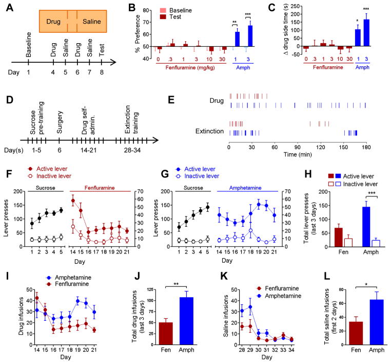
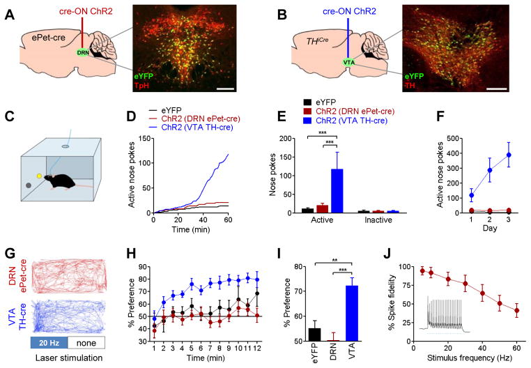
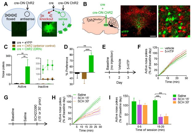
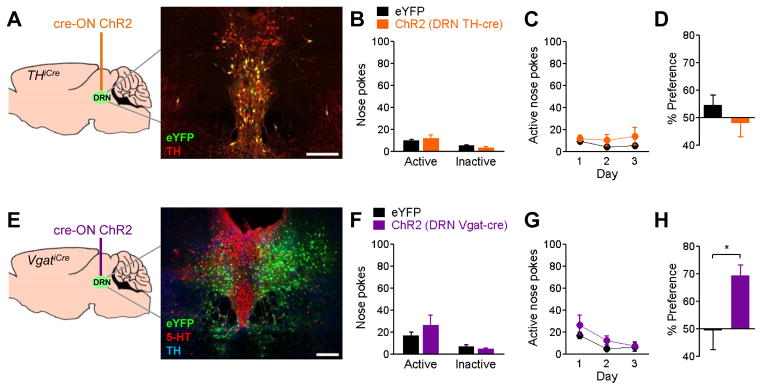
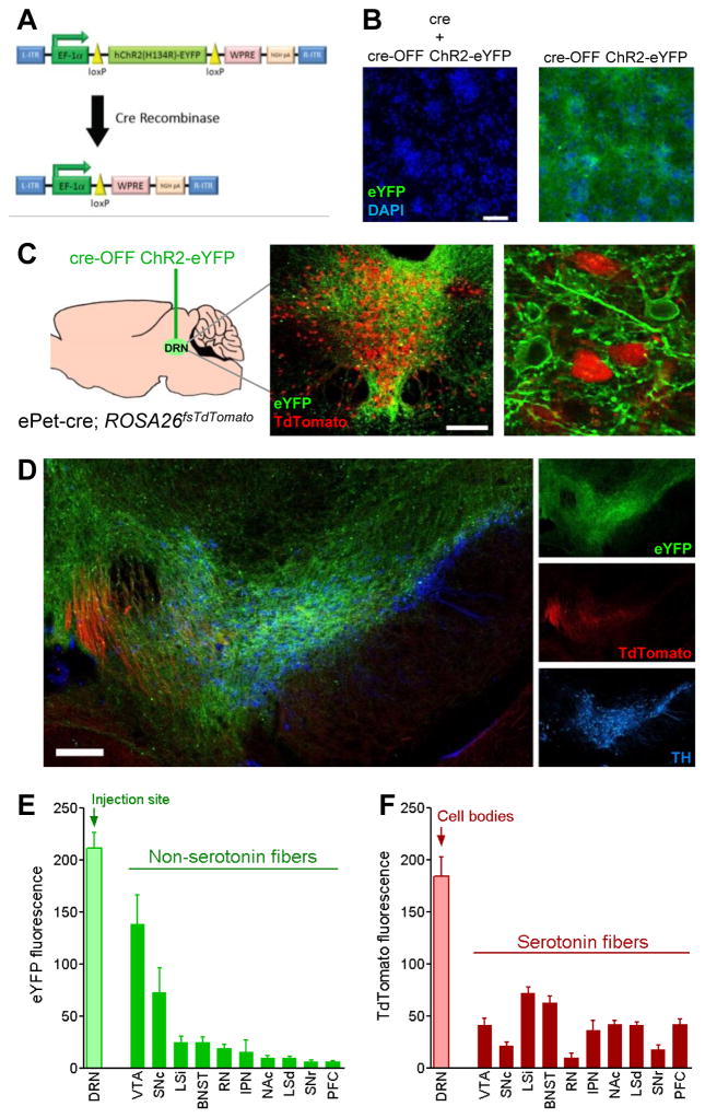
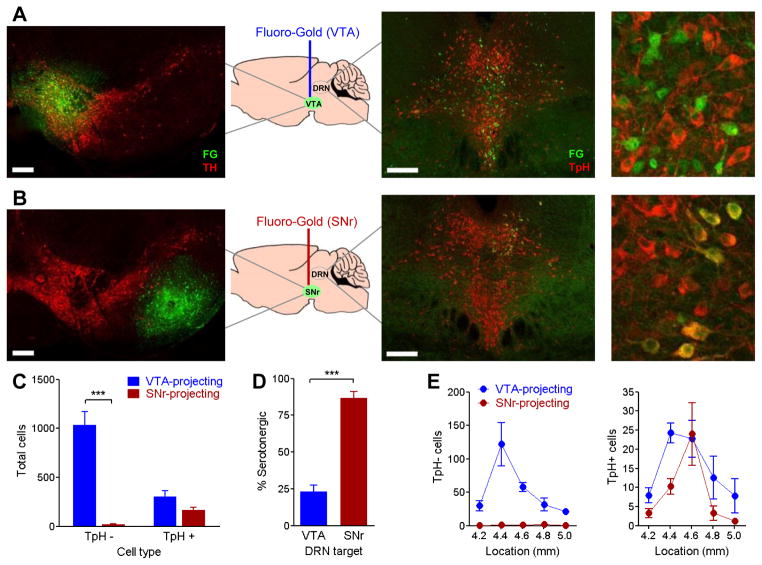
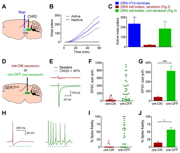
The dorsal raphe nucleus (DRN) contains the largest group of serotonin-producing neurons in the brain and projects to regions controlling reward. Although pharmacological studies suggest that serotonin inhibits reward seeking, electrical stimulation of the DRN strongly reinforces instrumental behavior. Here, we provide a targeted assessment of the behavioral, anatomical, and electrophysiological contributions of serotonergic and nonserotonergic DRN neurons to reward processes. To explore DRN heterogeneity, we used a simultaneous two-vector knockout/optogenetic stimulation strategy, as well as cre-induced and cre-silenced vectors in several cre-expressing transgenic mouse lines. We found that the DRN is capable of reinforcing behavior primarily via nonserotonergic neurons, for which the main projection target is the ventral tegmental area (VTA). Furthermore, these nonserotonergic projections provide glutamatergic excitation of VTA dopamine neurons and account for a large majority of the DRN-VTA pathway. These findings help to resolve apparent discrepancies between the roles of serotonin versus the DRN in behavioral reinforcement.


Conflict of interest statement

The authors declare no financial conflicts of interest.

Figures

Figure 1
Figure 1. Pharmacological stimulation of dopamine but not serotonin release reinforces behavior
A, Mice (n=9–18/group) were conditioned with injections of the serotonin-releasing agent fenfluramine (0–30 mg/kg, i.p.) or the dopamine-releasing agent amphetamine (1–3 mg/kg, i.p.). B, Percent of time spent in drug-paired chamber on baseline and test days. Repeated measures ANOVA (drug x day) interaction F(7,96)=4.166, p<0.001; ** p<0.01, *** p<0.001 post-hoc. C, Change in time spent on drug-paired chamber between test and baseline days. One-way ANOVA F(7,96)=5.318, p<0.0001; * p<0.05, *** p<0.001 Dunnett’s post-hoc vs. saline. D, Self-administration experiment in a separate cohort of mice. After pre-training for sucrose, mice were implanted with intravenous catheters and allowed to self-administer fenfluramine (0.03 mg/kg/infusion) or amphetamine (0.05 mg/kg/infusion), n=8/group. E, Sample data from individual self-administration sessions demonstrating timing of infusions for mice with access to fenfluramine (red) and amphetamine (blue), and during the first day of extinction training. F,G, daily lever-press counts during sucrose pre-training (left Y axis) and drug self-administration (right Y axis). These experiment phases are plotted on different scales because the number of maximally-allowed rewards differed. H, total number of lever presses during last three days of drug access. Two-way ANOVA (drug x lever) interaction F(1,28)=8.095, p<0.01; *** p<0.001 post-hoc. I, Daily drug infusions during drug self-administration phase. J, total number of drug infusions during last three days drug access, ** p<0.01. K, Daily counts of infusions of saline during extinction training. L, total number of saline infusions during the first two days of extinction, * p<0.05. Group data are presented here and in subsequent figures as mean ± SEM.
Figure 2
Figure 2. Optogenetic stimulation of VTA dopamine but not DRN serotonin cell bodies reinforces behavior
Selective targeting of gene expression was achieved by injecting cre-induced (“cre-ON”) vectors expressing ChR2-eYFP or eYFP alone into the (A) DRN of ePet-cre mice or (B) VTA of THiCre mice. Insets depict expression of eYFP (green), double-labeled in red with tryptophan hydroxylase (TpH) for DRN tissue or tyrosine hydroxylase (TH) for VTA tissue. Scale bars = 200 μm. C, Mice were trained to nose-poke into an active port to receive 3-second trains of 20 Hz laser stimulation; nose-pokes into an inactive port were not reinforced. D,E, Representative cumulative-activity graph and group mean nose-pokes made in first behavioral session for VTA-dopamine (n=11), DRN-serotonin (n=18), and a combined control group expressing eYFP in DRN or VTA (n=17). Two-way ANOVA (group x port) interaction F(2,86)=8.317, p<0.001; *** p<0.001 post-hoc. F, Active nose poke responding on three consecutive days of testing. G, Mice underwent a real-time place preference task in which presence in one half of a chamber triggered continuous 20 Hz laser stimulation. Example tracks for a DRN serotonin stimulated mouse (top; red) and a VTA dopamine stimulated mouse (bottom; blue). H, Minute-by-minute percent of time spent in the laser-paired half of the chamber. I, Overall preference for laser-paired side during 12-minute session. One-way ANOVA F(2,38)=12.05, p<0.0001; ** p<0.01, *** p<0.001 post-hoc. J, Percent of laser pulses in a 20 pulse train resulting in action potentials in ChR2+ DRN serotonin cell bodies, recorded ex vivo in whole-cell current clamp. Inset, sample trace with 20Hz stimulation. See also Figures S1 and S2.
Figure 3
Figure 3. Optogenetic stimulation of DRN cell bodies reinforces behavior in a dopamine-dependent, serotonin-independent manner
A, Left panel, schematic view of a cell transduced with cre-induced (“cre-ON”) viral vector. In the absence of cre recombinase, viral plasmid DNA remains in antisense orientation and does not express functional protein. Inset, lack of eYFP signal in mouse injected with cre-ON ChR2-eYFP. Right panel, co-injection of cre-ON and cre-expressing viral vectors results in knockout of floxed genomic DNA and rearrangement of viral plasmid DNA into sense orientation, resulting in expression of ChR2-eYFP. Inset, robust eYFP expression in mouse co-injected with cre-expressing and cre-ON viral vectors. B, Tph2lox/lox mice were co-injected with viral vectors expressing cre and cre-ON ChR2-eYFP or eYFP into DRN. Insets, whole DRN (scale bar = 200 μm) and detail of non-overlapping expression of eYFP (green) and tryptophan hydroxylase (TpH, red). Thus, cells with ChR2 lack the enzyme necessary for serotonin synthesis. An additional anatomical control group was co-injected with cre and cre-ON ChR2 0.7mm anterior to the DRN. C, Nose pokes during first day of self-stimulation testing for non-serotonergic DRN stimulation (n=10), anterior controls (n=8), and eYFP controls (n=7). Two-way ANOVA (group x nose port) interaction F(2,44)=4.482, p<0.05; ** p<0.01 post-hoc. Inset, active nose pokes on 3 consecutive days of testing. D, Percent of time spent on laser side in a real-time place preference task. One-way ANOVA F(2,22)=11.24, p<0.001; ** p<0.01, *** p<0.001 post- hocs. E, Non-serotonin DRN stimulated mice (n=6) were tested for nose-poke optical self-stimulation 30 minutes after injection of vehicle or 5-hydroxytryptophan (5-HTP; 40 mg/kg i.p.), the intermediate in the serotonin synthesis pathway. 5-HTP is the product of the enzyme tryptophan hydroxylase, which is knocked out in ChR2-positive cells of these mice. F, Cumulative-activity graph of nose pokes in test sessions after injection of vehicle or 5-HTP. Individual data points were normalized to percent of nose pokes achieved during a 30-minute baseline session on day 1. G, Non-serotonin DRN stimulated mice (n=6) were tested after injection of the dopamine D1 receptor antagonist SCH23390 (SCH; 30 μg/kg, i.p.) at either 15 or 30 minutes before testing. H, Cumulative-activity graph of active nose pokes in 30-minute sessions following injection of saline or SCH. I, Active nose pokes during 5-minute bins at the beginning or in the middle of test depicted in panel H. Individual data points were normalized to percent of responses during baseline day. Repeated-measures ANOVA (drug x epoch) interaction F(2,15)=4.560, p<0.05; *** p<0.001 Dunnett’s post-hoc vs saline. See also Figures S3 and S4.
Figure 4
Figure 4. Optogenetic stimulation of dopaminergic or GABAergic DRN cell bodies fails to reinforce nose-poke self-stimulation
A, DRN dopamine neurons were targeted by injecting cre-induced (“cre-ON”) vectors expressing ChR2-eYFP (n=10) or eYFP (n=14) into the DRN of THiCre mice. Inset shows eYFP (green) double-labeled with tyrosine hydroxylase (TH, red). B, Nose pokes in the first day of testing. C, Active nose pokes on three consecutive days of testing. D, Percent of time spent on laser side in real-time place preference task. E, DRN GABA neurons were targeted by injecting cre-ON ChR2-eYFP (n=8) or eYFP (n=4) into the DRN of VgatiCre mice. Inset shows eYFP (green) cell bodies in the lateral DRN, which do not co-label for serotonin (5-HT, red) or tyrosine hydroxylase (TH, blue). Laser stimulation did not reinforce nose poke self-stimulation (F,G) but did induce a real-time place preference (H), p<0.05. Scale bars = 200 μm.
Figure 5
Figure 5. Unlike serotonergic neurons, non-serotonergic DRN neurons preferentially project to the VTA
A, Schematic of cre-silenced (“cre-OFF”) DNA construct containing loxP-flanked ChR2-eYFP coding region. B, Transduction of primary cultured rat neurons with cre-OFF ChR2-eYFP viral vector produces eYFP fluorescence (left) that is abolished in cells co-transduced with a vector expressing cre recombinase (right); DAPI nuclear staining (blue) is unaffected. Scale bar = 100 μm. C, Transgenic mice co-expressing cre and TdTomato in serotonergic neurons (ePet-cre; ROSA26fsTdTomato, n=4) were injected with cre-OFF ChR2-eYFP into the DRN. Inset depicts whole DRN tissue (scale bar = 200 μm) and detail demonstrating segregation of TdTomato and eYFP fluorescence into separate populations of cells. D, Serotonergic (red) and non-serotonergic (green) axons are visible in the VTA, identifiable by tyrosine hydroxylase immunoreactivity (TH, blue). Scale bar = 200 μm. E,F Quantitation of (E) eYFP and (F) TdTomato fluorescence intensity in brain regions with conspicuous eYFP expression. Abbreviations: ventral tegmental area (VTA), substantia nigra pars compacta (SNc), intermediate portion of the lateral septum (LSi), bed nucleus of the stria terminalis (BNST), red nucleus (RN), interpeduncular nucleus (IPN), nucleus accumbens (NAc), dorsal portion of the lateral septum (LSd), substantia nigra reticulata (SNr), prefrontal cortex (PFC).
Figure 6
Figure 6. The majority of DRN cell bodies that project to VTA are non-serotonergic
The retrograde tracer Fluoro-Gold was iontophoretically infused into the (A) VTA or (B) substantia nigra reticulata (n=4/group). Left panels, Fluoro-Gold (green) at infusion site, double-labeled with tyrosine hydroxylase (TH) to label dopamine neurons (red). Right panels, retrograde-labeled cells in DRN, double-labeled with tryptophan hydroxylase (TpH) to label serotonin neurons (red). Scale bars = 200 μm. C, Number of Fluoro-Gold-labeled cells in DRN tissue from mice injected with Fluoro-Gold in VTA or substantia nigra reticulata. Fluoro-Gold cells were grouped by presence or absence of tryptophan hydroxylase double-label (TpH+, TpH−). Two-way ANOVA (region x TpH label interaction) F(1,12)=34.11, p<0.0001; *** p<0.001 post-hoc. D, Percent of Fluoro-Gold labeled cells double-labeling for tryptophan hydroxylase. *** p<0.0001. E, Number of TpH− (left) and TpH+ (right) Fluoro-Gold labeled cells across the rostrocaudal axis of the DRN. X-axis indicates location of DRN tissue, in millimeters posterior to bregma.
Figure 7
Figure 7. DRN-VTA projections reinforce behavior and provide synaptic glutamatergic excitation of VTA dopamine neurons primarily via non-serotonergic projections
A, Mice (n=11) were injected with non-specific ChR2 viral vector in the DRN, and implanted with fiber optic cables in the VTA. B, Cumulative activity-graph of nose pokes into active and inactive ports on the first day of training. Total number of responses was greater into the active port (p<0.01). C, Total number of active nose pokes on day 1 from DRN-VTA mice. For comparison, data is reconstituted from previous experiments stimulating serotonergic and non-serotonergic DRN cell bodies. D, Serotonergic and non-serotonergic DRN projections were targeted by injecting cre-induced (“cre-ON”) (n=6) or cre-silenced (“cre-OFF”) (n=4) vectors expressing ChR2-eYFP in SERTcre mice. E, Representative voltage-clamp traces of VTA dopamine neurons showing optically-evoked glutamatergic excitatory post-synaptic current (EPSC) resulting from stimulation of terminals of the serotonergic (top trace) or non-serotonergic (bottom trace) DRN-VTA pathway. F, EPSC amplitudes in response to optical stimulation. Graph includes cells that did not respond to light (plotted as 0 pA). G, Average amplitude of light-responsive EPSCs, ***, p<0.0001. H, Representative current-clamp traces of a VTA dopamine neuron spiking in response to 20 Hz laser stimulation of DRN-VTA serotonin (left) or non-serotonin (right) pathways. I, Individual spike fidelity measurements; represented as percent of laser pulses during a 0.5 second, 20 Hz train that resulted in action potentials. J, Average spike fidelity in cells that responded to light with at least one action potential, p<0.05.

References

    1. Adamantidis AR, Zhang F, Aravanis AM, Deisseroth K, de Lecea L. Neural substrates of awakening probed with optogenetic control of hypocretin neurons. Nature. 2007;450:420–424. - PMC - PubMed
    1. Bang SJ, Commons KG. Forebrain GABAergic projections from the dorsal raphe nucleus identified by using GAD67-GFP knock-in mice. J Comp Neurol. 2012;520:4157–4167. - PMC - PubMed
    1. Boye SM, Rompre PP. Mesencephalic substrate of reward: axonal connections. J Neurosci. 1996;16:3511–3520. - PMC - PubMed
    1. Britt JP, Benaliouad F, McDevitt RA, Stuber GD, Wise RA, Bonci A. Synaptic and behavioral profile of multiple glutamatergic inputs to the nucleus accumbens. Neuron. 2012a;76:790–803. - PMC - PubMed
    1. Britt JP, McDevitt RA, Bonci A. Use of channelrhodopsin for activation of CNS neurons. In: Crawley Jacqueline N, et al., editors. Current protocols in neuroscience. Unit2. Chapter 2. 2012b. p. 16. - PMC - PubMed

Publication types

LinkOut - more resources