Review
. 2015 Aug 27:302:174-203.
doi: 10.1016/j.neuroscience.2014.09.027. Epub 2014 Sep 19.

The neuroimmunology of degeneration and regeneration in the peripheral nervous system

Affiliations
Review

The neuroimmunology of degeneration and regeneration in the peripheral nervous system

A DeFrancesco-Lisowitz et al. Neuroscience. .

Abstract

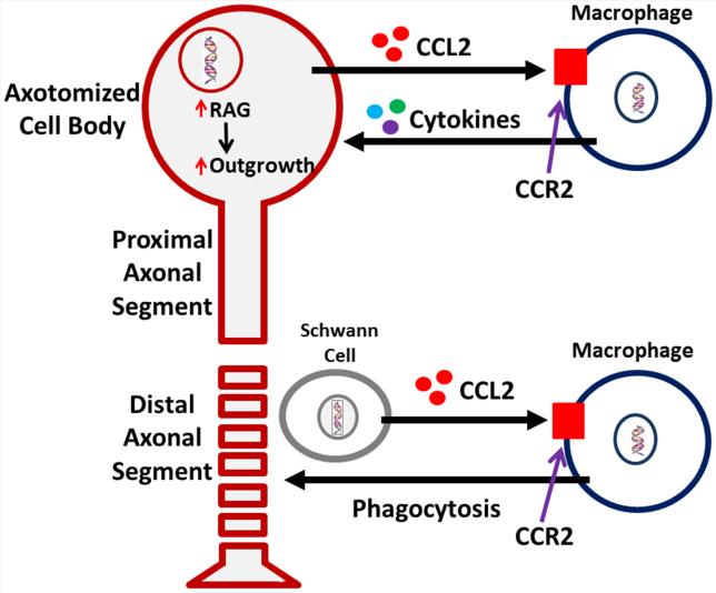
Peripheral nerves regenerate following injury due to the effective activation of the intrinsic growth capacity of the neurons and the formation of a permissive pathway for outgrowth due to Wallerian degeneration (WD). WD and subsequent regeneration are significantly influenced by various immune cells and the cytokines they secrete. Although macrophages have long been known to play a vital role in the degenerative process, recent work has pointed to their importance in influencing the regenerative capacity of peripheral neurons. In this review, we focus on the various immune cells, cytokines, and chemokines that make regeneration possible in the peripheral nervous system, with specific attention placed on the role macrophages play in this process.

Keywords: axotomy; chemokine; conditioning lesion; cytokine; dorsal root ganglion; macrophage.


Figures

Figure 1
Figure 1
A novel perspective on the response of macrophages to peripheral axotomy and their involvement in the regenerative response. Monocyte entry into a nerve, distal to the site of injury, is mediated through upregulation and release of the major monocyte chemokine, CCL2, by Schwann cells. Once the monocytes enter, beginning around 48 h post-injury, they phagocytose axonal and myelin debris resulting from the ongoing degenerative process. Removal of the debris, which is inhibitory to regenerating axons, is a known prerequisite for successful regeneration in vivo. Macrophages also enter into peripheral ganglia following nerve injury. Recent work has now suggested that macrophage presence around axotomized cell bodies helps mediate the conditioning lesion response. CCL2 is hypothesized to be the primary chemokine responsible for monocyte entry into the dorsal root ganglion. Under conditions in which macrophages do not enter the dorsal root ganglia, there is no enhanced growth after a conditioning lesion. Thus, there are two sites of action for macrophages in response to a peripheral nerve injury, in the distal nerve and around injured neuronal cell bodies.
Figure 2
Figure 2
Efficiency of myelin clearance in the distal sciatic nerve of CCR2 −/− mice is comparable to that in WT mice despite the total absence of axotomy-induced macrophage infiltration into the distal nerve in the knockout animals (see Niemi et al., 2013). Seven days after the sciatic nerve was unilaterally transected, changes in reactivity for myelin proteins were determined in nerves from WT, Wlds, and CCR2 −/− mice by staining with LFB. The distal nerve segments from WT and CCR2 −/− mice showed significantly less myelin staining compared with contralateral nerves, whereas axotomized nerves from Wlds mice retained >80% of myelin reactivity compared with contralateral nerves (a). The micrographs represent sections from the ipsilateral (e-g) and contralateral (b-d) nerves from WT, Wlds, and CCR2 −/− mice, respectively. Infiltrating macrophages have been considered necessary for the phagocytosis and clearance of myelin debris after injury; however the reduction in this cell population at the distal sciatic nerve does not hinder the clearance mechanism. *p<0.05, **p<0.001. Scale bar, 20 μm. (Niemi et al., 2013)
Figure 3
Figure 3
The cells and molecules involved in WD in peripheral nerve. Within minutes of axonal injury, Ca2+ enters the proximal and distal nerve and initiates axonal breakdown. The resulting DAMPs stimulate the dedifferentiation of Schwann cells through the upregulation of JUN and erbB2. Through TLR signaling, dedifferentiated Schwann cells upregulate proinflammatory molecules (blue inset). Increased TNF-α and IL-1β activity leads to upregulation of both MMP-9 and CCL2. Schwann cells also respond to IL-6 from fibroblasts, which through gp130 signaling, triggers an autocrine cascade of LIF and CCL2 upregulation. Fibroblasts respond within hours by upregulating IL-6 and GM-CSF, the latter of which leads to gal-3 expression by macrophages and Schwann cells (green inset). Neutrophils infiltrate the injury site briefly within the first day after injury. Endogenous antibodies respond to nerve injury by activating complement. Three days post injury, macrophages have begun to infiltrate the nerve. Activated macrophages seem to play a dual role in WD evident by the factors they secrete (purple inset). Initially, they contribute to the inflammatory state by their production of TNF-α and IL-1β. CR3 on macrophages binds complement on degenerating myelin inducing phagocytosis. There is controversy over the subtype of macrophage involved; M1 markers have been shown at 1 and 3 d after injury, yet M2 markers have been found as early as 3 d and remain elevated for at least 14 d. It is thought that M2 macrophages are involved in regulating the inflammatory response in WD by upregulating the anti-inflammatory cytokine IL-10 leading to downregulation of pro-inflammatory cytokines once myelin degradation is complete.
Figure 4
Figure 4
Macrophage accumulation around axotomized DRG neurons is significantly reduced in CCR2 −/− mice leading to no effect of a CL. The sciatic nerve was unilaterally transected in WT and CCR2 −/− mice. Seven days post-injury, the ipsilateral and contralateral L5 DRGs were removed for analysis of macrophage accumulation using an antibody to CD11b (a-e) or placed in explant cultures for measuring neurite outgrowth in response to a CL (f-i). In response to nerve injury, A 4-fold increase in staining is evident in the axotomized WT L5 DRG (a,c). Axotomized DRGs from CCR2 −/− mice show significantly diminished macrophage accumulation compared to WT (a). Representative micrographs show very little CD11b+ staining in contralateral WT (b) or CCR2 −/− (d) ganglia, or in ipsilateral CCR2 −/− ganglia (e). DRGs from CCR2 −/− mice also show a complete lack of the CL response at 24 and 48 h in explant culture (f,g). Representative micrographs of neurite outgrowth at 48 h in vitro for WT (h) and CCR2 −/− (i). *p<0.05, **p<0.001. Scale bar, 20 μm (IHC); 100 μm (explant). (Modified from Niemi et al., 2013)
Figure 5
Figure 5
The cell body response and the CL response. (a) The cell body response describes the morphological and molecular changes that occur in neurons after axotomy. Two types of signals reach the cell body from the site of injury. First, there is a very rapid Ca2+ wave, and, later, there are signals that have been retrogradely transported along microtubules via dynein and adaptor proteins. These different signals lead to changes in RAG expression in the axotomized cell body. In addition (though not shown), the accumulation of macrophages and the activation of satellite glial cells near the cell bodies leads to the release of extrinsic signals that also alter neuronal gene expression, for example, the secretion of gp130 cytokines by satellite glial cells in the SCG triggers an increase in galanin expression (Sun et al., 1994; Habecker et al., 2009; Zigmond, 2012b). Finally, in addition to the cell body response there is an alteration of protein translation in the axons themselves. (b) When a CL is made prior and also distal to a test lesion and neurite outgrowth is measured from the test lesion either in vivo or in vitro, growth is accelerated compared to when only a test lesion is made. As discussed in the text, macrophage accumulation in the region of axotomized cell bodies is required for a normal CL response.
Figure 6
Figure 6
The role of gp130 cytokines in the CL response in the mouse SCG. To remove the influence of all of the gp130 cytokines, a conditional knock out (CKO) of the receptor gp130 was produced in sympathetic neurons using a dopamine beta hydroxylase (DBH) Cre. Seven days following unilateral SCG axotomy in WT(C57) and gp130DBHcre CKO mice, ipsilateral (Ax) and contralateral sham-operated (Sh) SCGs were removed and maintained in explant culture. Neurite outgrowth from SCGs was imaged at 24 and 48 h, and the 20 longest neurites from each ganglion were measured. WT (C57) mice showed a significant CL response at both 24 (a) and 48 h (b) in culture. gp130 CKO mice showed no CL effect at either time point. *p<0.05. (Modified from Hyatt-Sachs et al., 2010)

References

    1. Aamar S, Saada A, Rotshenker S. Lesion-induced changes in the production of newly synthesized and secreted apo-E and other molecules are independent of the concomitant recruitment of blood-borne macrophages into injured peripheral nerves. J Neurochem. 1992;59:1287–1292. - PubMed
    1. Aigner L, Arber S, Kapfhammer JP, Laux T, Schneider C, Botteri F, Brenner H-R, Caroni P. Overexpression of the neural growth-associated protein GAP-43 induces nerve sprouting in the adult nervous system of transgenic mice. Cell. 1995;83:269–278. - PubMed
    1. Ambron RT, Zhang XP, Gunstream JD, Povelones M, Walters ET. Intrinsic injury signals enhance growth, survival, and excitability of Aplysia neurons. J Neurosci. 1996;16:7469–7477. - PMC - PubMed
    1. Anegon I, Moreau JF, Godard A, Jacques Y, Peyrat MA, Hallet MM, Wong G, Soulillou JP. Production of human interleukin for DA cells (HILDA)/leukemia inhibitory factor (LIF) by activated monocytes. Cell Immunol. 1990;130:50–65. - PubMed
    1. Anthony RM, Rutitzky LI, Urban JF, Jr., Stadecker MJ, Gause WC. Protective immune mechanisms in helminth infection. Nat Rev Immunol. 2007;7:975–987. - PMC - PubMed

Publication types

MeSH terms